%20abutment.jpg)
|
|
|
|
|
|
|
|
|
|
|
|
||
Immediate Implant Threads Covered by Bone Graft
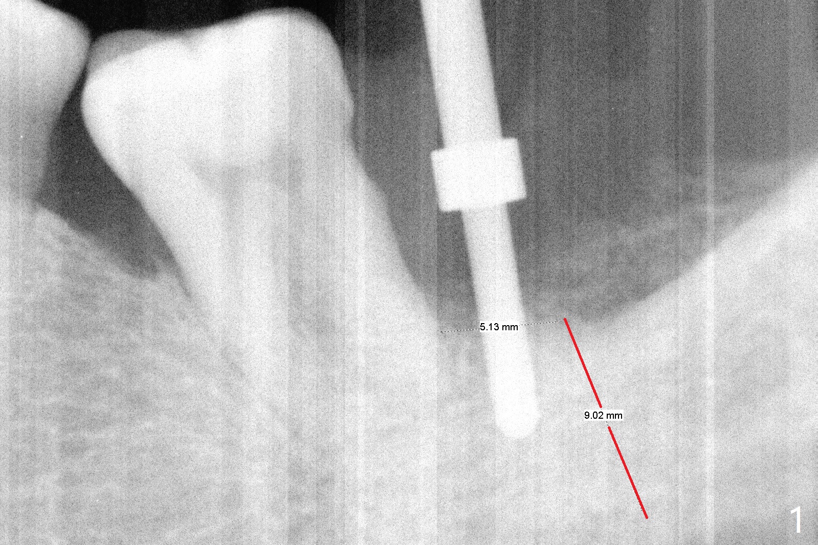
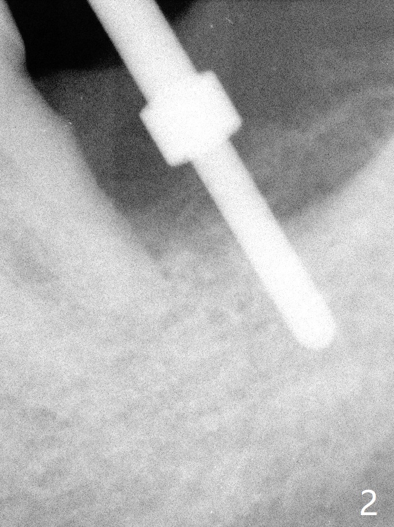
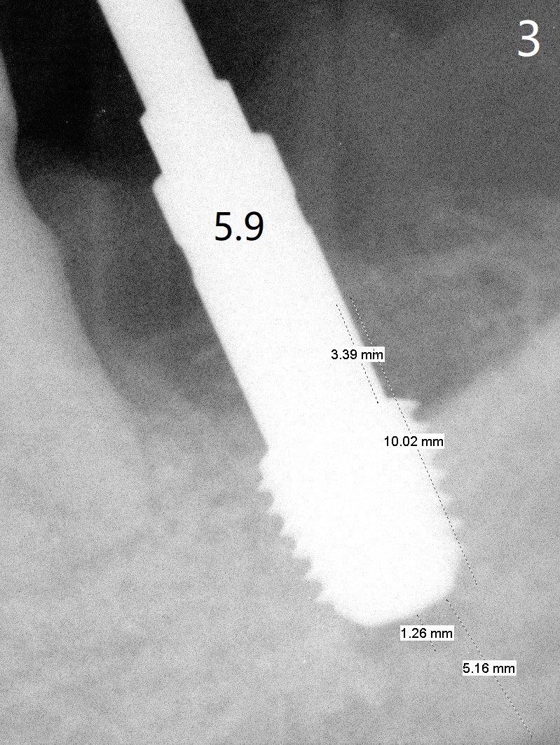
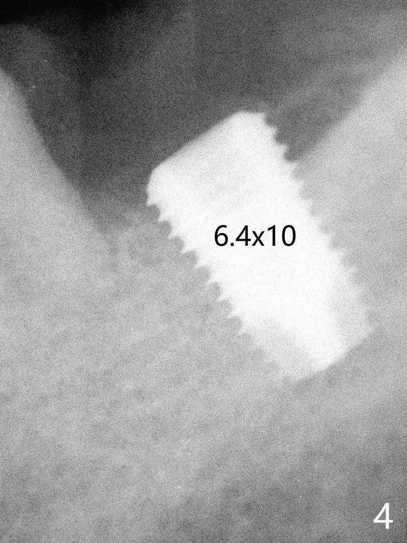
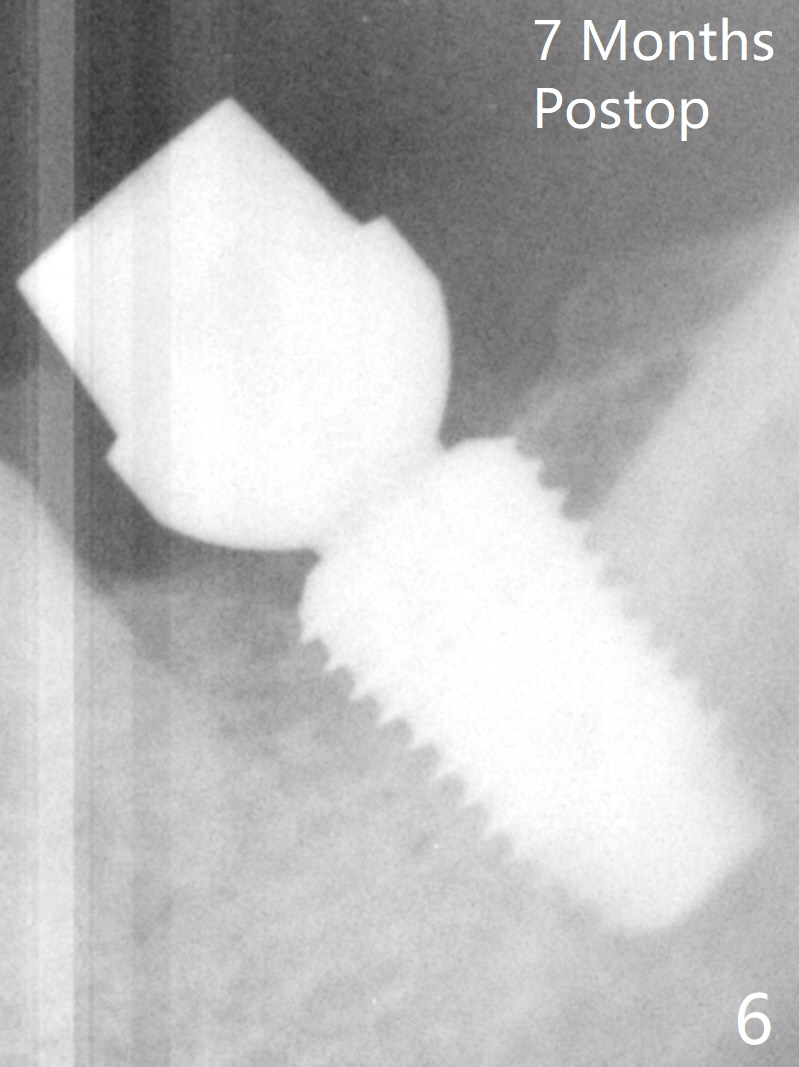
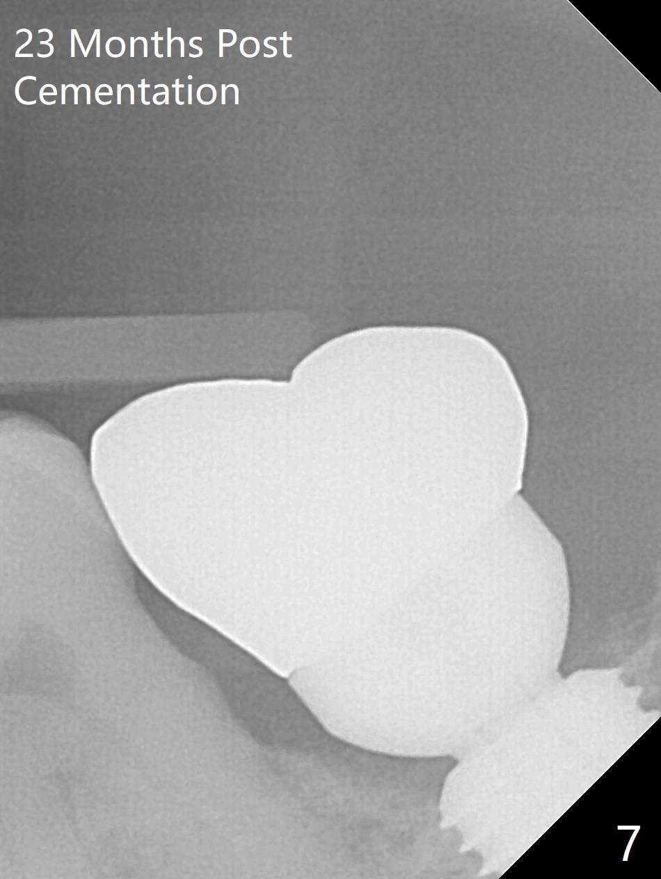
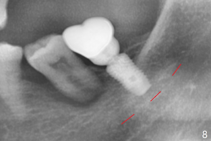
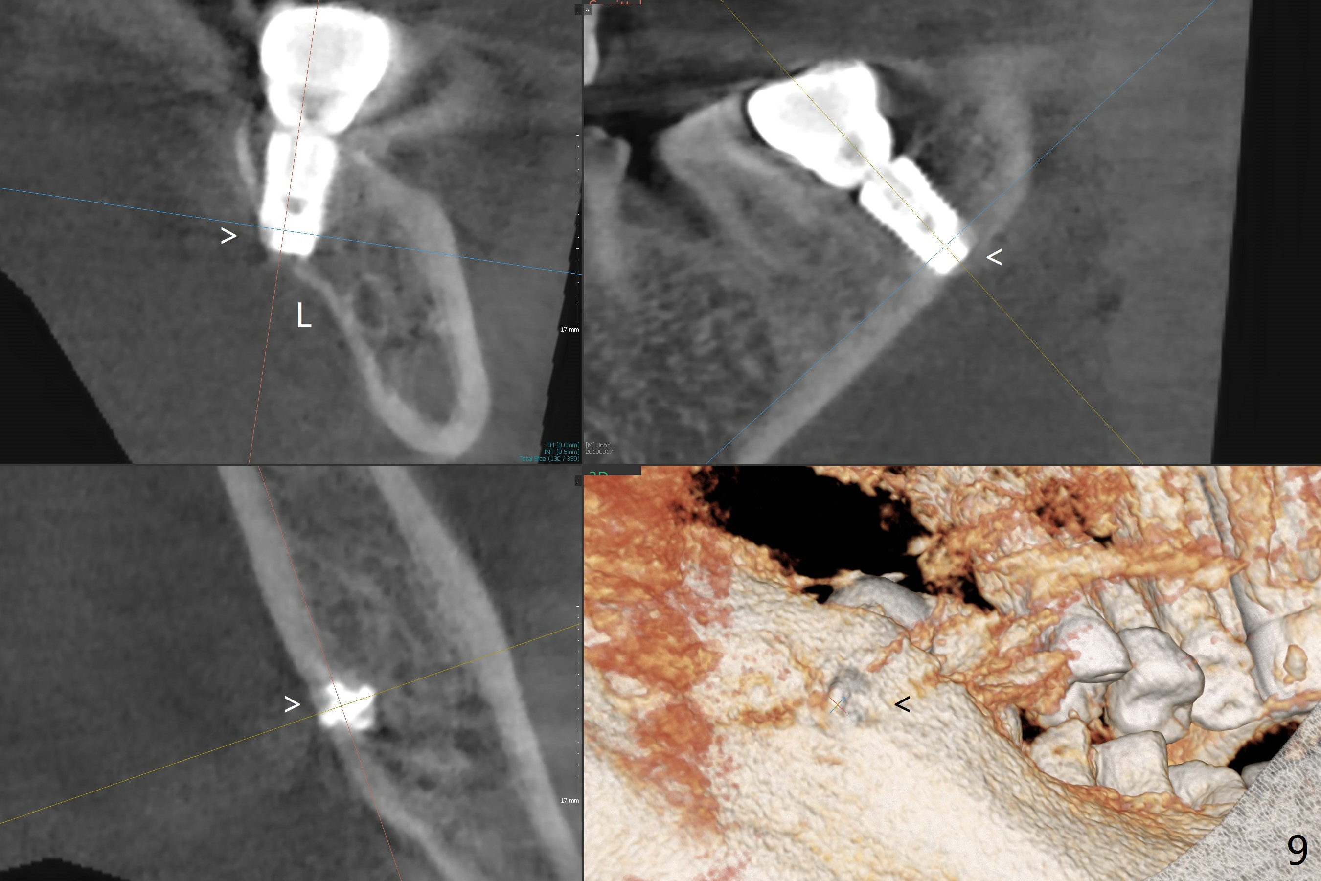
After extraction, curettage and Clindamycine gauzes in the sockets of the tooth #18 for 3 times, a 2 mm pilot drill makes initial osteotomy in the socket from 8 to 14 mm (Fig.1 (gingival level)). It appears that the osteotomy should move distal (Fig.1: red line, Fig.2 (17 mm)). After 5.9x10 mm drill, a 5.9 mm tap is placed (Fig.3). There is 4 mm clearance. Following 6.4x10 mm drill for 2 mm deeper, a 6.4x10 mm implant is placed with 60 Ncm. The implant plateau is level with the lingual crest, while the mesiobuccodistal bone is low. Autogenous bone mixed with Osteogen is placed in the defect area, followed by insertion of a 6.8x4(4) mm abutment (Fig.5). Collagen dressing is placed on the top of the graft. An immediate provisional is fabricated to close the remaining socket. Impression is taken 7 months postop (Fig.6). There seems to be no bone loss 23 months post cementation without opposing teeth (Fig.7) in spite of severe periodontitis at #19 (Fig.8 (25 months post cementation)). More surprising is the asymptomatic lingual (L) plate perforation, revealed by CT (Fig.9 arrowheads).
Return to Lower Molar Immediate Implant, 3,12,14,15, 30 Xin Wei, DDS, PhD, MS 1st edition 03/12/2015, last revision 03/24/2018