|
|
|
|
||
|
|
|
|
||
|
|
|
|
||
|
|
|
|
|
|
Socket Preservation
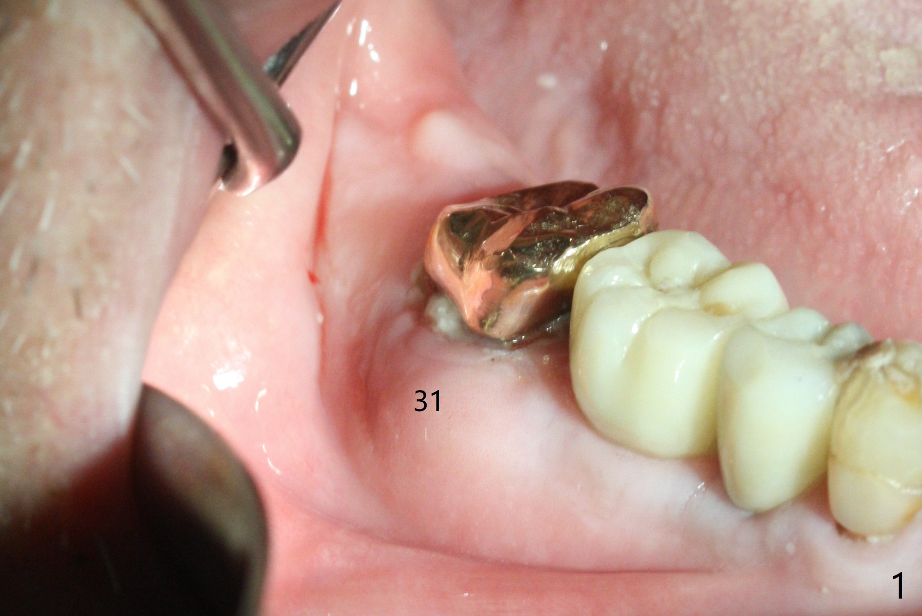
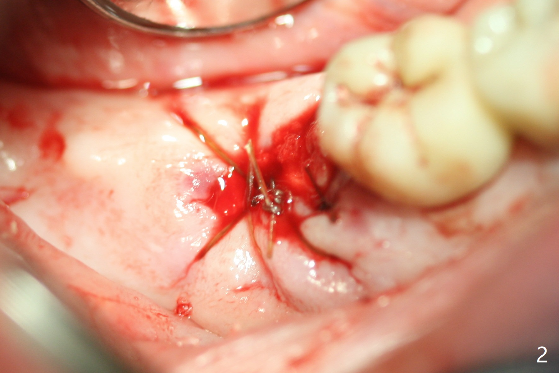
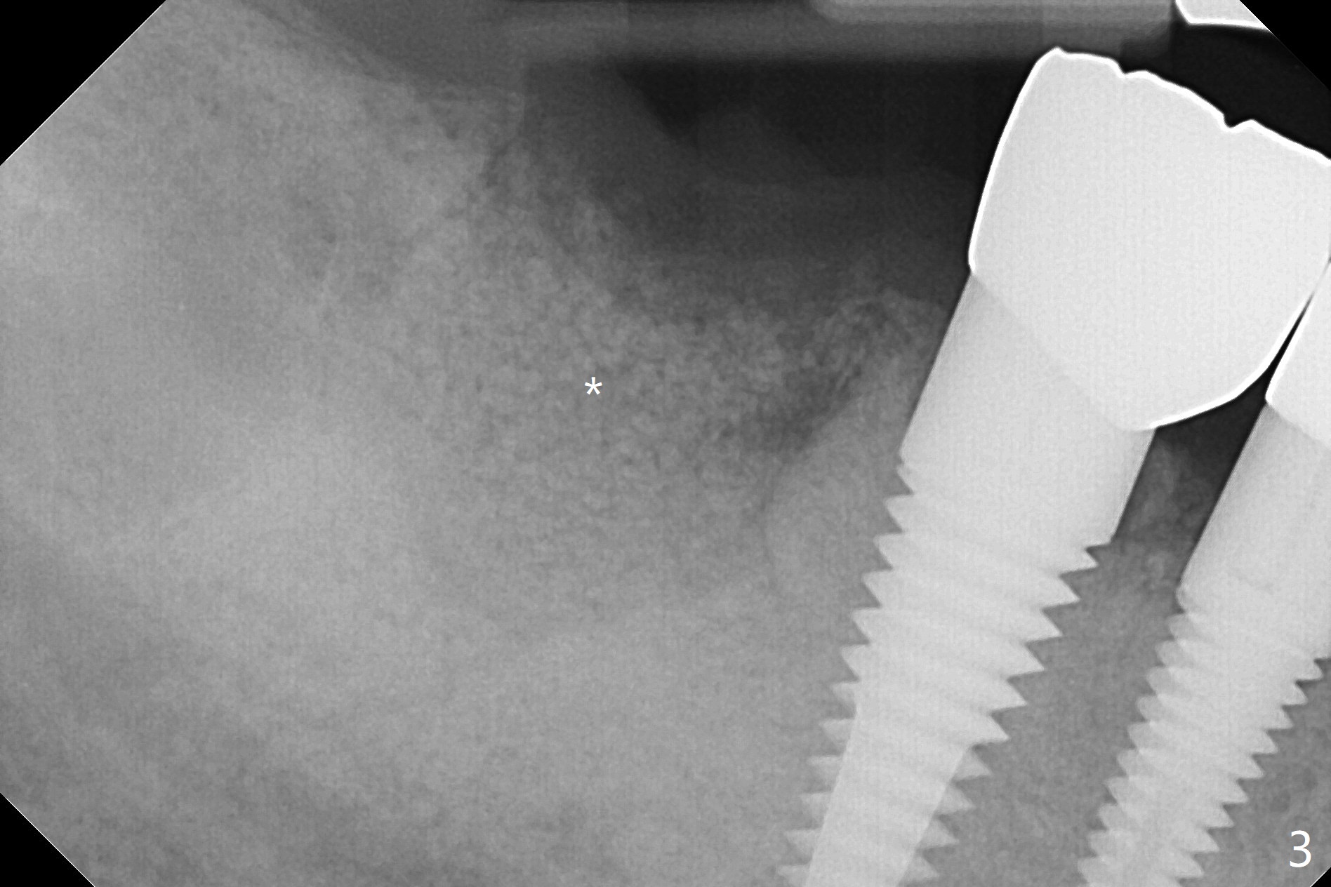
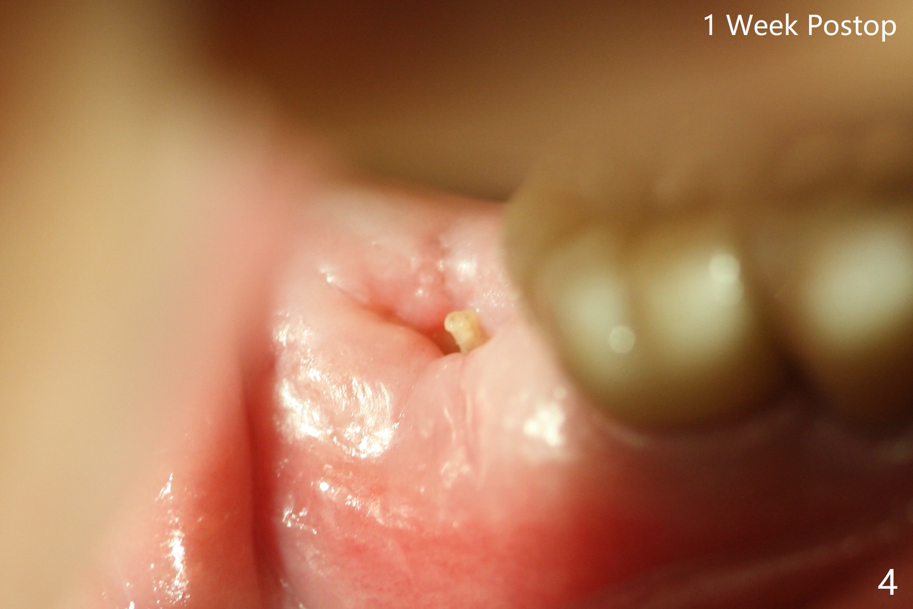
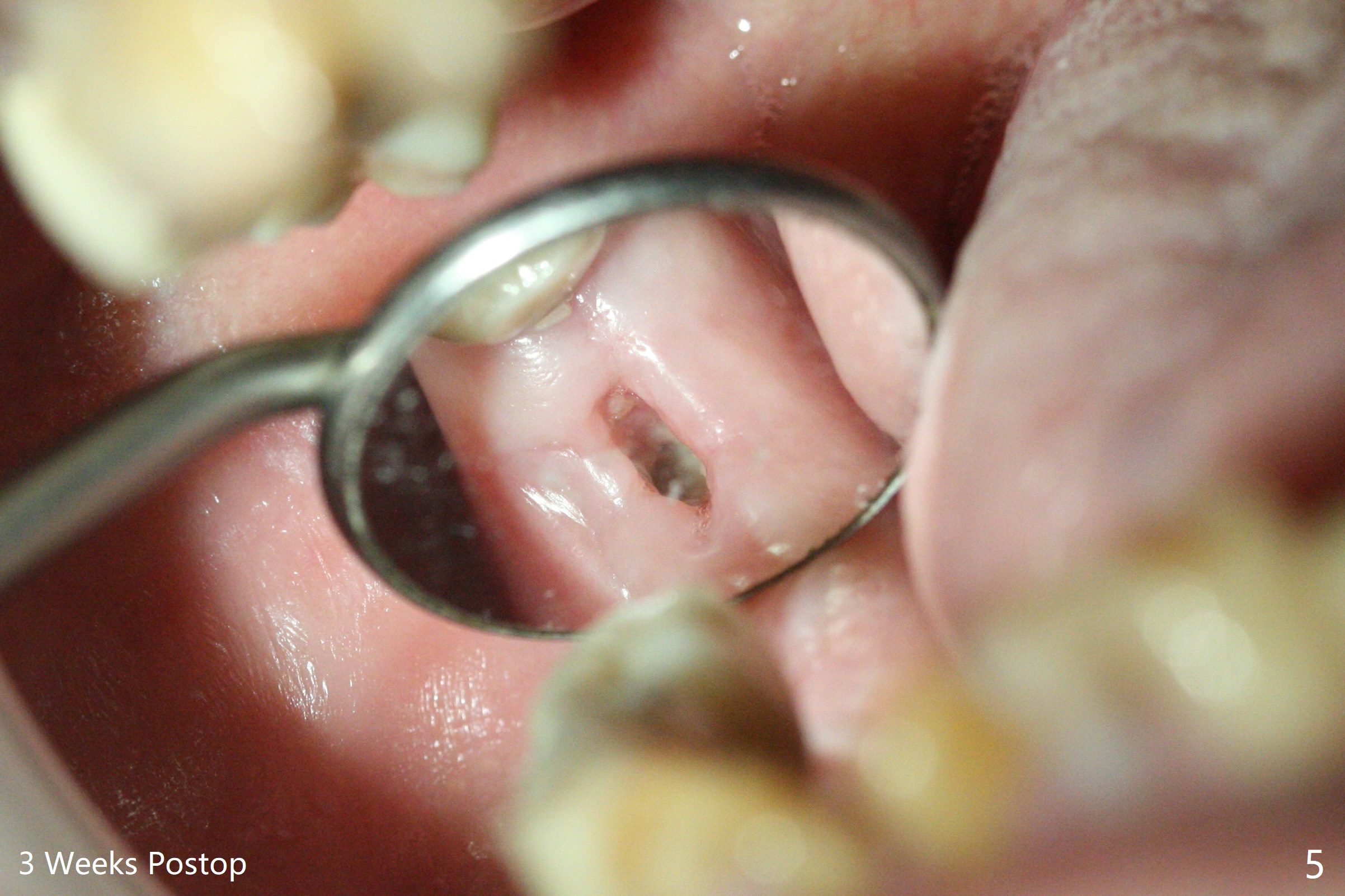
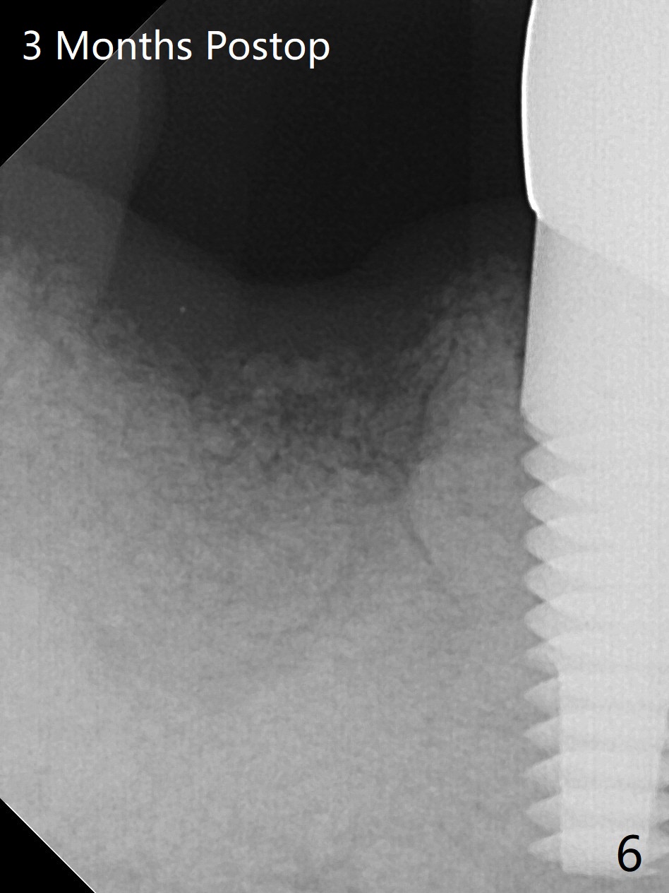
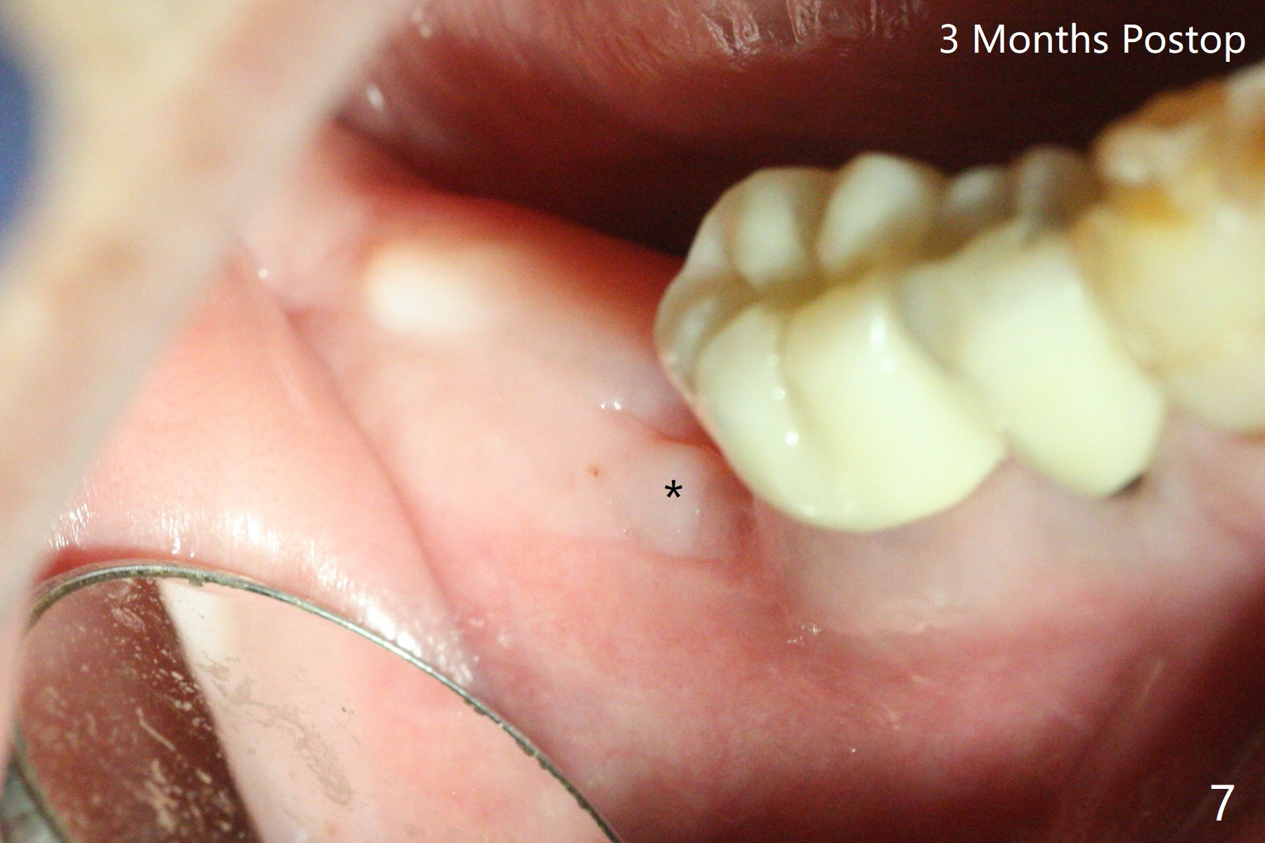
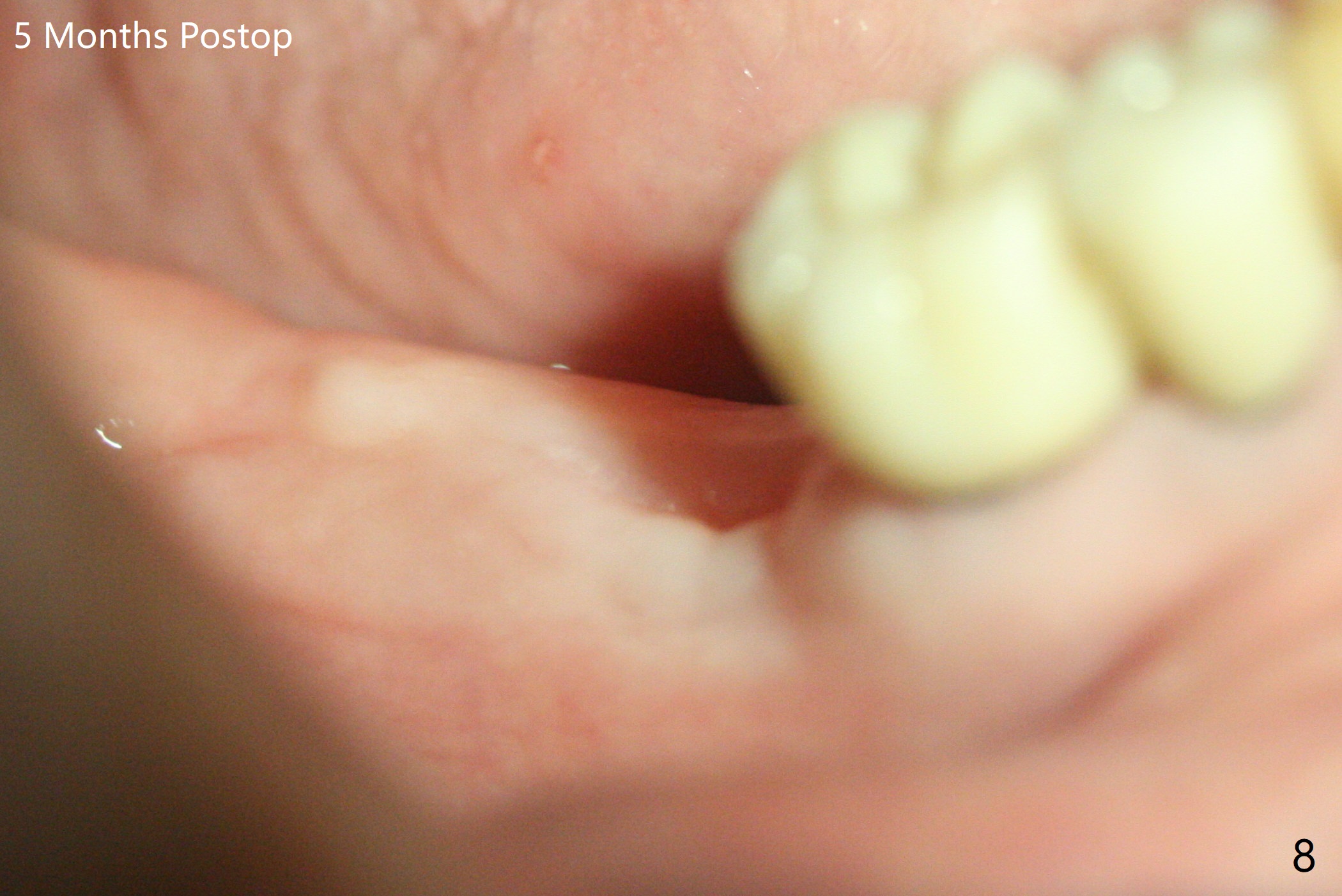
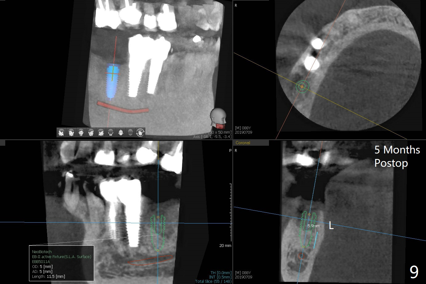
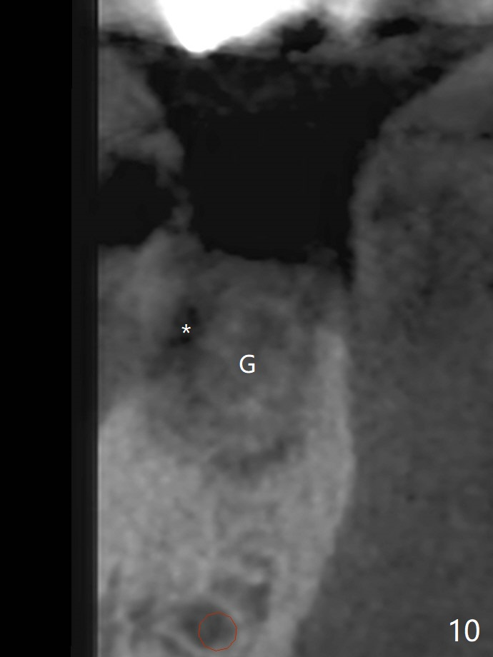
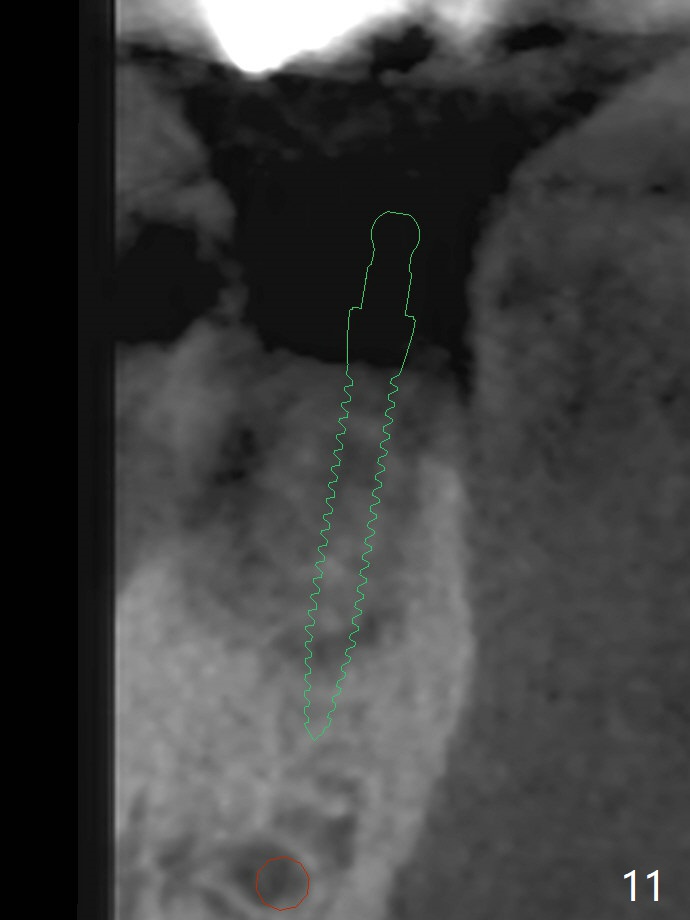
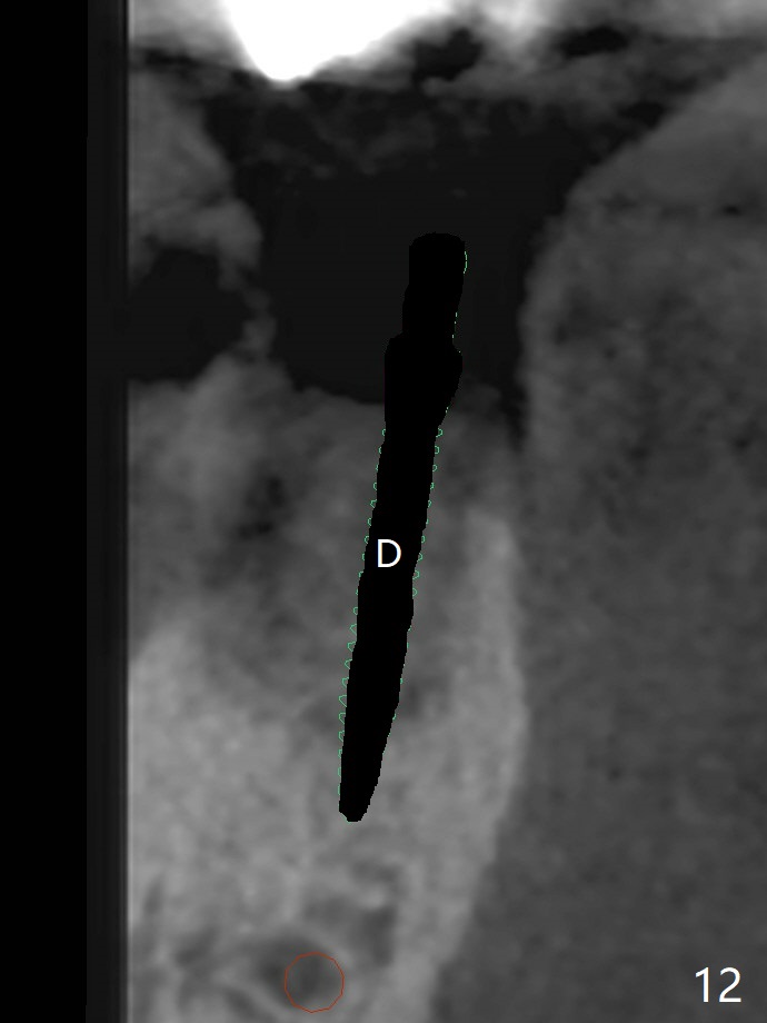
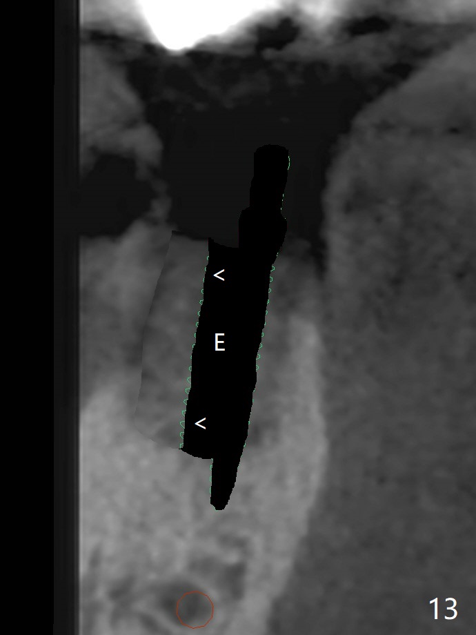
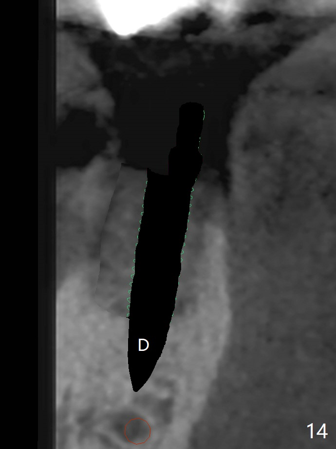
A 88-year-old man returns with pain and swelling (Fig.1), one year after diagnosis of #31 mesial root fracture. After discussion, the tooth is extracted with socket preservation (Vanilla graft mixed with Osteogen, covered with Osteogen plug and Collagen plug, Fig.2,3). The buccal crest is much lower than the lingual one. The socket opening reduces with resolution of 4-0 Chromic gut suture 1 week postop (Fig.4). The wound is wider with foul odor 2 weeks postop (data not shown). The socket and ridge shrink with loss of the bone graft 3 weeks postop (Fig.5). The bone volume reduces with buccal plate collapse 3 months postop (Fig.6,7). Immediate implant and provisional should be able to help restore the lost buccal plate. The buccal plate remains concave 5 months postop (Fig.8,9). The coronal section shows that there is space (Fig.10 *) buccal to the graft (G). A 2.0 mm pilot drill is used to create an initial osteotomy through the graft zone and in the beginning of the native bone (Fig.11,12). Use bone expanders (Fig.13 E) to push (arrowheads) and condense the graft bone and close the buccal gap. The apical portion of the osteotomy requires regular drills (Fig.14 D).
Return to
Lower
Molar Immediate Implant,
Trajectory
II
#15 Acrylic Dressing
Xin Wei, DDS, PhD, MS 1st edition
02/11/2019, last revision
07/27/2019