
|
|
|
|
|
|
|
|
|
|
Use Lower RPD as Surgical Stent
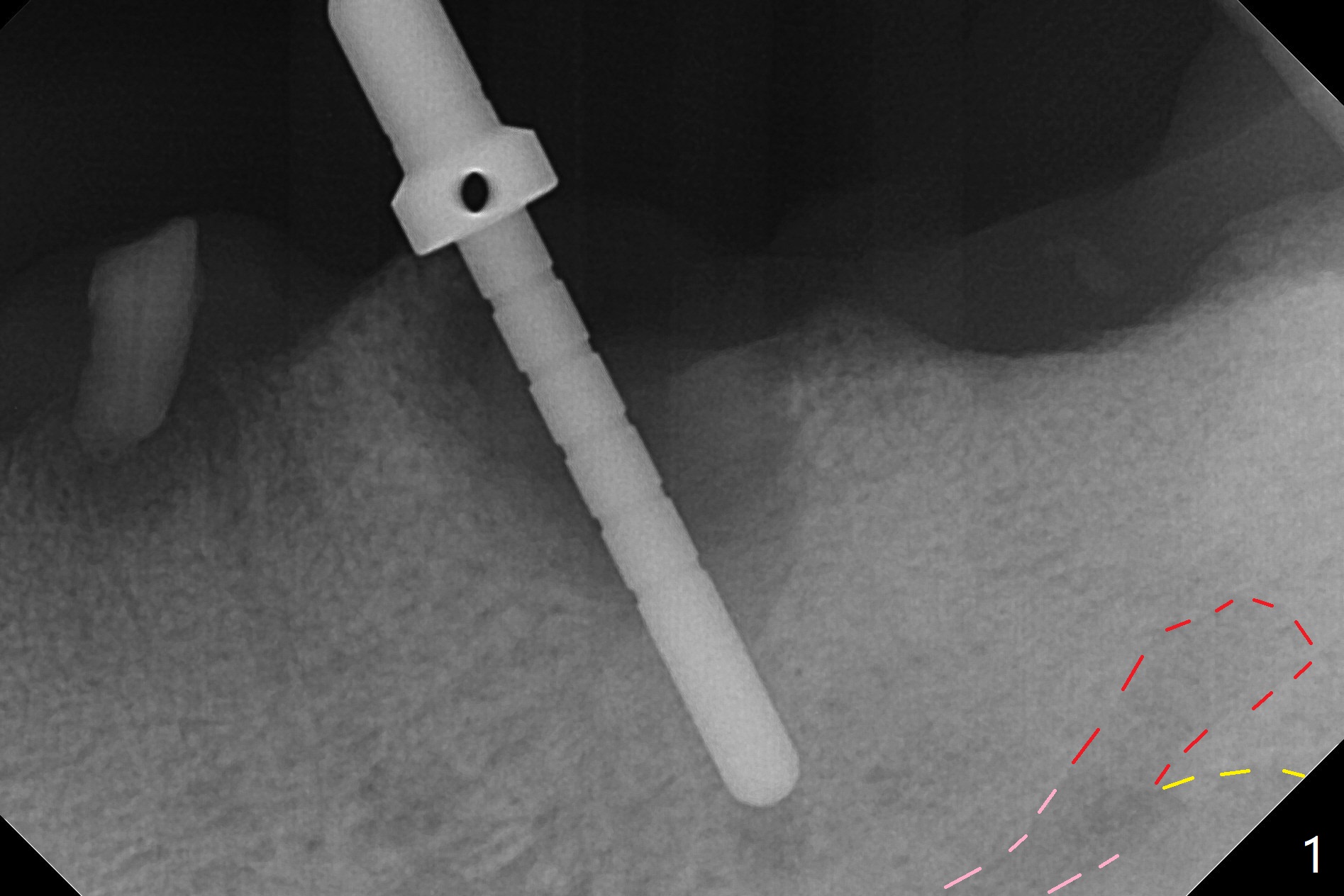
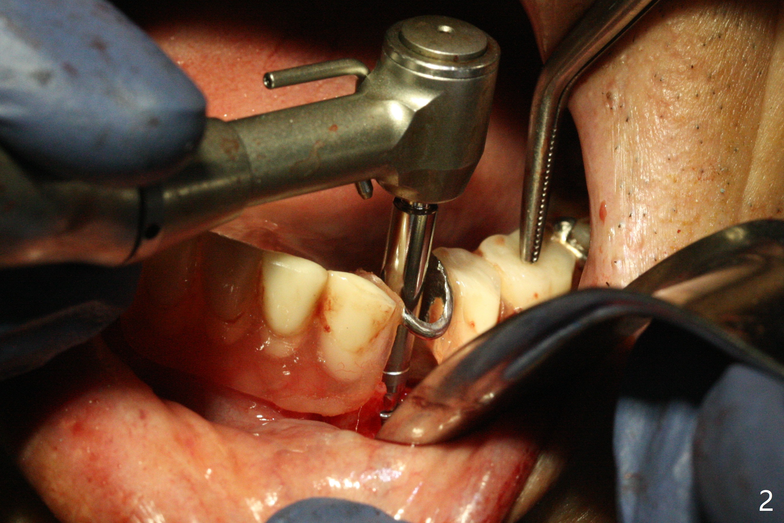
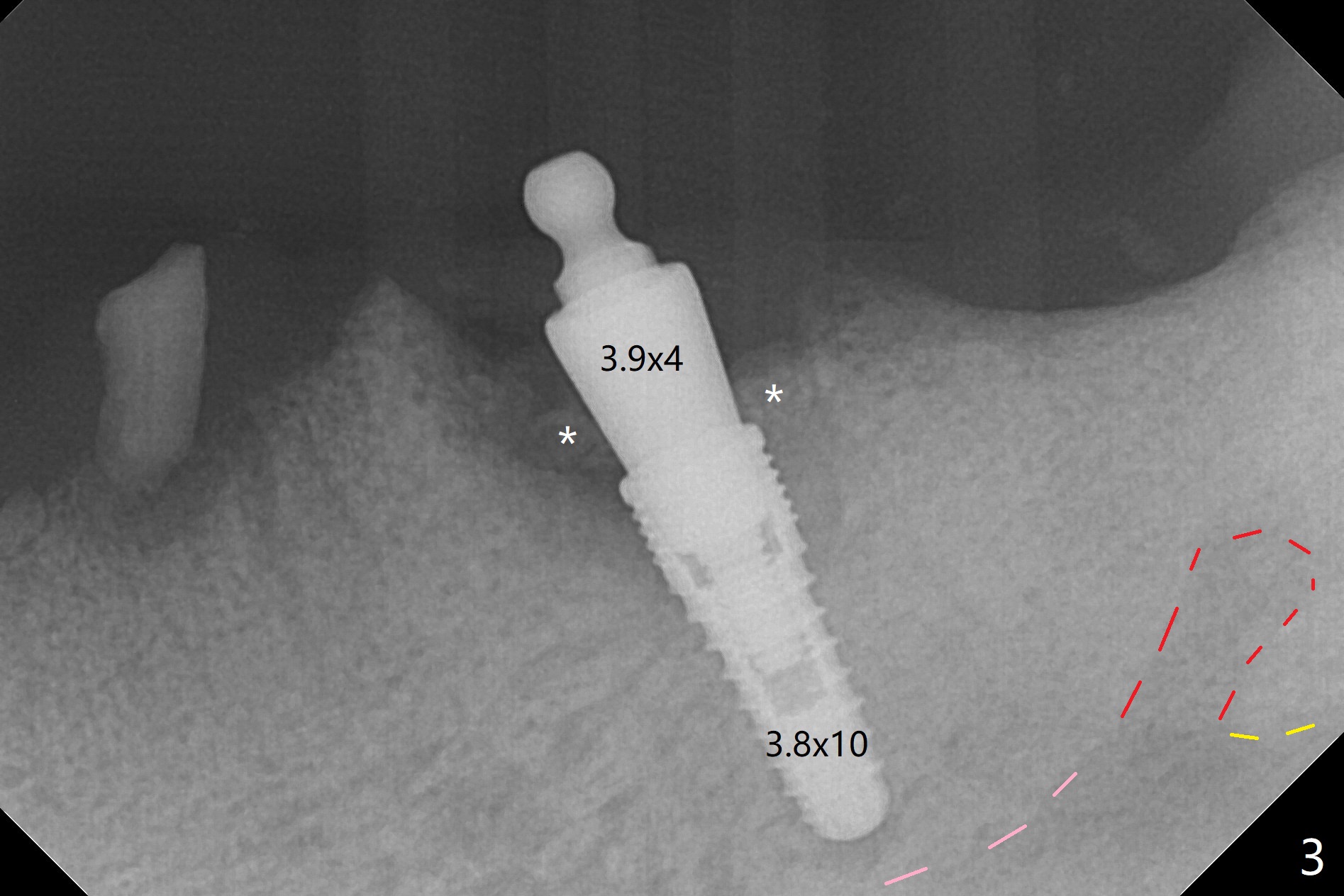
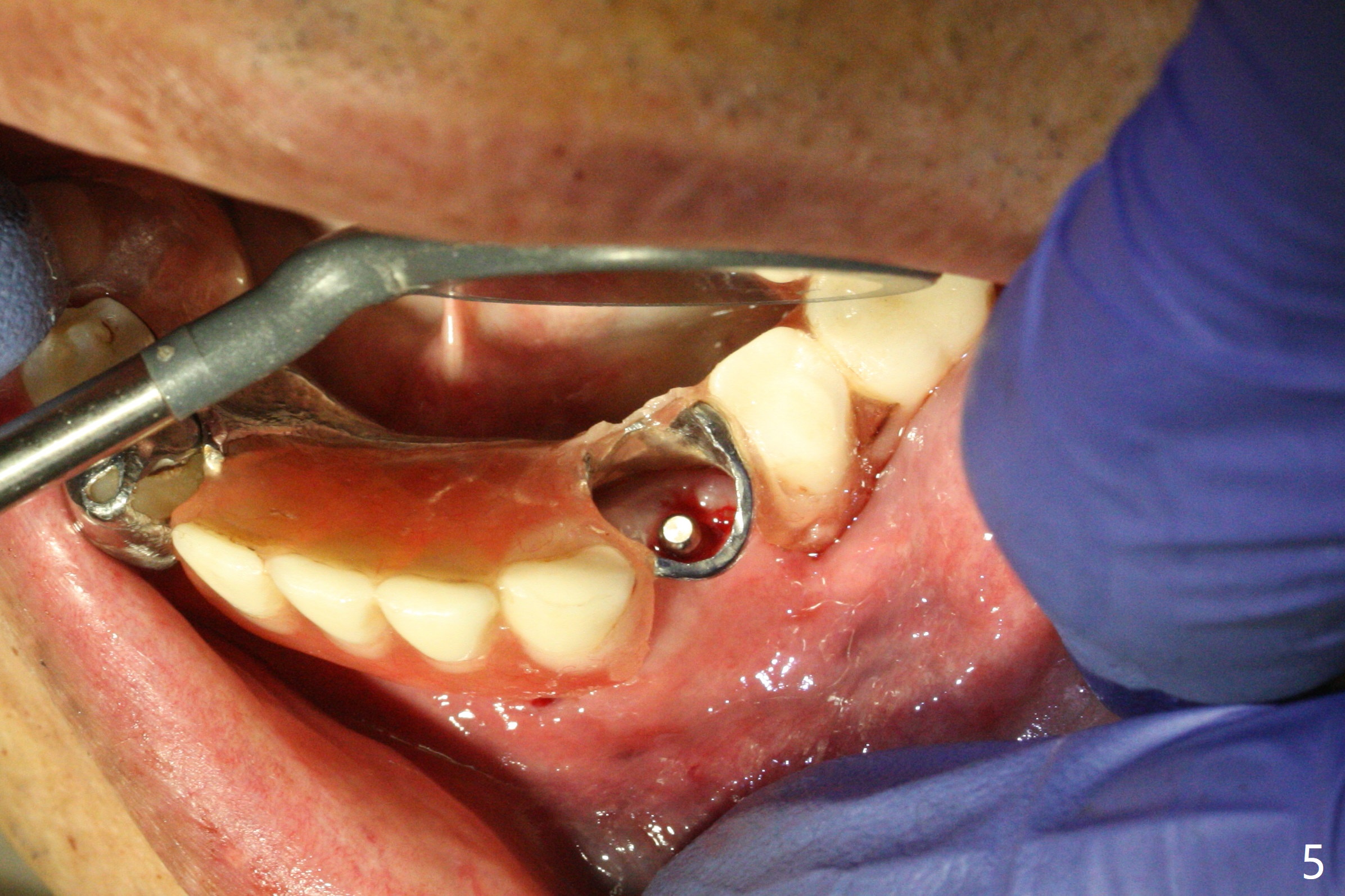
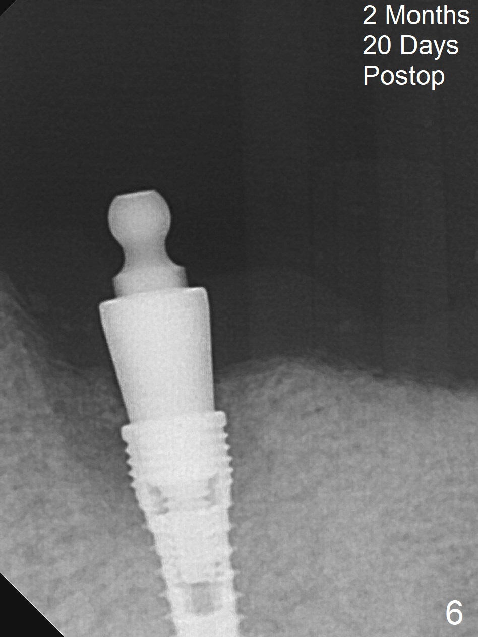
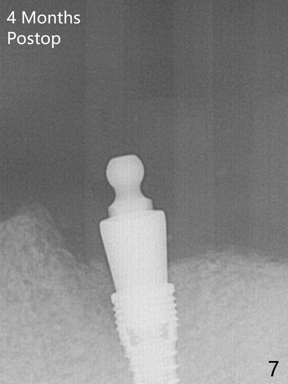
After making incision around the healed socket at #21, osteotomy is initiated free hand (Fig.1: 12 mm), double checked with the lower RPD. When a 3.8x10 mm implant is partially placed, the RPD is seated as a surgical stent (Fig.2). Therefore the trajectory of the implant is tightly controlled (Fig.3-5). After placement of a ball abutment (3.9x4 mm), Vera graft is packed buccally (Fig.3 *), followed by collagen dressing. The bone density apparently increase in the mesial and distal sockets 2 months 20 days postop (Fig.6). The implant becomes functional 4 months postop (Fig.7) with placement of a 5x4 mm ball cap.
Return to
Lower
Premolar Immediate Implant, Armaments
11/13
Xin Wei, DDS, PhD, MS 1st edition 03/02/2018, last revision 06/17/2020