|
|
|
||
|
|
|
|
|
Labial Plate Repair
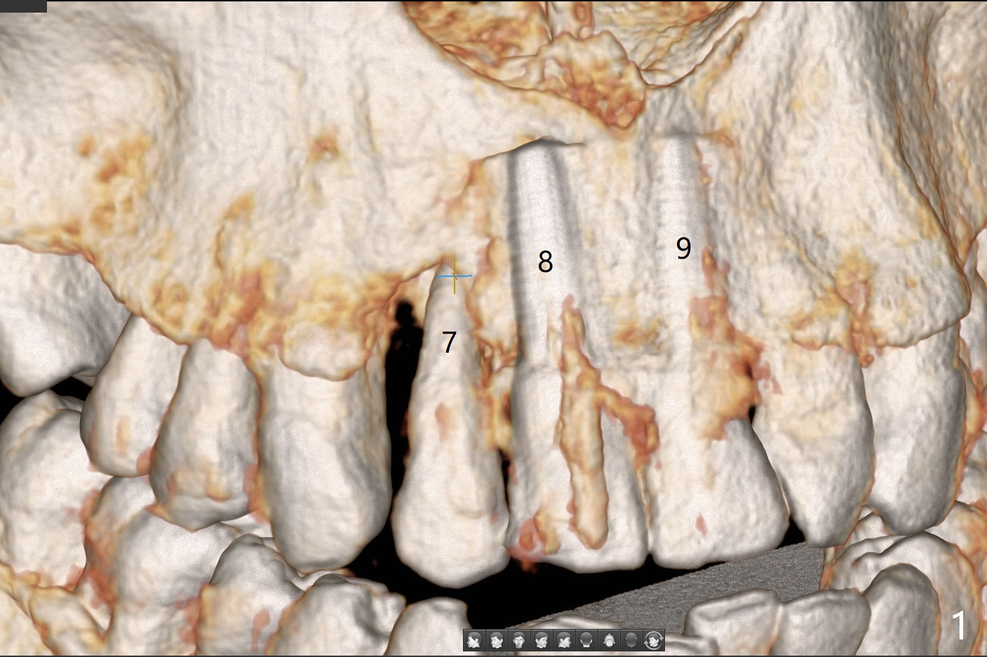
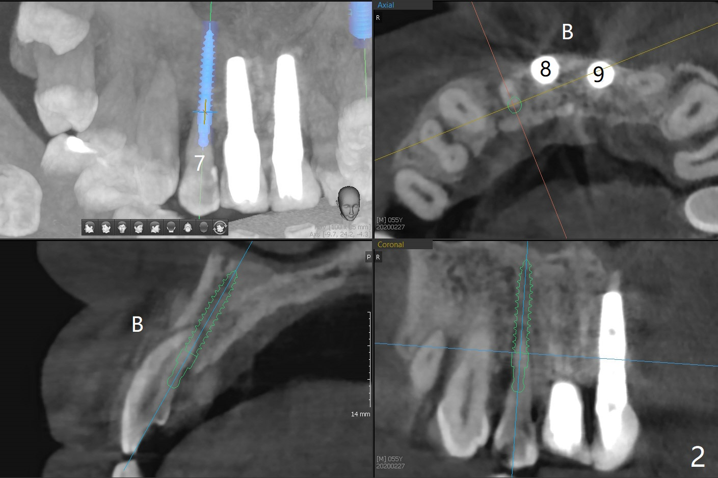
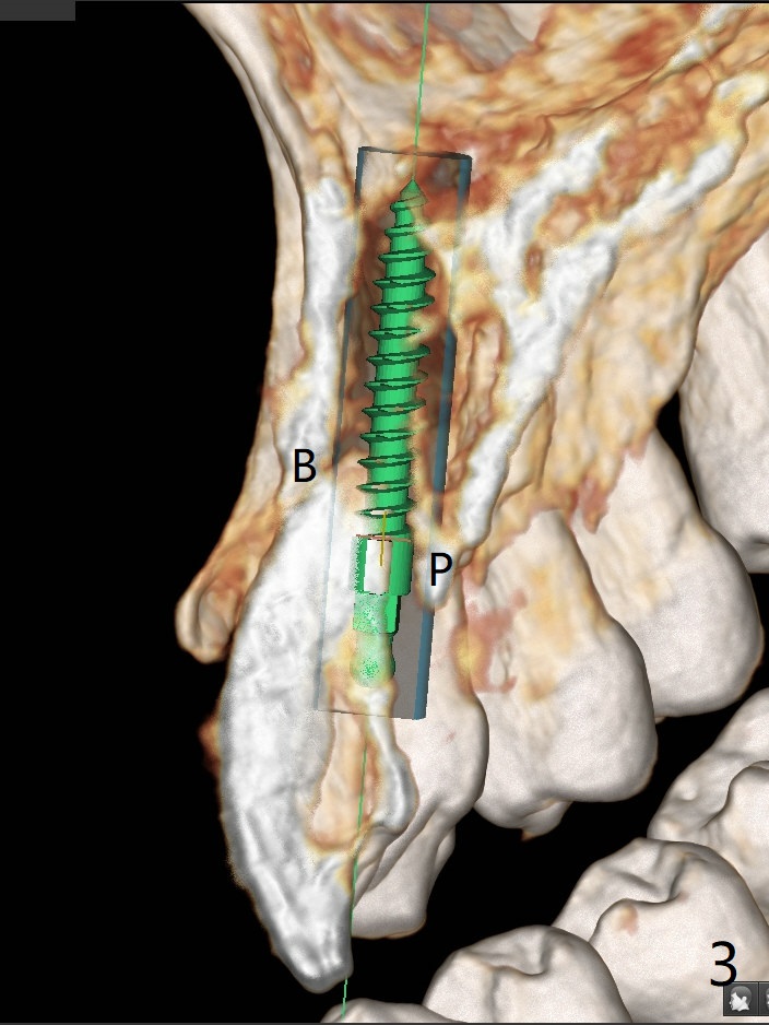
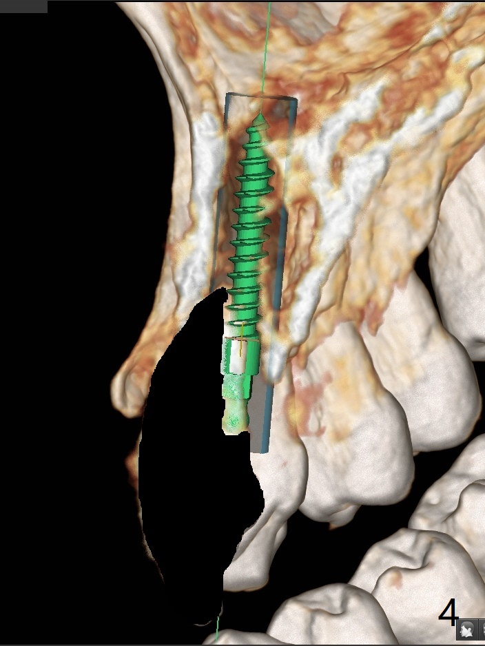
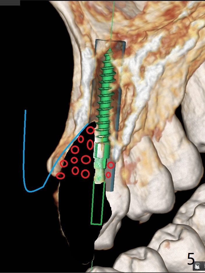
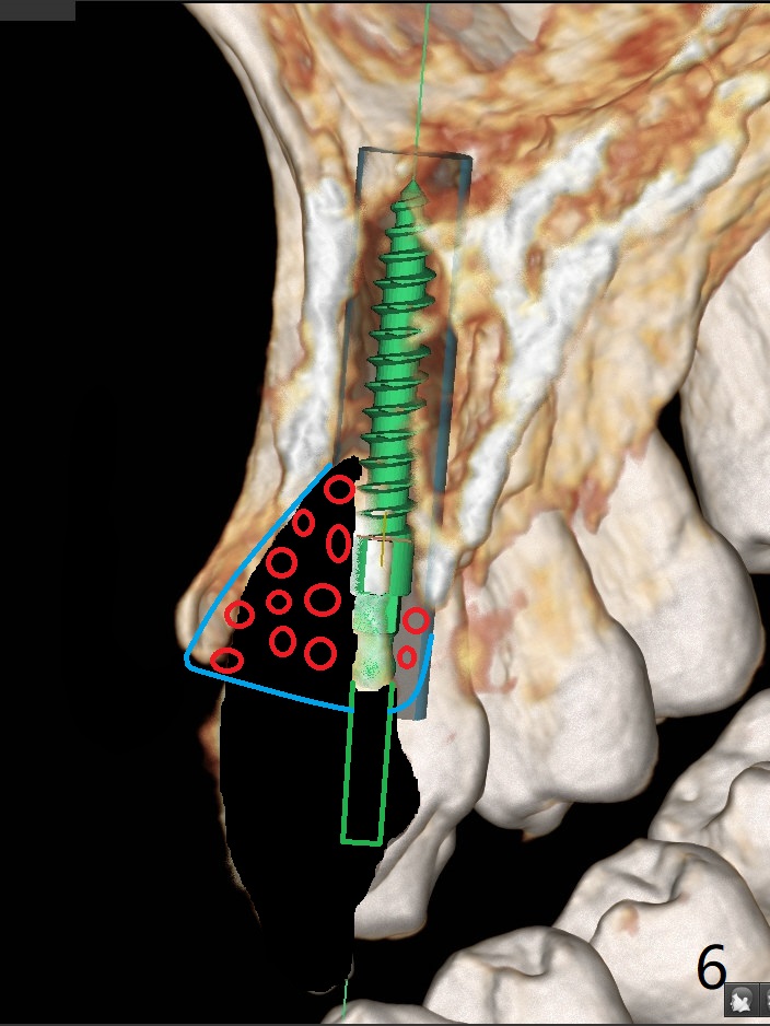
A 58-year-old man recently had sudden pain at #7 with labial plate loss (Fig.1 CT taken 3 years earlier). To avoid the labial placement of the large implants at #8, and 9, the implant at #7 will be smaller and palatally inserted (Fig.2). Fig.3 is a coronal section of 3D image of #7, showing the labial (B) and palatal (P) crests. After extraction (Fig.4 black), the narrow implant is placed between the crests (green). To repair the coronal labial plate, a piece of PRF membrane (Fig.5 blue) is placed inside the socket, followed by sticky bone (red circles). The portion of PRF membrane outside the socket will be flipped palatal, inserted into the abutment with a pre-punched hole and finally tucked underneath the palatal gingiva.
Return to Protect Graft Clindamycin Metronidazole No Antibiotic 15
Xin Wei, DDS, PhD, MS 1st edition 08/14/2021, last revision 08/14/2021