|
|
|
|
|||
|
|
|
|
|
|
|
|
|
|
||||
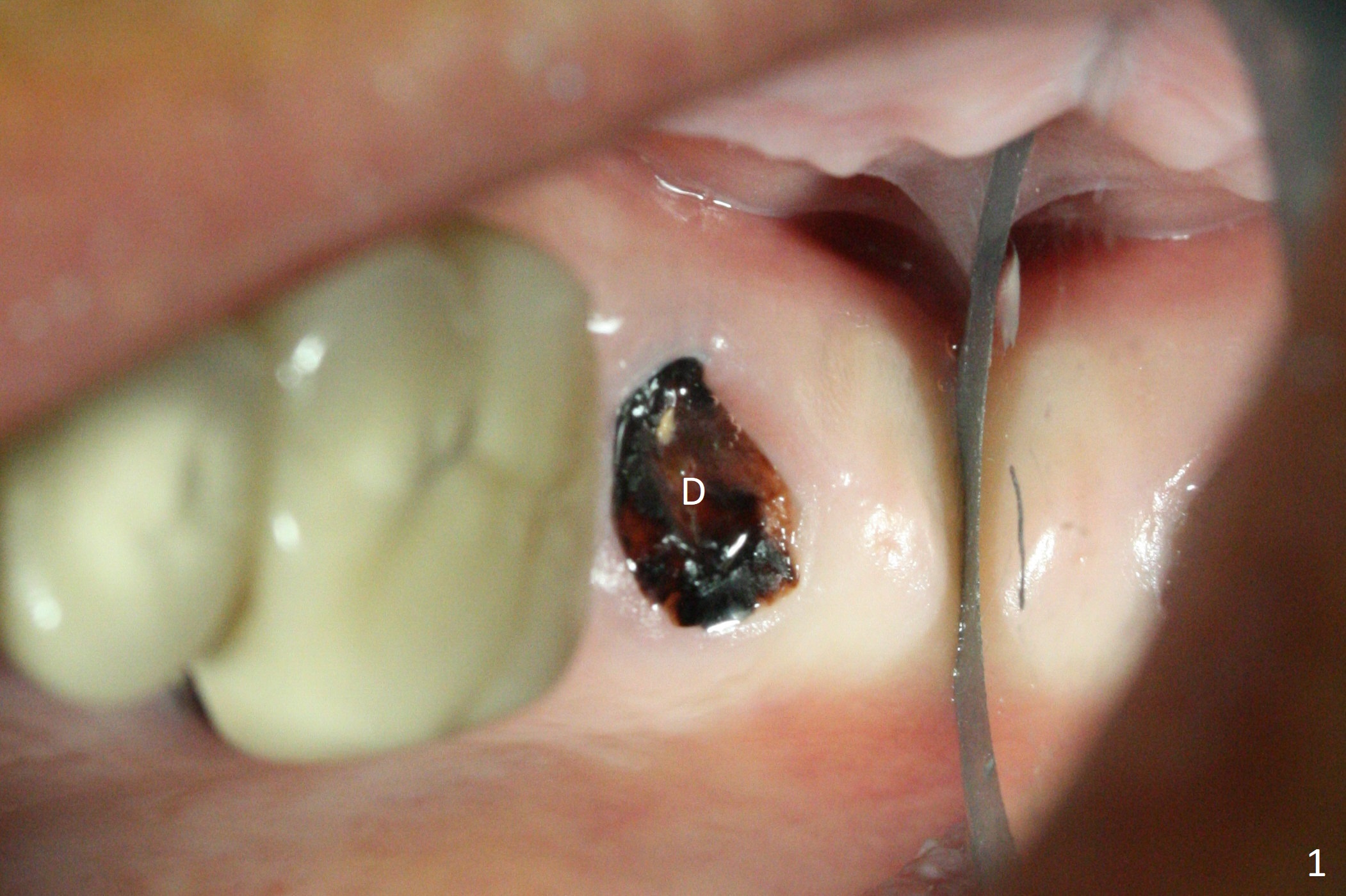
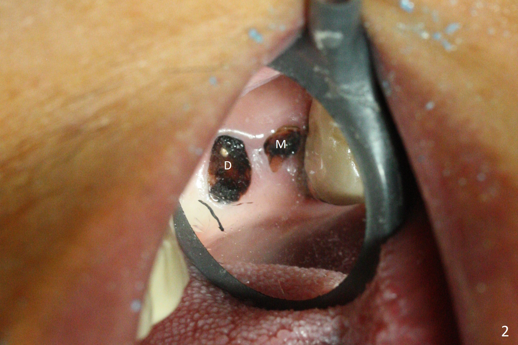
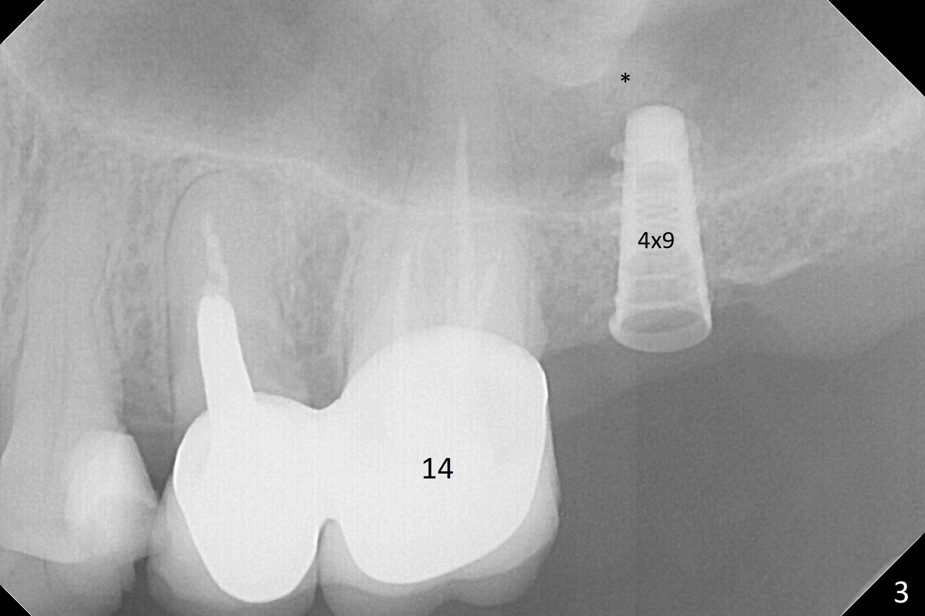
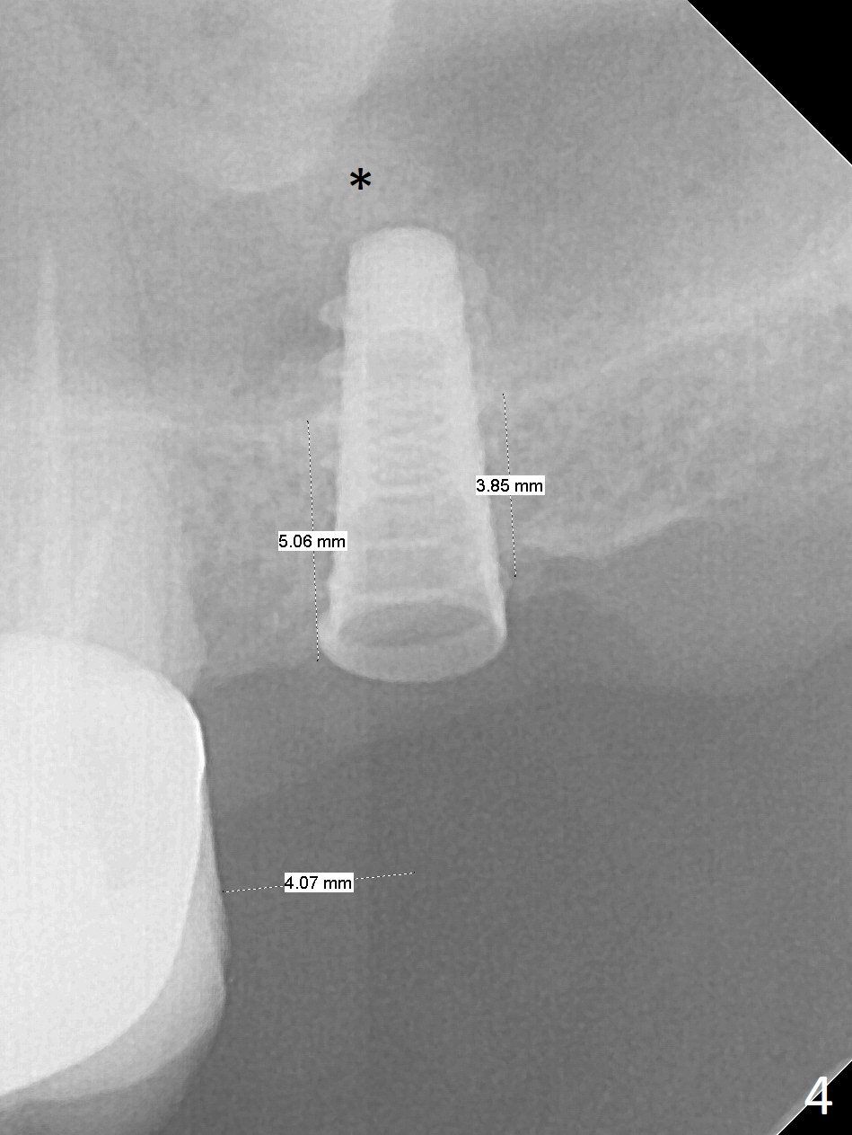
Sinus Lift M
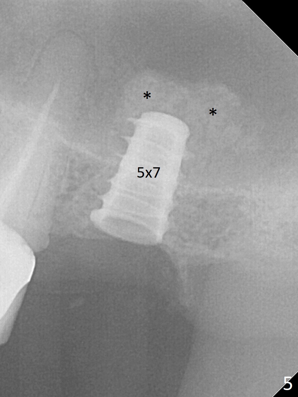
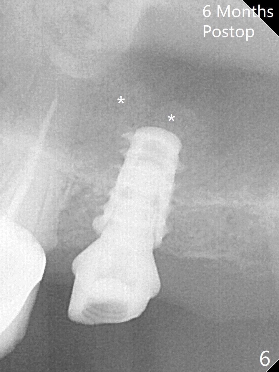
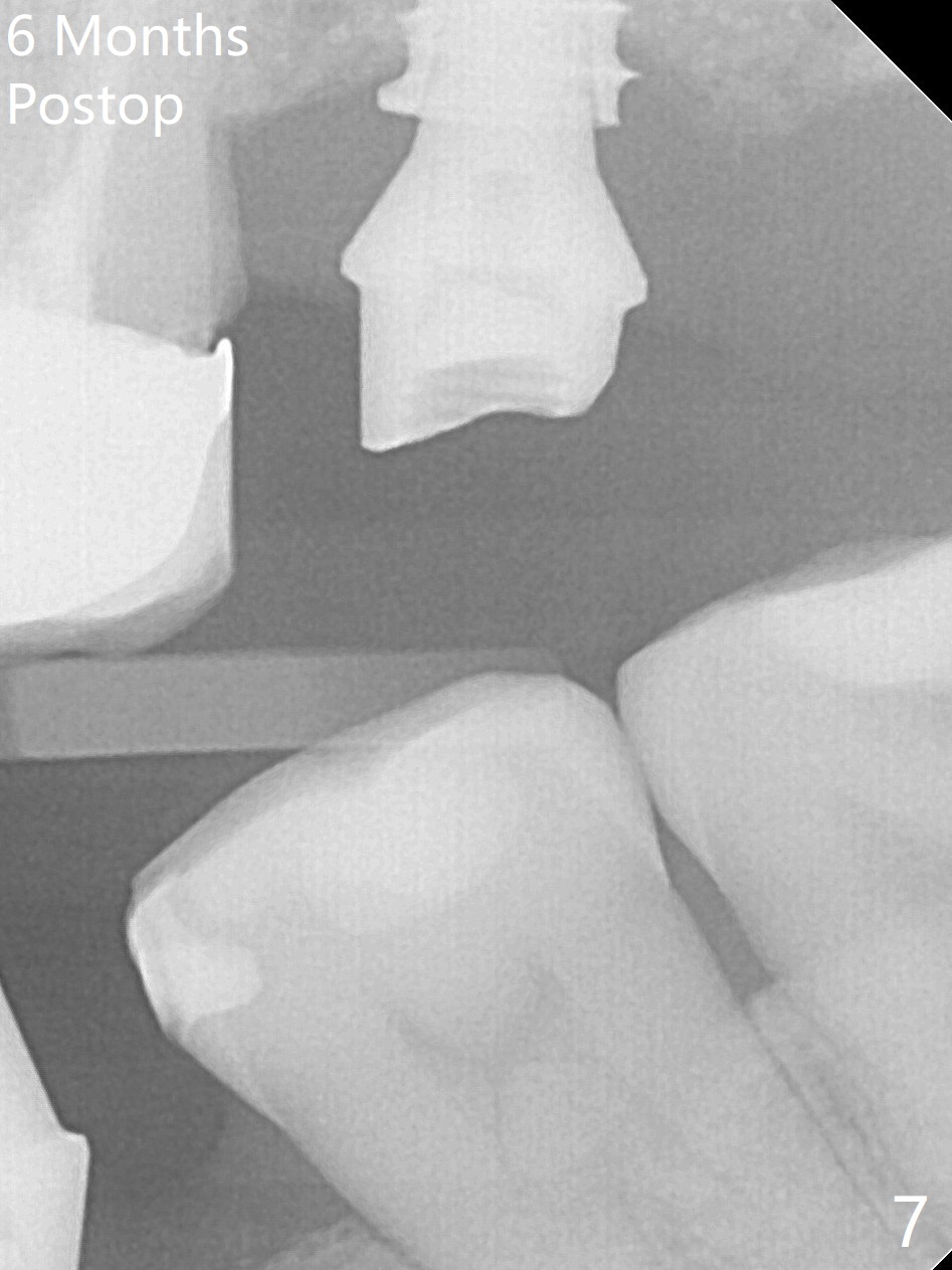
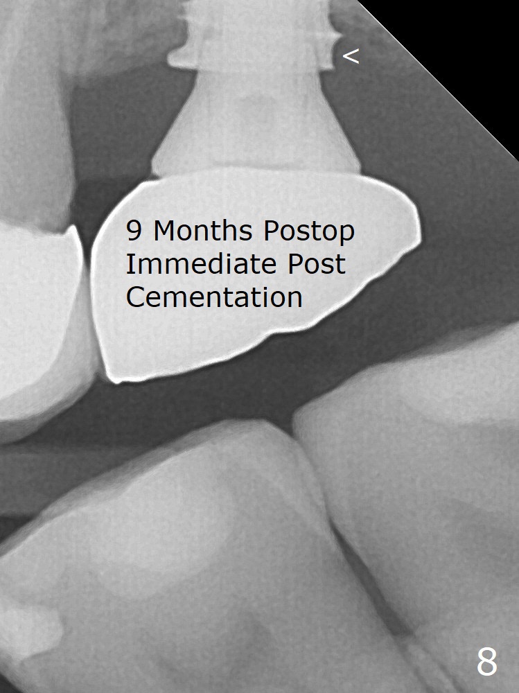
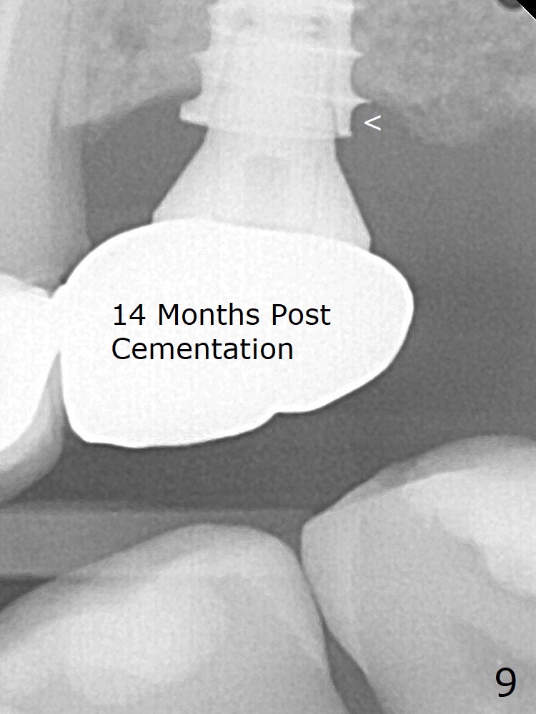
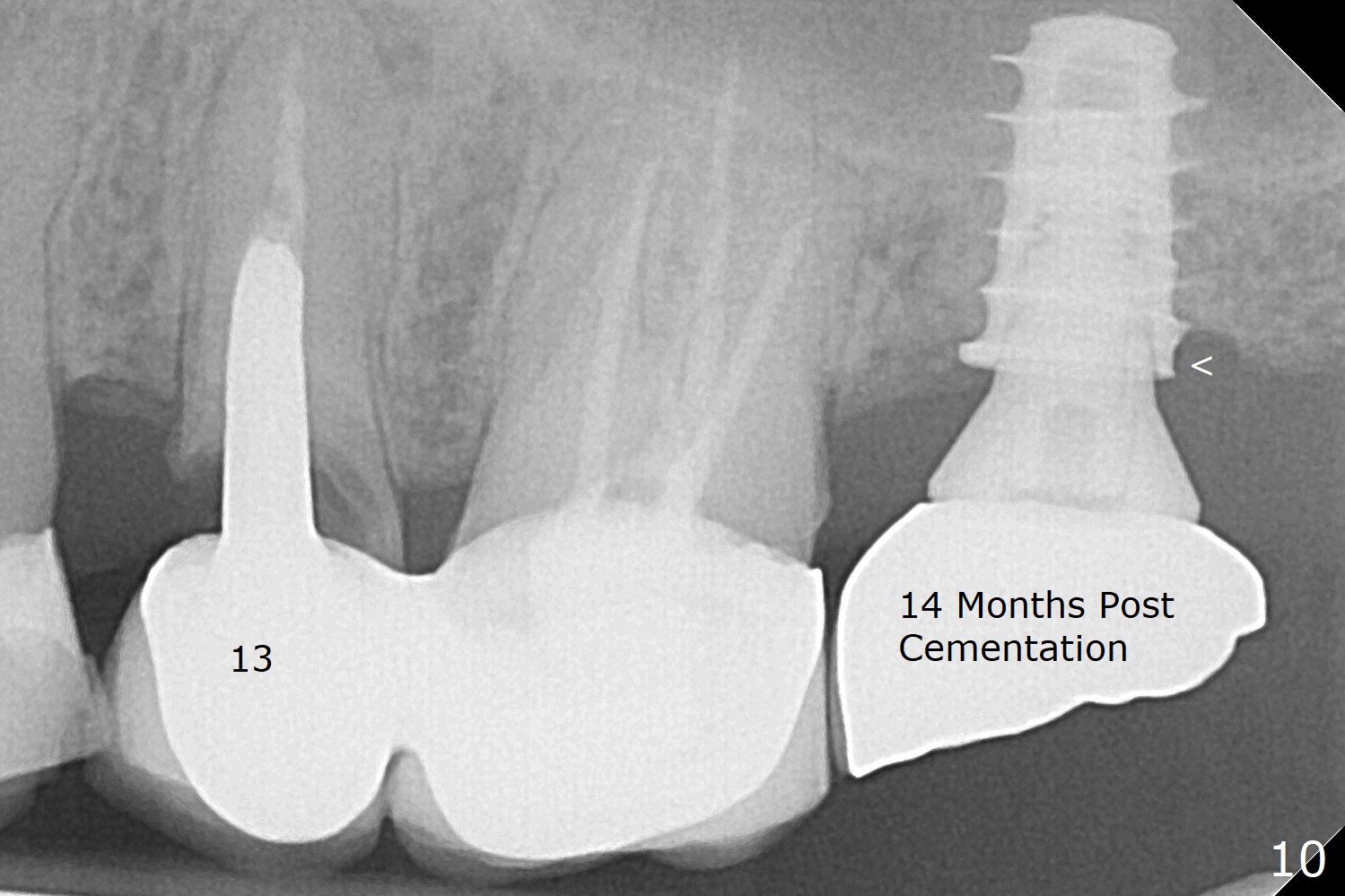
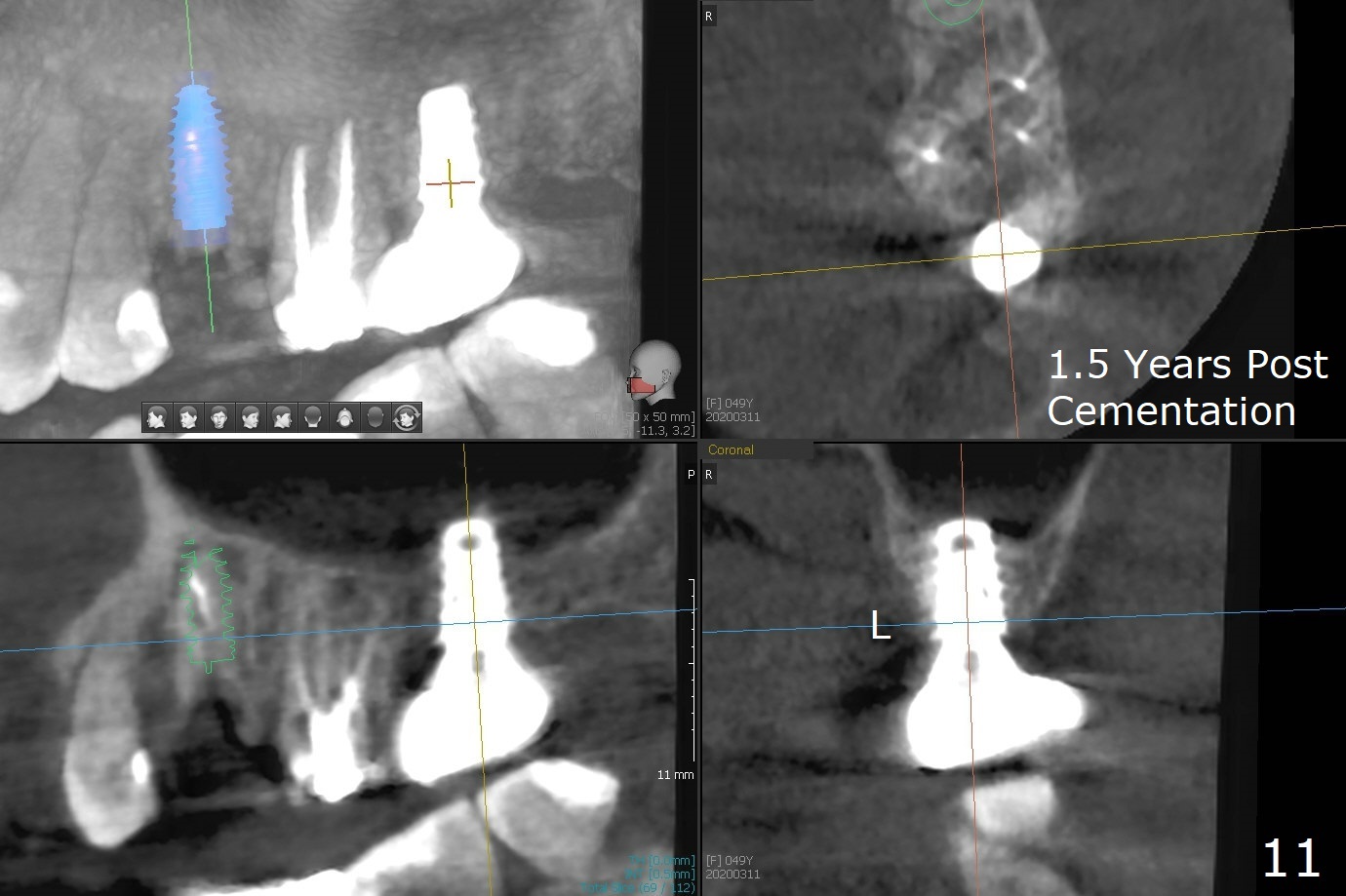
After removal of the mesial (M) and distal (D) residual roots of the tooth #15 (Fig.1,2), Magic Split is used to test bone density (high), followed by Magic Drills from Magic Sinus Lift Kit (for 4 mm) and Magic Surgical Kit (for 5 mm). It appears that the sinus floor has been perforated with the intact sinus membrane. Following minimal use of Magic Lifter, Vanilla Graft is inserted (Fig.3,4 *) and a 4x9 mm dummy implant is placed. After placement of more allograft (Fig.5 *), a 5x7 mm implant is placed with ~ 35 Ncm. With placement of a 5.5x4(2) mm abutment, an immediate provisional is fabricated to close the socket. Six months postop, the bone graft remains in the sinus around the apical end of the implant (Fig.6 *), while there seems no bone loss coronally (Fig.7). In fact there is, as shown later (Fig.8-11 < and lingual (L)). The crown is recemented 6 months post cementation (due to short abutment). The abutment seems to be incompletely seated. When the crown at #14 is reprep following #13 implant, the abutment screw is being untightened, the crown dislodges first. The abutment is confirmed short with more than enough occlusal clearance. After use of 5.5 and 6.0 mm bone profile drills, a 5x4(3) mm abutment is placed with complete seating.
Return to
Upper
Molar Immediate Implant, Prevent Molar Periimplantitis (Protocols,
Table),
Armaments, #19
13
Xin Wei, DDS, PhD, MS 1st edition 12/28/2017, last revision 05/21/2020