
%20with%20prep%20later.jpg)
,%205.5m%20postop,%2015%20mesial%20gap.jpg)
|
|
|
|
|
|
|
|
|
|
|
|
No Space for Sheath
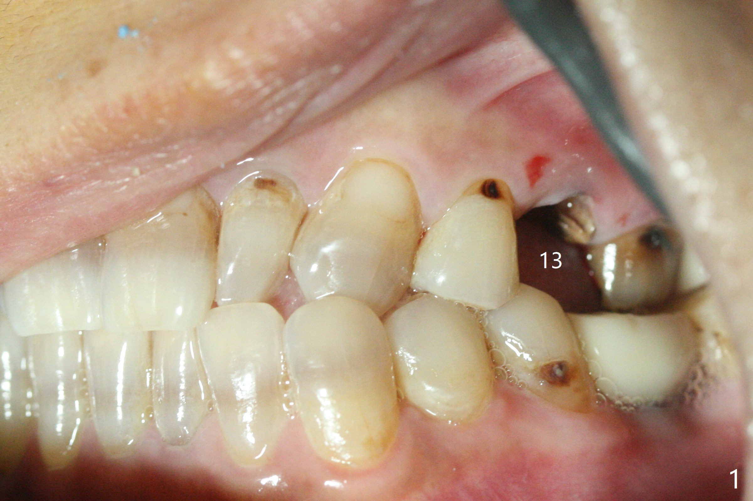
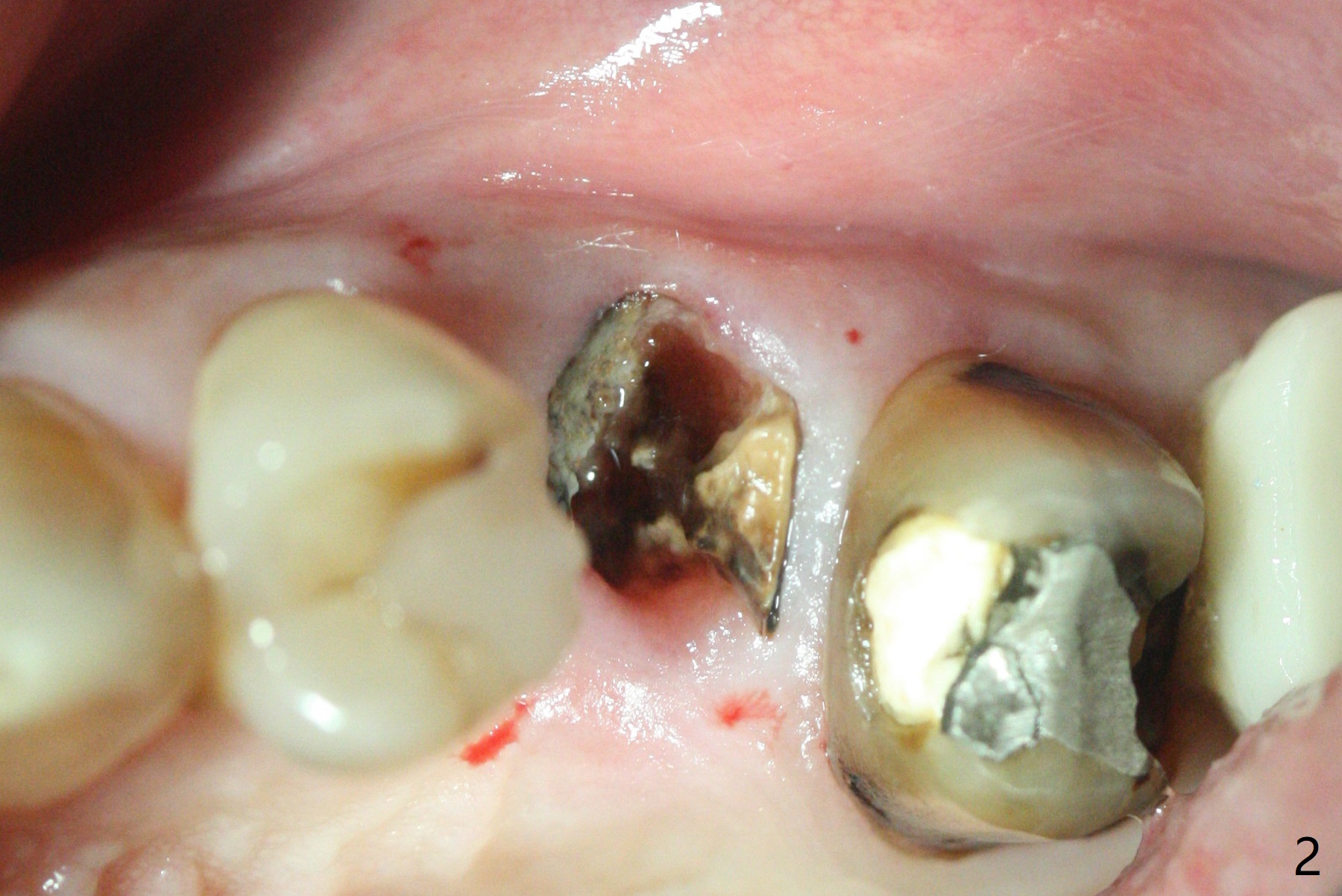
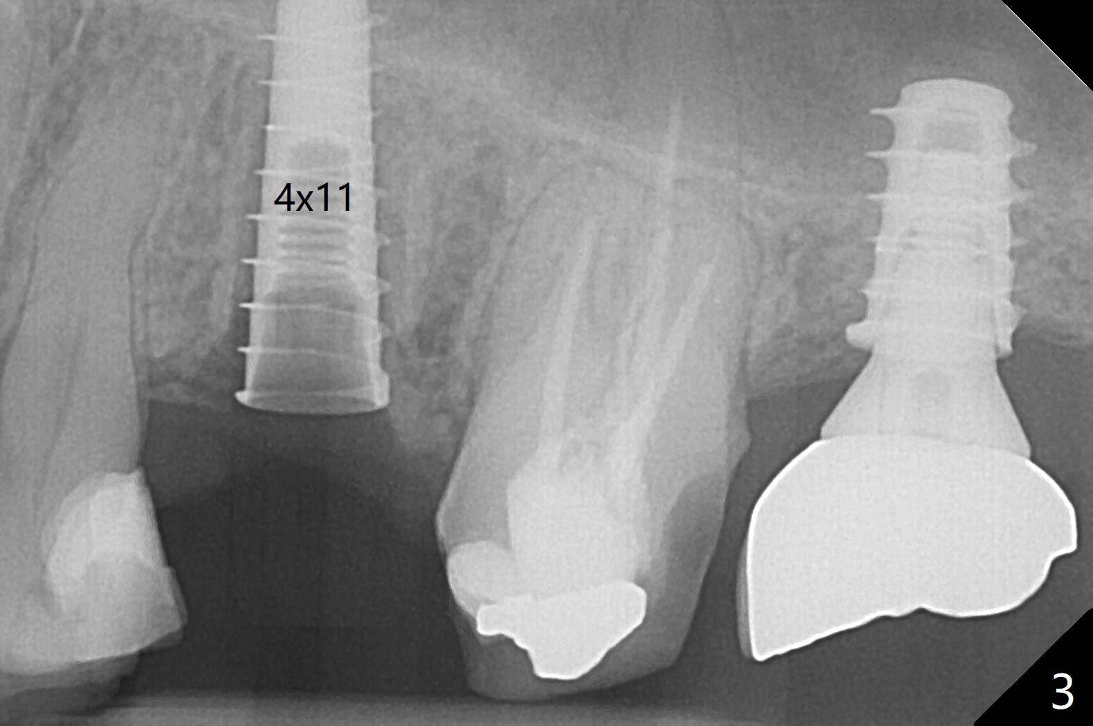
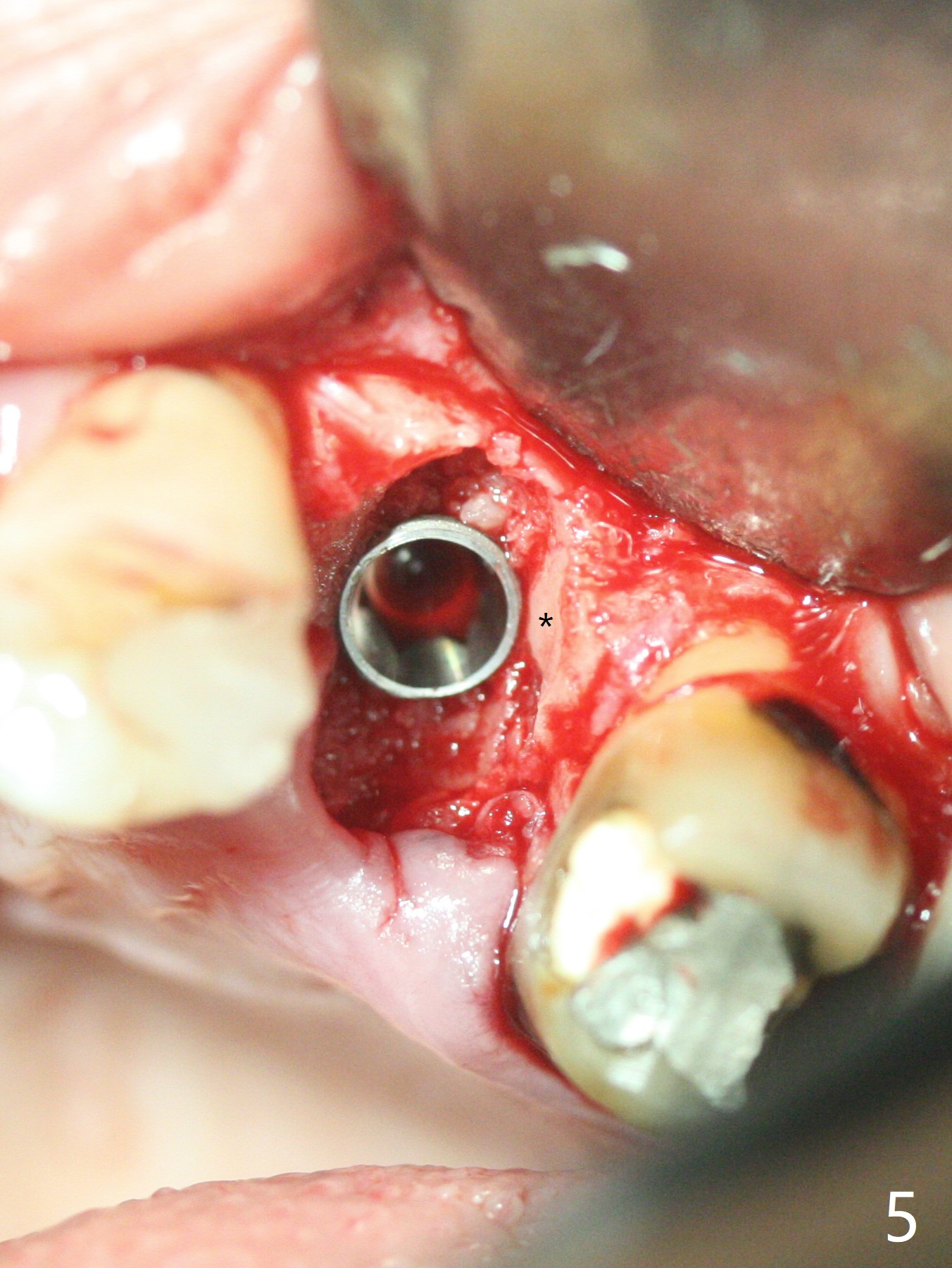
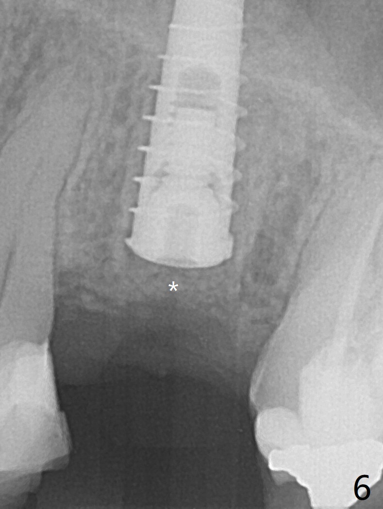
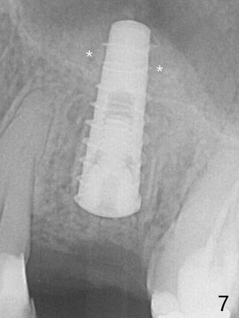
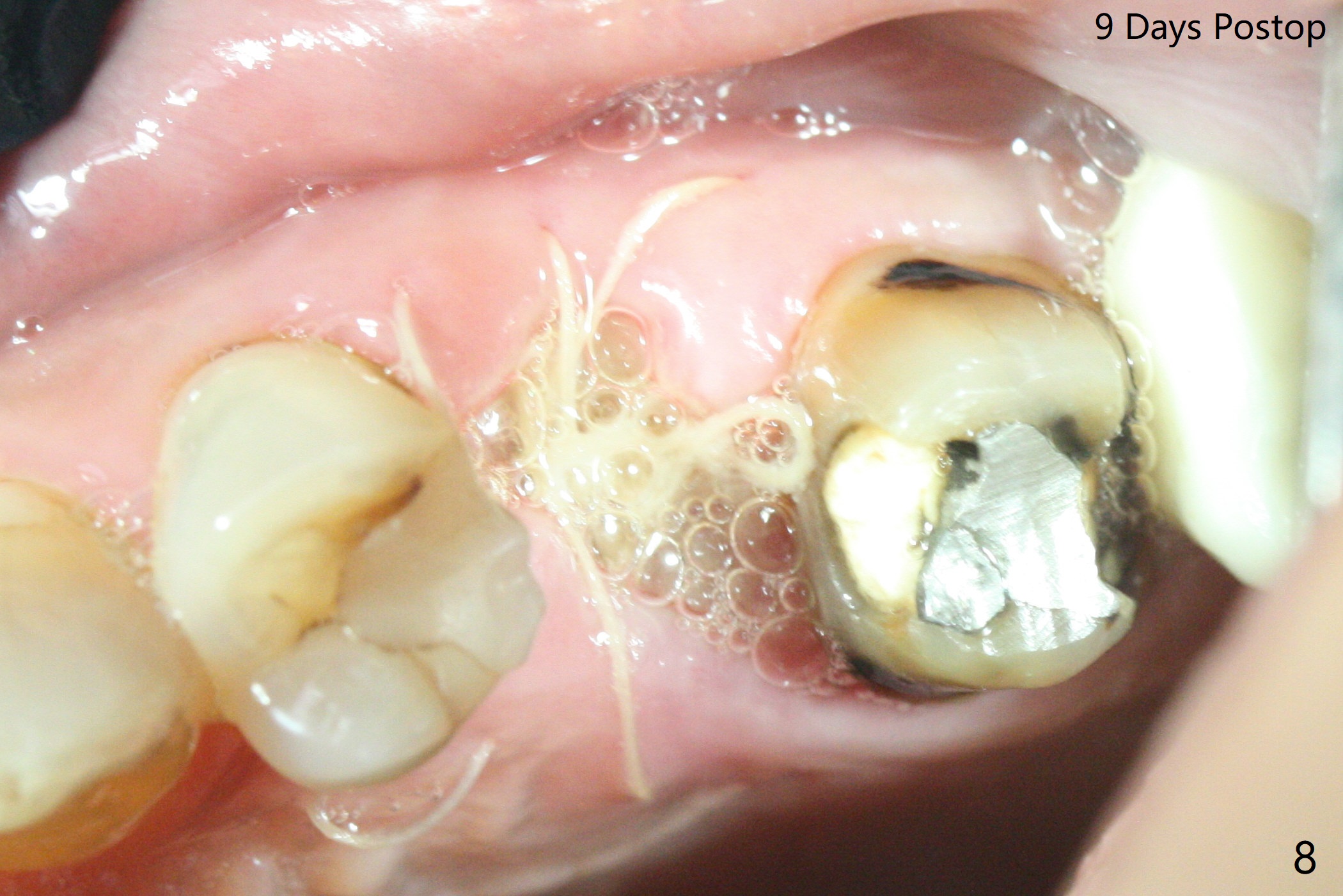
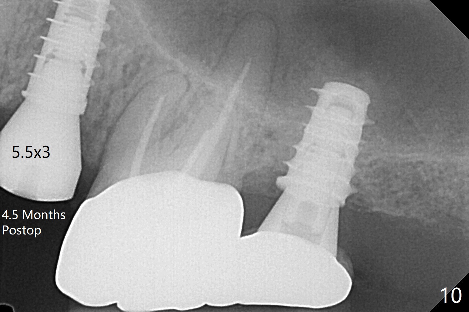
The patient with the anterior cross bite at the left lateral and canine and incipient Class V caries returns to clinic for #13 residual root extraction (Fig.1,2). There is no quality root structure (Fig.2) or space (Fig.3) for socket sheath. Two amalgam carriers of Vanilla is inserted and pushed by the 4x11 mm dummy implant for sinus lift (Fig.4 *). When the same sized final implant (4x11 mm) is placed, a 5.5 mm bone profile drill is used to trim the proximal crests (Fig.5 * sticky bone packed in the buccal and palatal gaps). In fact the implant turns when a 4.5x4(3) mm pair abutment is tightened. The implant is backed up; with a healing screw, sticky bone is placed (Fig.6 *) and then covered with a piece of PRF and a part of GEM cap. The wound is sutured with 4-0 PGA; periodontal dressing is applied. The implant is somewhat over-seated without the bone graft covering the apical end of the implant (Fig.7, as compared to Fig.4). A 4.5 mm implant should be placed. The GEM Cap appears to be absent 9 days postop, while the periodontal dressing dislodged in 1 week (Fig.8). It appears that the existing abutment at #15 is incompletely seated (Fig.3, 5.5x4(2) mm). After use of 5.5 and 6.0 mm bone profile drills, a 5x4(3) mm abutment is placed with 35 Ncm torque. In fact the bone graft coronal to the implant remains (Fig.9 *) in spite of apparent loss of the overlying PRF and GEM cap (Fig.8). After use of a 4.6 mm profile drill, a 5.5x3 mm healing abutment is placed 4.5 months postop (Fig.10). Without a provisional at #13 for ~ 4 months, there is a gap between crowns at #14 and 15 (not shown due to angulation). Impression is taken for #13 crown fabrication 5.5 months postop and for #15 crown repair (addition of porcelain for the mesial contact, Fig.11). It appears critical for a provisional to maintain the position of the neighboring tooth.
Return to
No Deviation
Shield
Next Case
Xin Wei, DDS, PhD, MS 1st edition
05/11/2020, last revision
10/30/2020