|
|
|
|
|
|
|
|
Oral Scanner Raises Diagnosis to Another Level
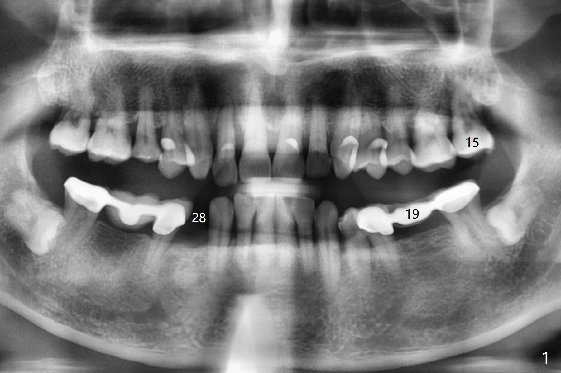
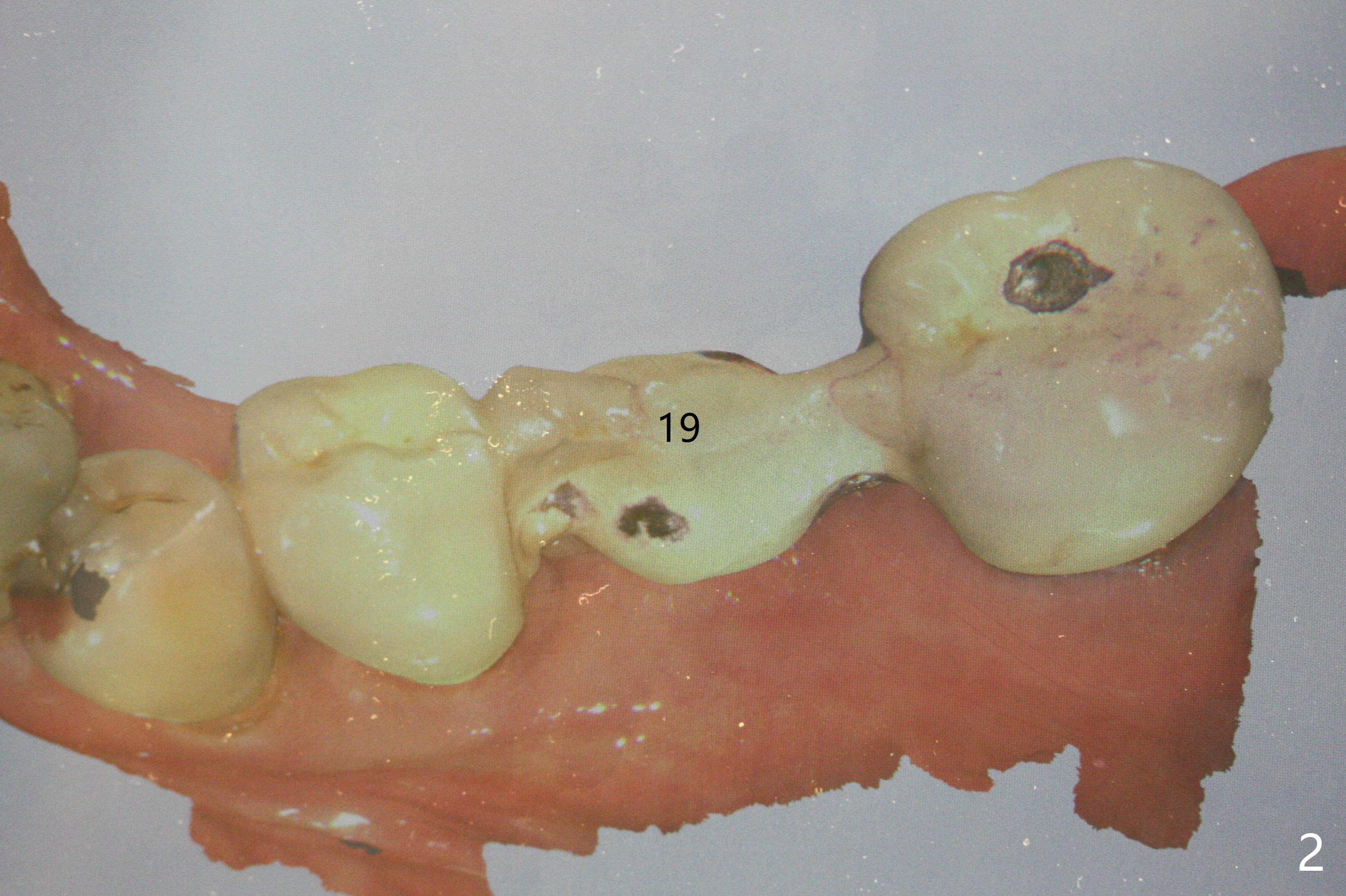
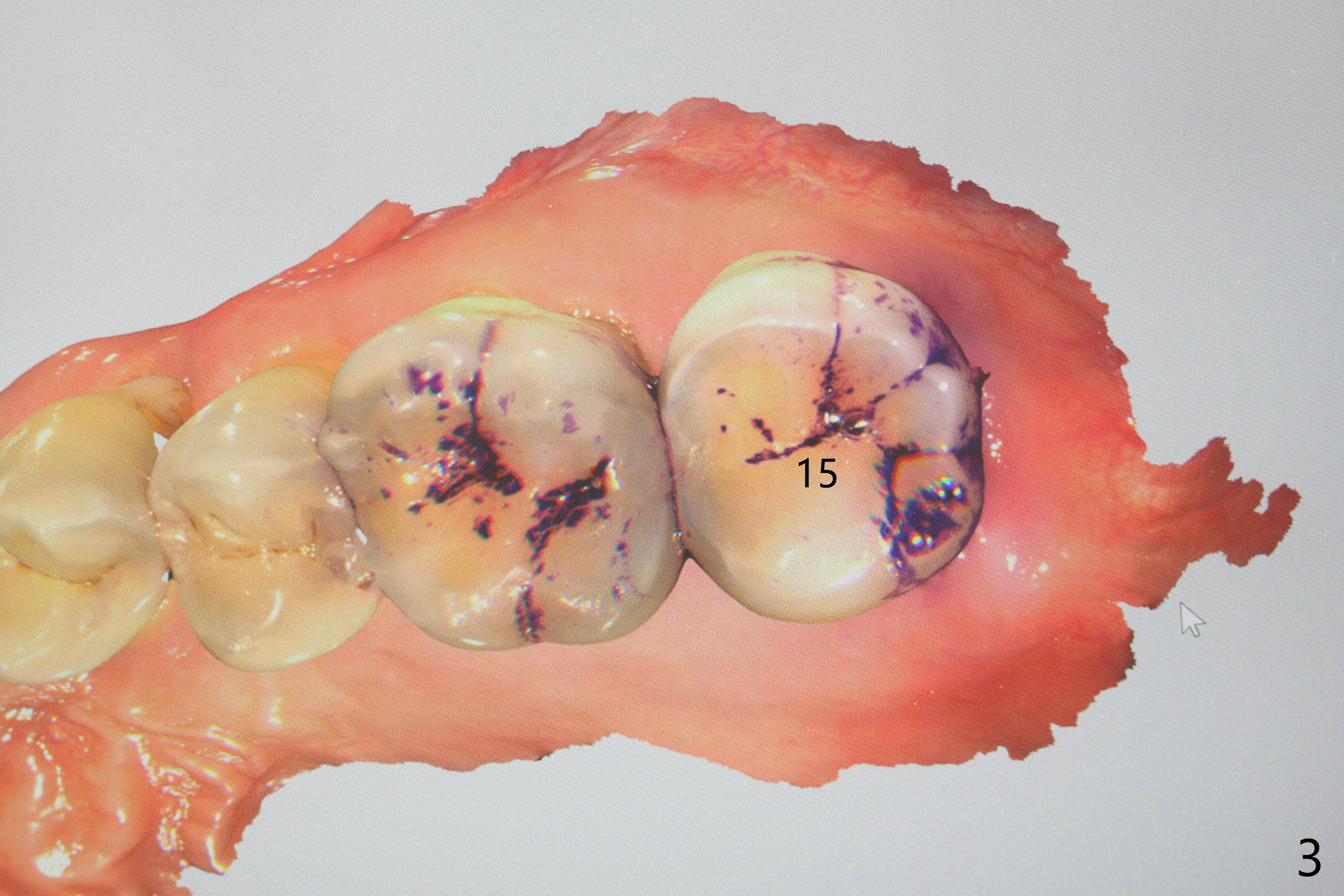
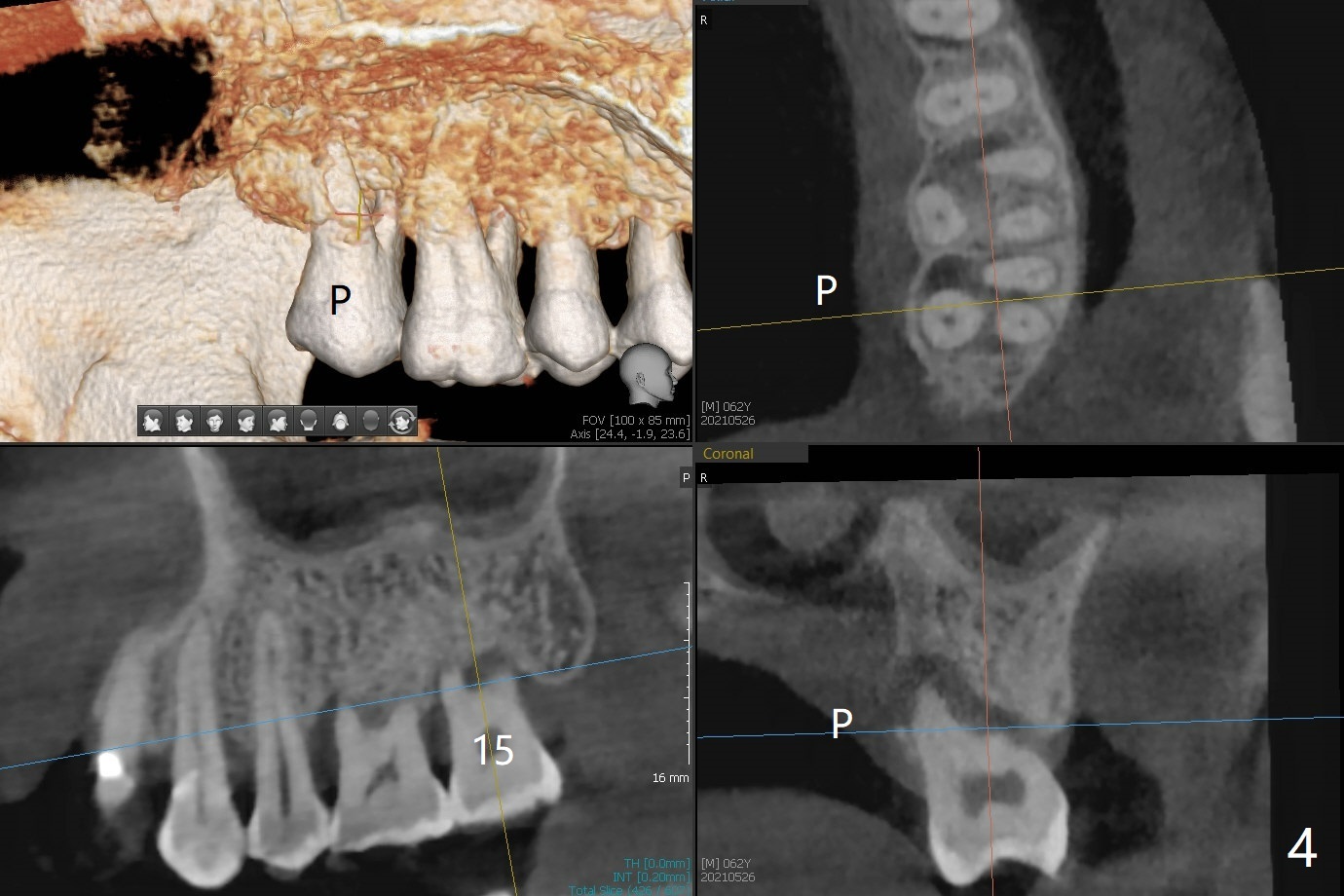
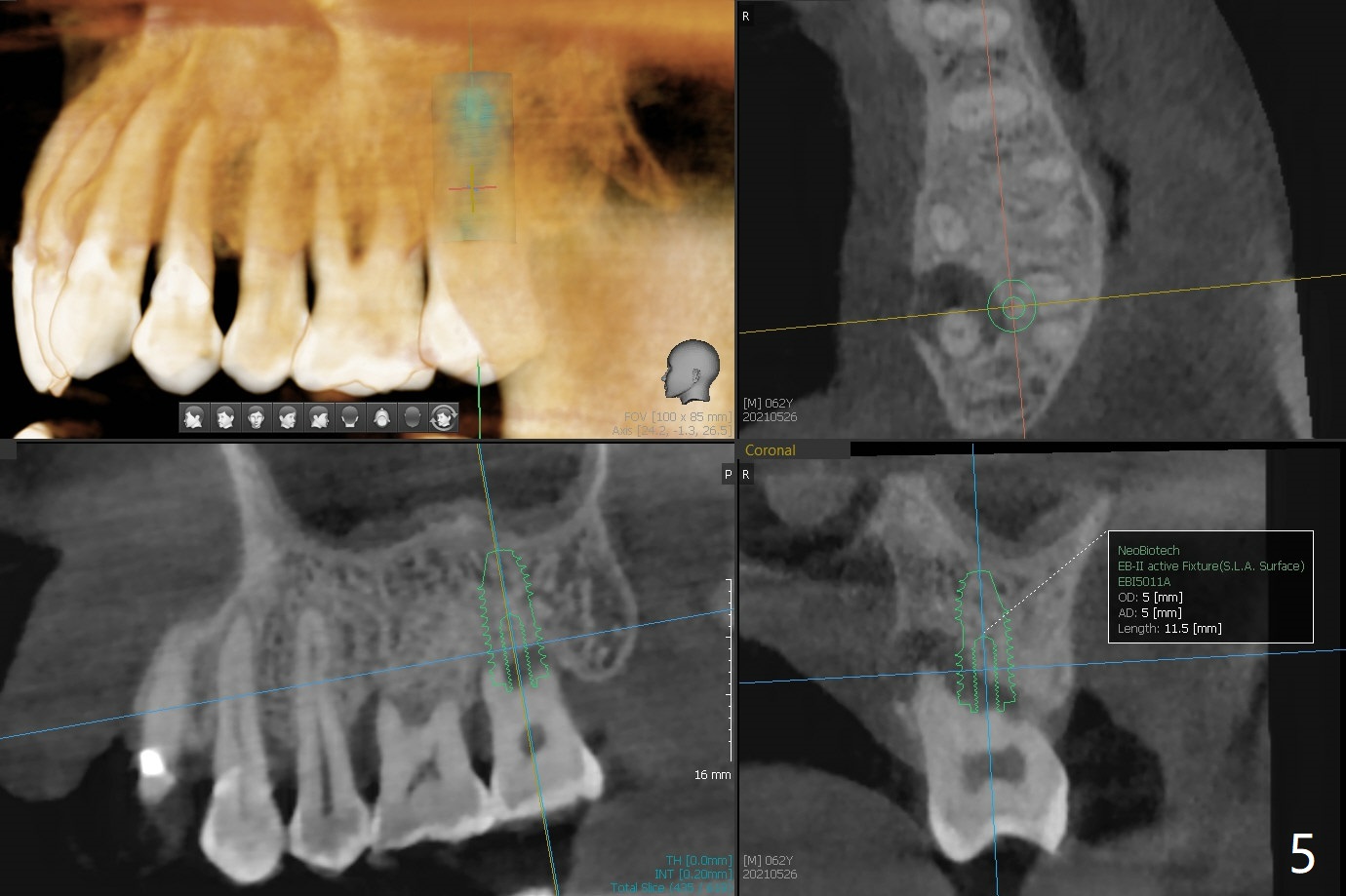
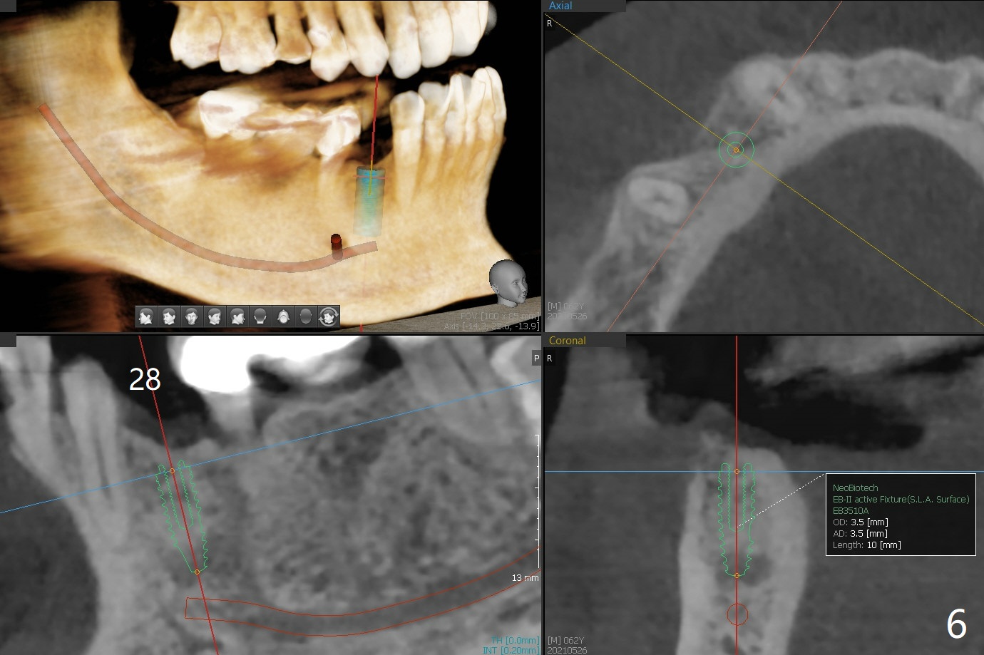
A 62-year-old man with sign of bruxism (#19 porcelain chip (Fig.2), 28 fracture/extraction) complains of severe mastication pain at #15 (Fig.1). More severe pain is elicited when the buccal cusps bite on a bite stick than the palatal one (Fig.3 with suspicious crazing lines). CT shows bone loss around the palatal root (Fig.4 P). A 5x11.5 mm implant will be placed immediately (Fig.5). The bone at #28 is able to hold a 3.5x13 mm implant (Fig.6). It is extremely difficult to take photos for 2nd molar crack line with a regular camera (Fig.3). The Shining Oral Scanner acts additionally as an intraoral camera. As a busy clinician, I do not have time to take photos for #19 porcelain chip. My assistants take over the task. After work, I am able to have bumper harvest. With the information just mentioned, I will be in a better position to present a more comprehensive treatment plan to the patient when treatment at #15 is finished: remove #18-20 bridge, place an implant at #19 and fabricate new crowns at #18 and 20.
Return to Prevent Molar Periimplantitis (Protocols, Table) Protect Graft Metronidazole Oral Scanner Surgery Xin Wei, DDS, PhD, MS 1st edition 05/26/2021, last revision 07/15/2021