|
|
|
|
|
|
|
|
|
|
|
|
|
|
|
|
|
|
Sinus Lift Post Osteotomy
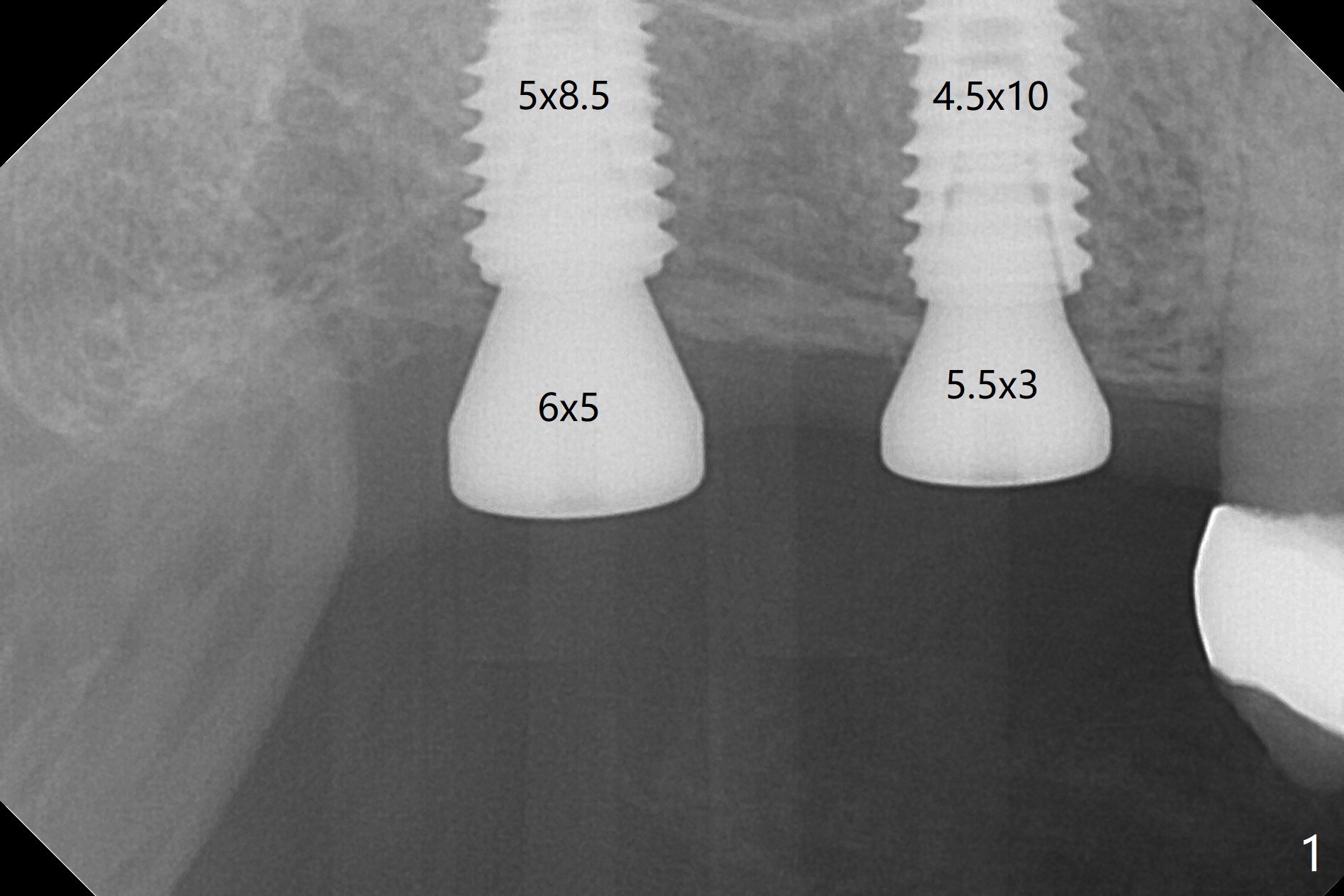
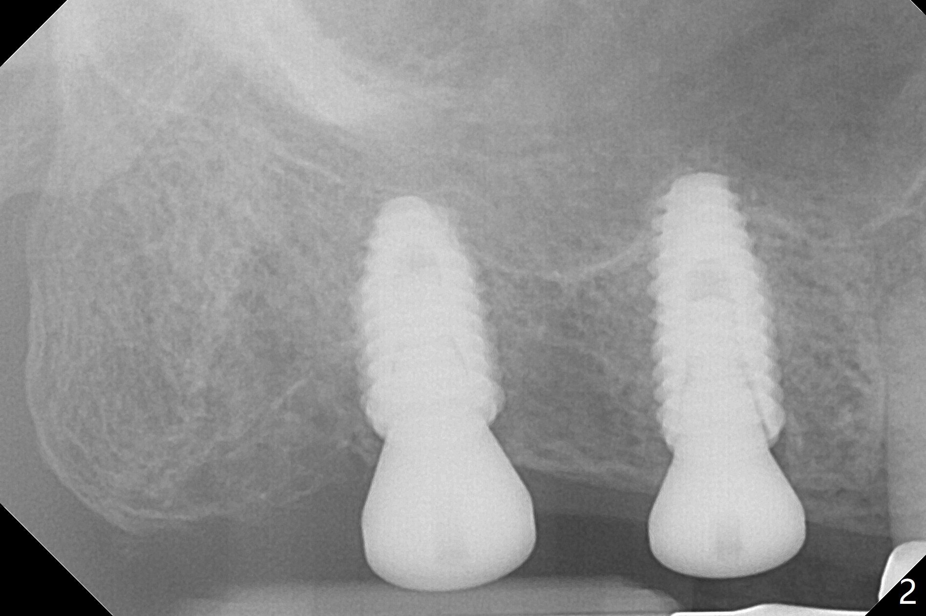
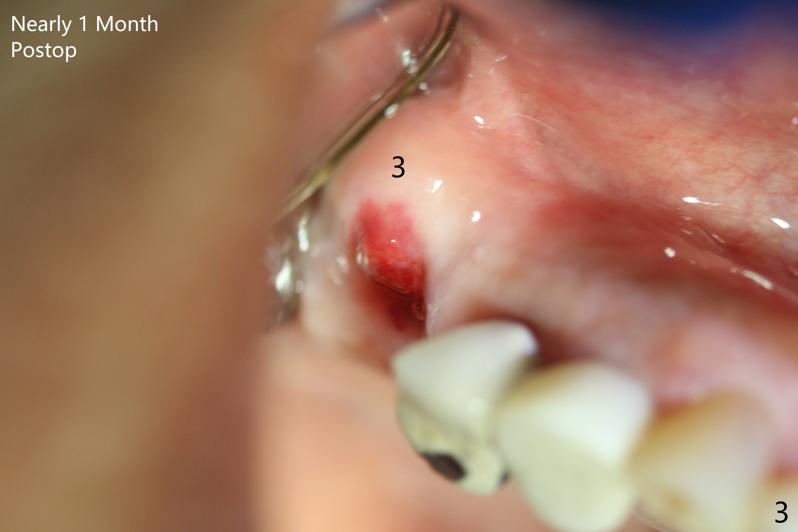
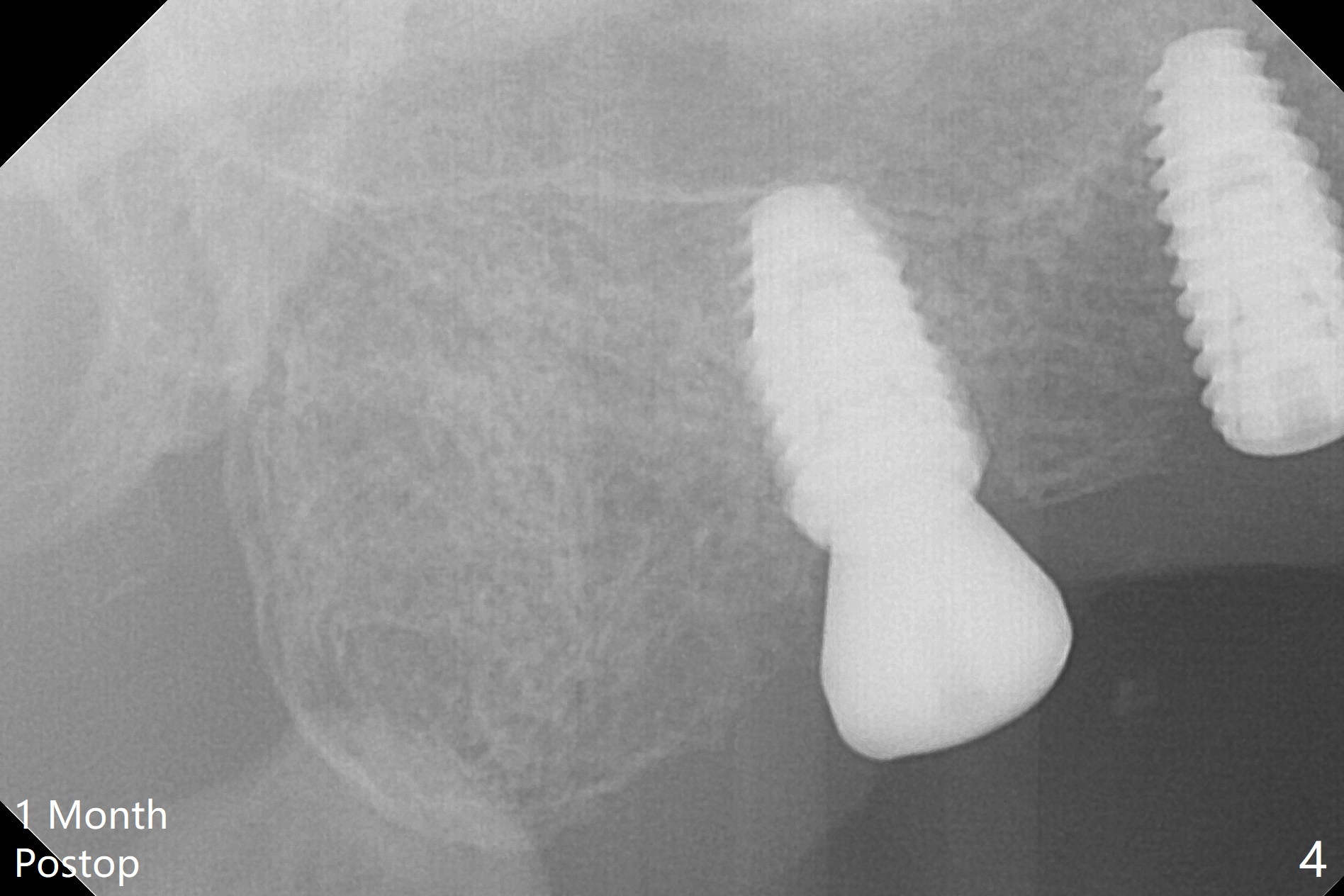
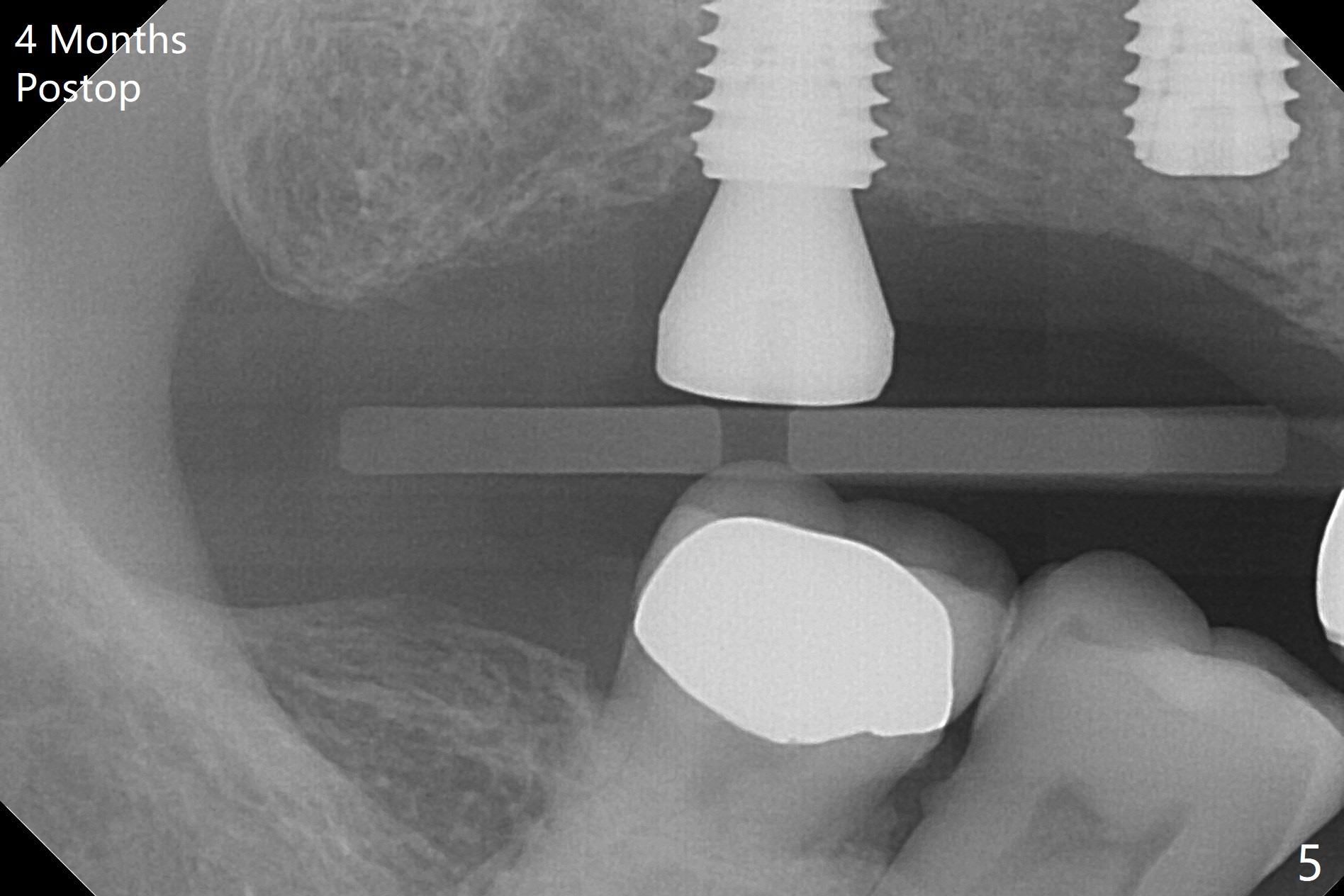
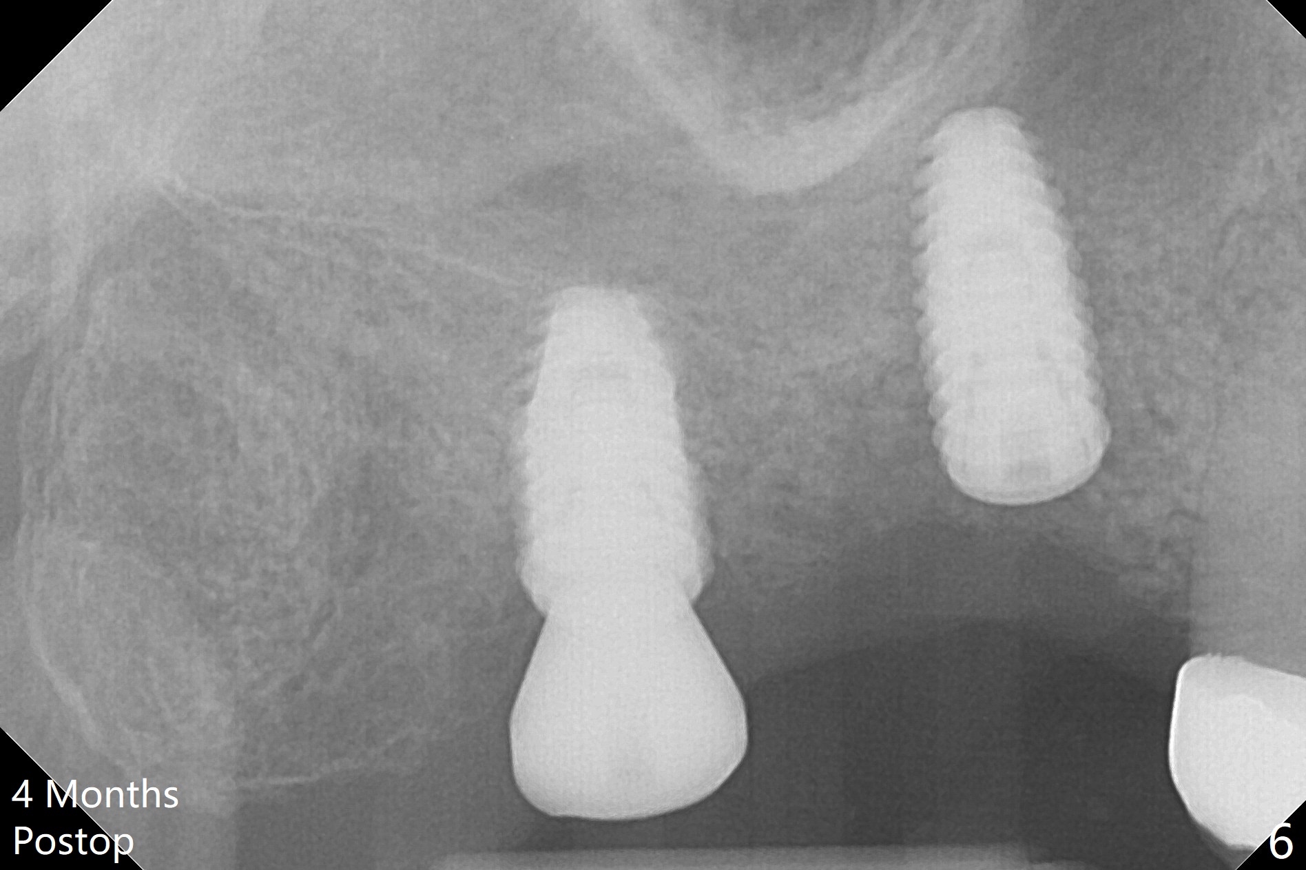
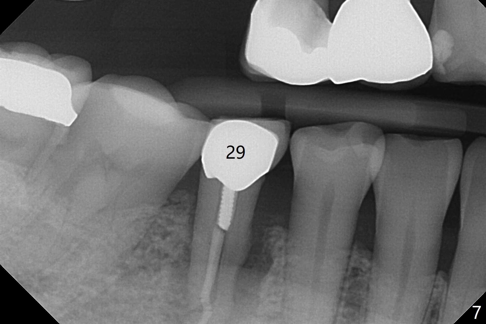
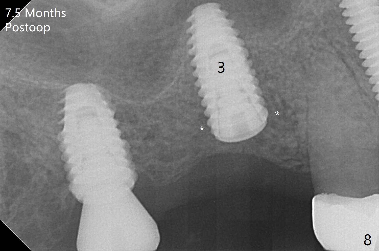
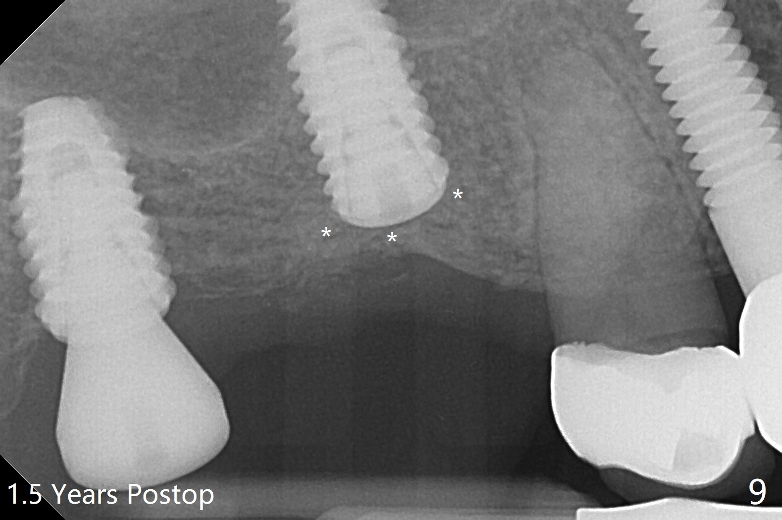
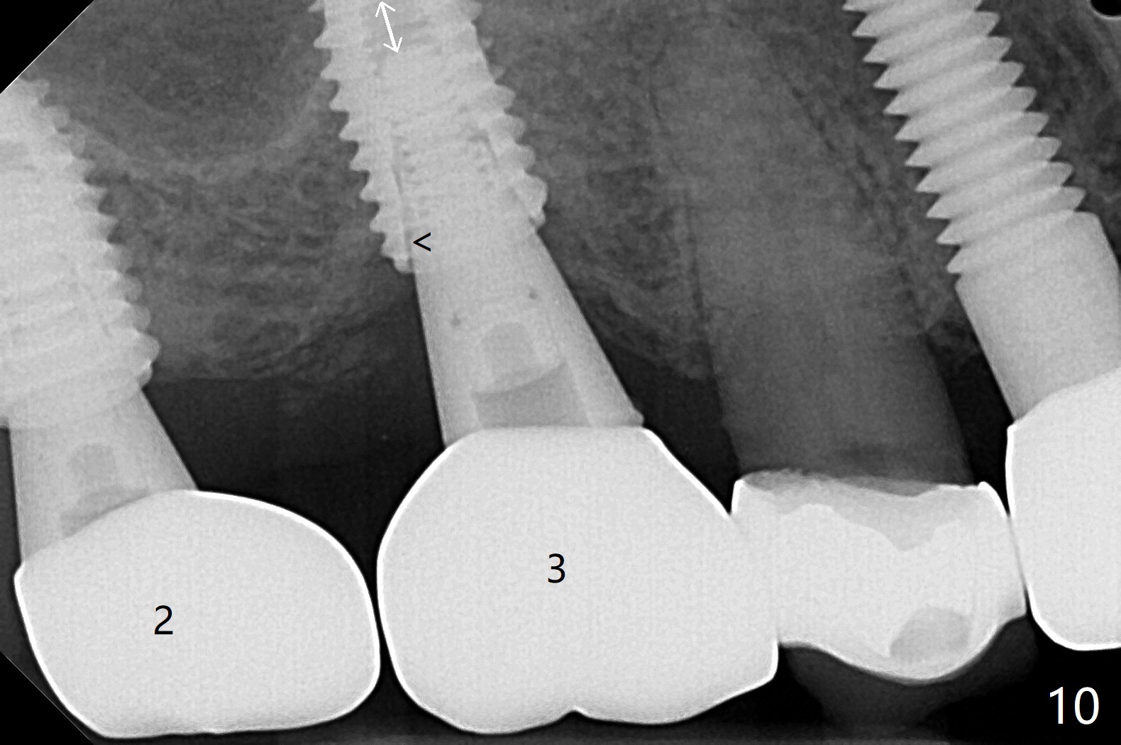
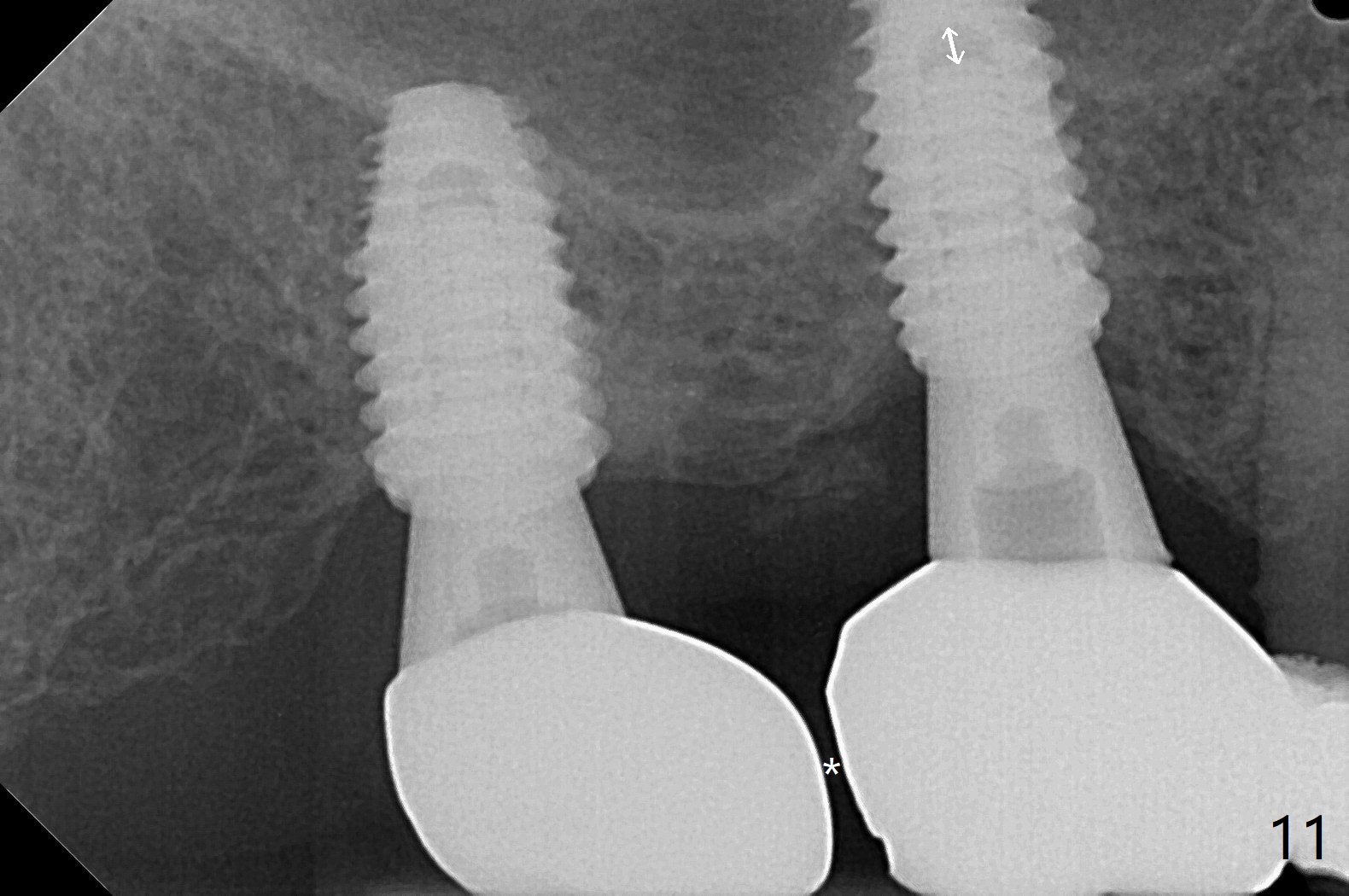
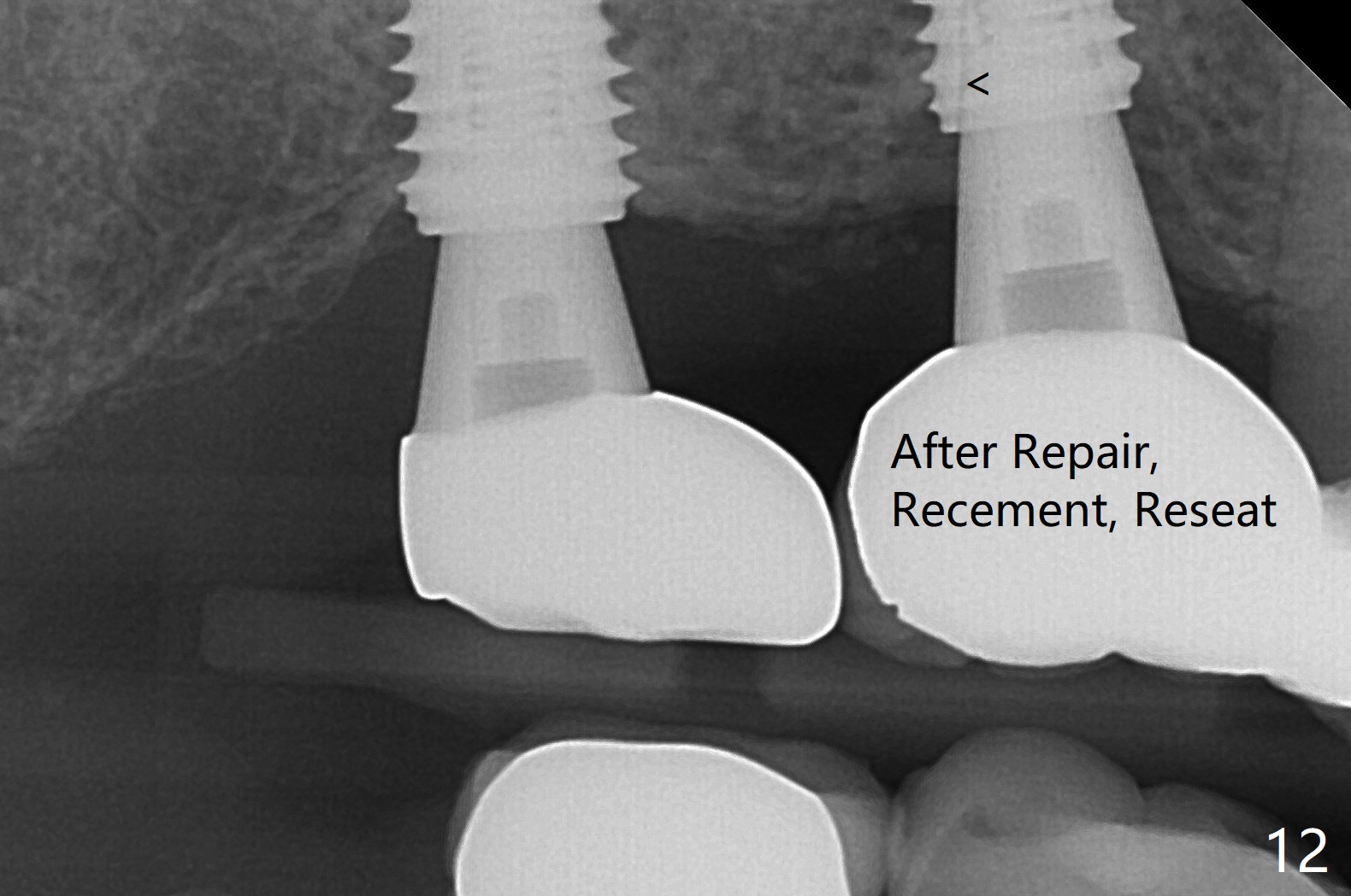
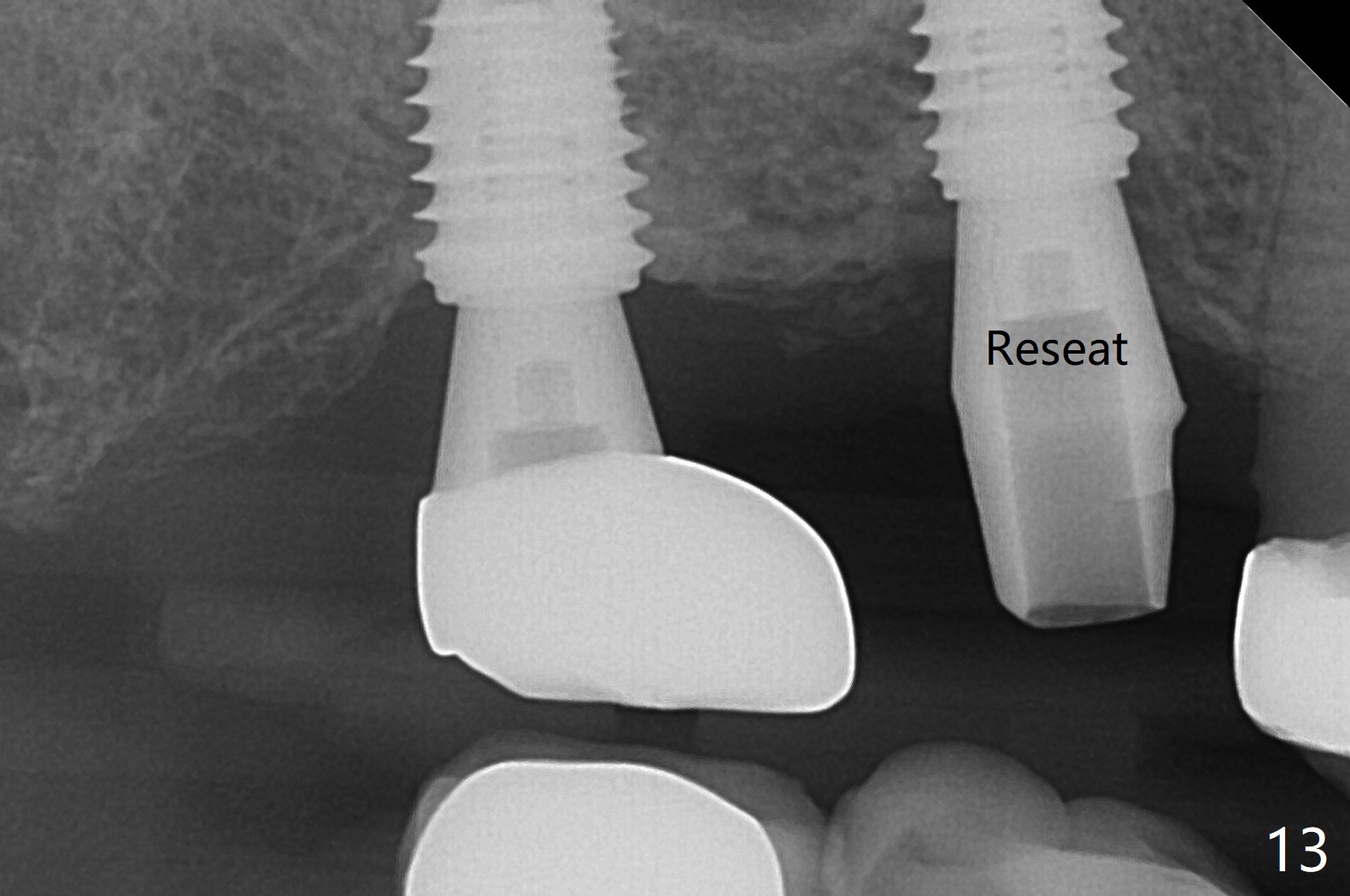
Osteotomy at the sites of #2 and 3 goes on as planned. Sinus lift using DIO 2.8 mm round bur with 6 and 7 mm stoppers (respectively) is carried on without certainty, because it is difficult to feel the stop through the osteotomy. The crest may be uneven or soft. It appears that the soft tissue landmark as a stop may be easier to identify. In fact a 3 mm IBS Magic Expander (an osteotome) was used for sinus lift at #3. The 2 implants are placed with 20 and 30 Ncm (Fig.1,2). Healing abutments are inserted. At 2-week follow-up, the patient reports "pain a few days earlier, took a pill of antibiotic, pain gone. UR metal is sharp". She does not take antibiotic regularly. Exam reveals that there is heavy plaque around #2,3 healing abutments. The buccal edge of #2 abutment is trimmed for comfort. Nearly 1 month postop, #3 healing abutment dislodges with buccal gingival erythema and edema with purulent discharge (Fig.3). Healing screw is placed at #3 with Amoxicillin and Chlorhexidine prescribed. One week later, the implant at #3 turns when the healing screw is retightened (Fig.4). After debridement, Vanilla graft is placed. There is no infection at #2 or 3 four months postop (Fig.5,6); the lingual gingiva is erythematous and edematous at #29 with mobility II (Fig.7 (vertical root fracture)). Uncover is conducted at the site of #3; there is no infection superficial 7.5 months postop (Fig.8). There is a large buccal defect upon incision with dark hemorrhage. In fact the bone density is low crestally (Fig.8 *). Bone graft is placed for the 2nd time. Eleven months later (1.5 years post implant placement), the bone regrows crestal (Fig.9 *). The crown at #3 is loose 1 year 5 months post cementation (Fig.10). After proximal trimming (Fig.11 *), the abutment at #3 is seated completely. After lab repair, crown oral cement, crown/abutment removal for excess cement removal, the crown/abutment cannot be torqued >25 Ncm (Fig.12, 30 Ncm). PA shows incomplete seating (Fig.12 <). Then the crown is sectioned so that the abutment has more freedom to be seated completely with pressure against the gingiva (the patient feels pain, Fig.13). Torque is 30 Ncm. After crown cementation, the abutment will not be removed for cement removal.
Return to Upper Molar Immediate Implant, Armaments 导板与提升 Torque
Xin Wei, DDS, PhD, MS 1st edition 03/28/2018, last revision 04/19/2021