|
|
|
|
|
|
|
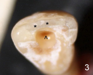
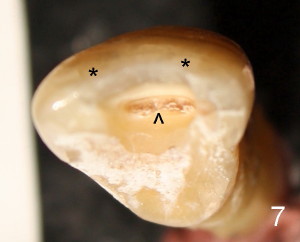
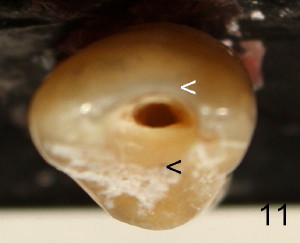
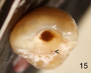
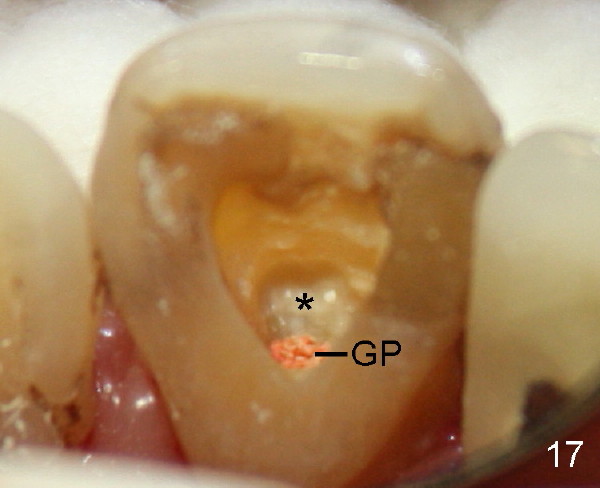
Initial access shows that the labial portion of the recessed pulpal horn (brown, < in Fig3 (occlusal view) is not fully exposed, while that the most lingual aspect of the incisal edge (between **) has been removed. Further access appears to be necessary to expose the obliterated pulpal chamber (Fig.7 <) and more of the incisal edge has been violated ( between **). With good exposure, the canal is easily found around the arrowhead (<) in Fig.7. Fig.11 shows the access after application of Gates-Glidden files. Before rotary files, the access is enlarged further both labially and lingually with diamond/carbide endo access burs (Fig.15). Traditional way to get access actually sacrifices more lingual structure (Fig.11,15 black <) than the labial one (white <) or creates a ledge (Fig.17 (mirror view) *) labial to the orifice (gutta percha (GP). Return to main article
Xin Wei, DDS, PhD, MS 1st edition 10/02/2011, last revision 10/10/2011