|
|
|
|
|
|
Pathogenesis of Globulomaxillary Cyst
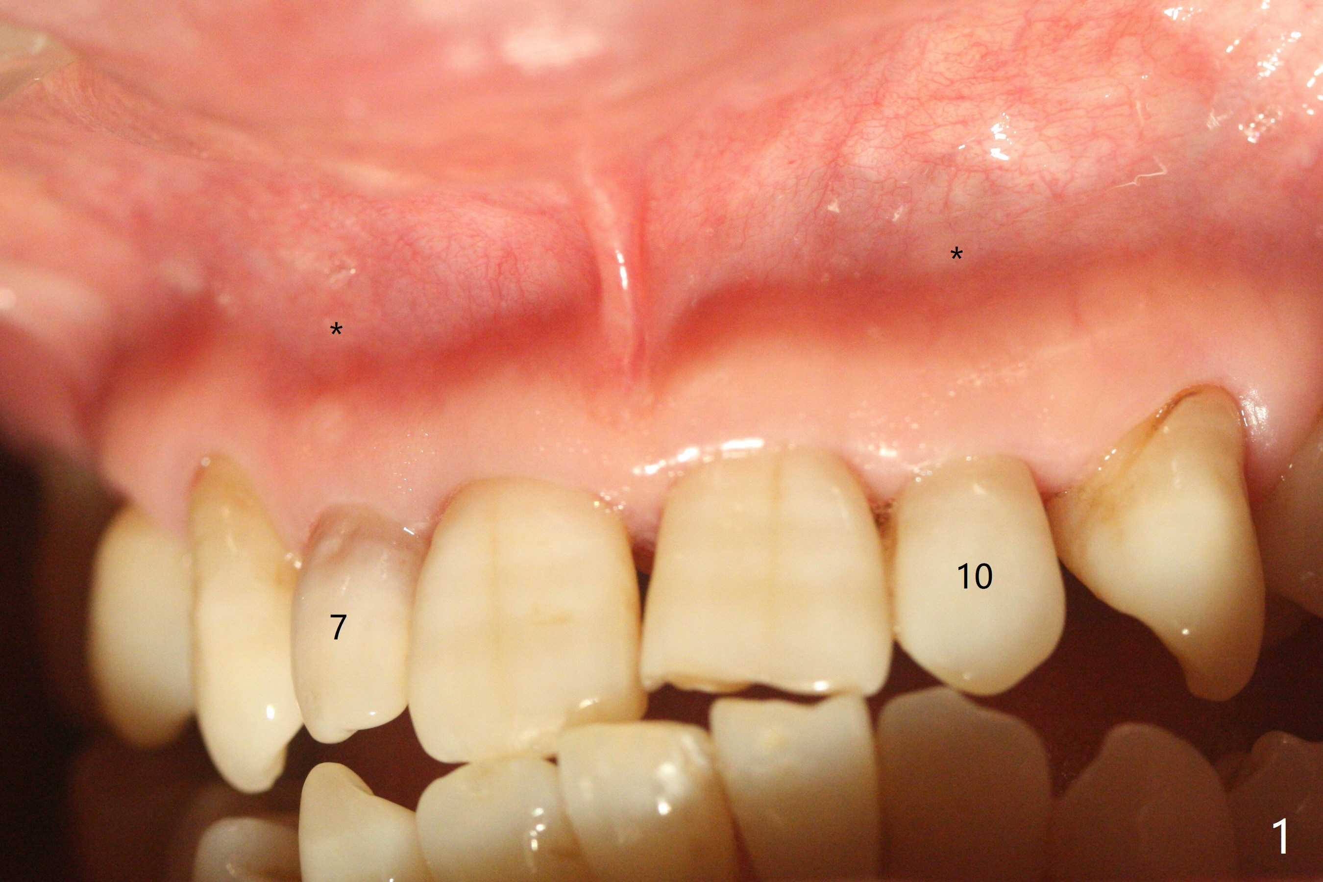
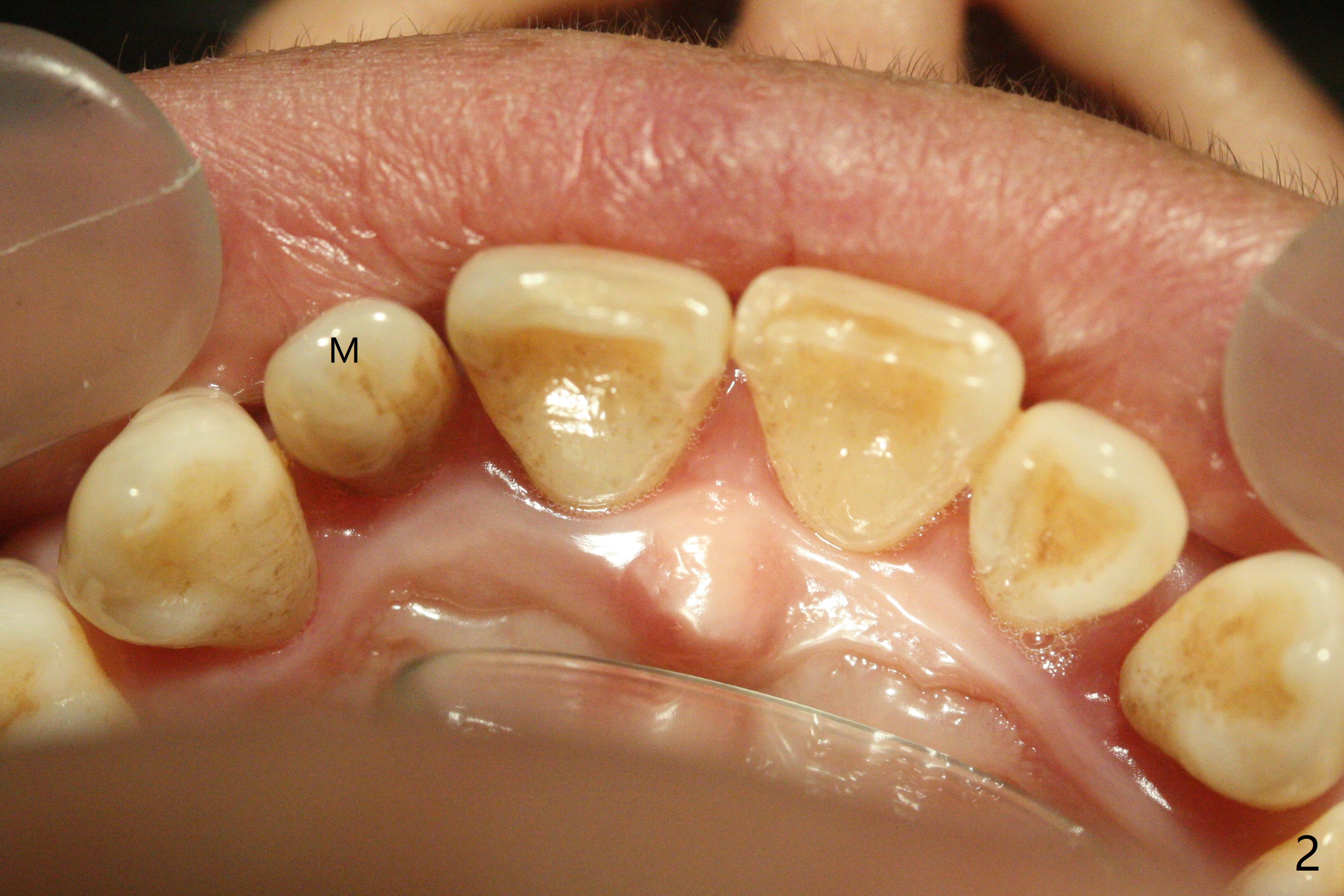
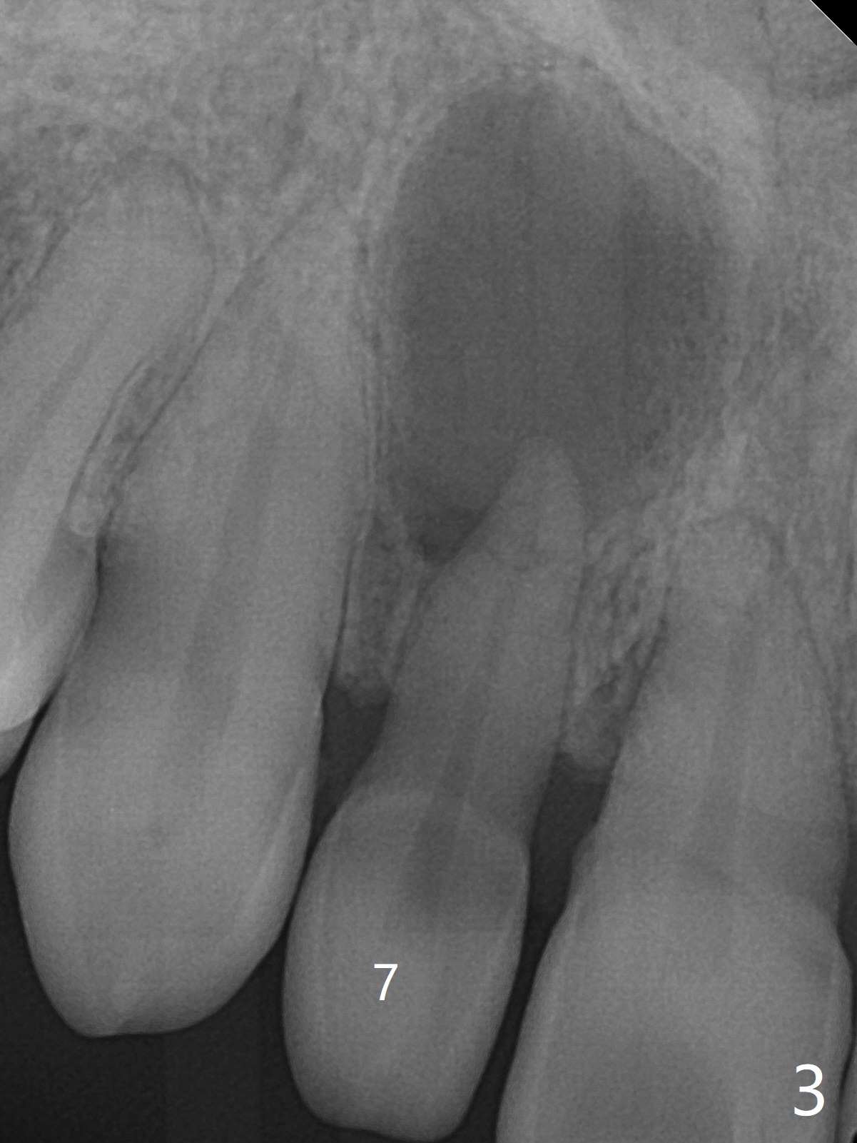
A 45-year-old woman presented to office for new patient examination. The latter reveals discoloration (Fig.1 necrosis on pulpal test; * labial concavity), microdontia with possible root canal malformation (Fig.2: M) and large inverted pear-shaped cyst (Fig.3) of the tooth #7. Guarded prognosis will be emphasized prior to treatment.
Return to
Professionals
1st
Case
Xin Wei,
DDS, PhD, MS 1st edition 02/27/2021, last revision
03/01/2021