|
|
|
|
|
|
|
|
Composite After RCT
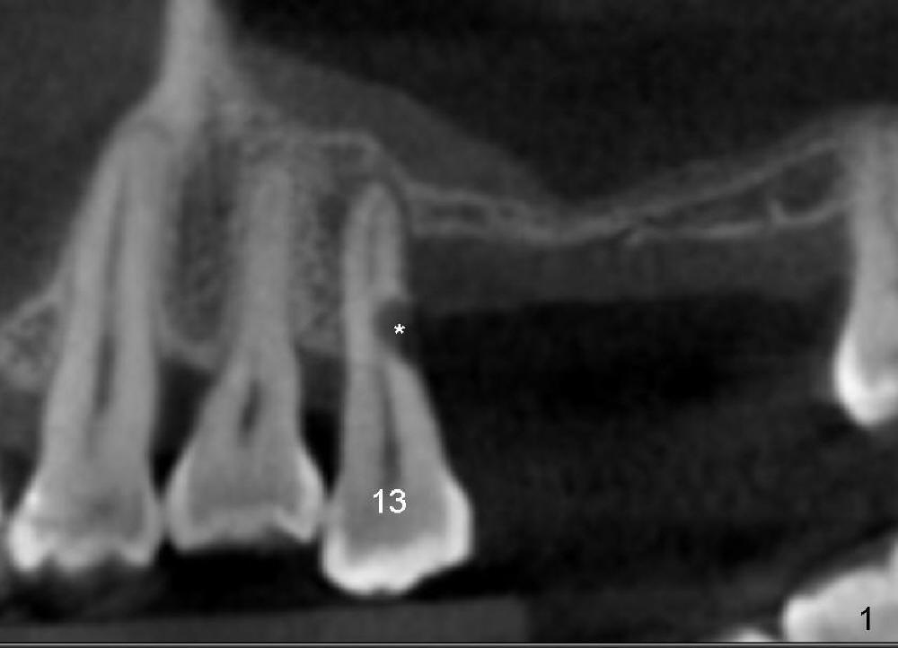
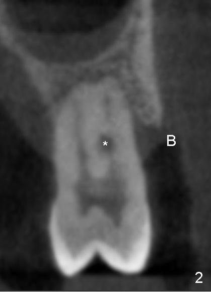
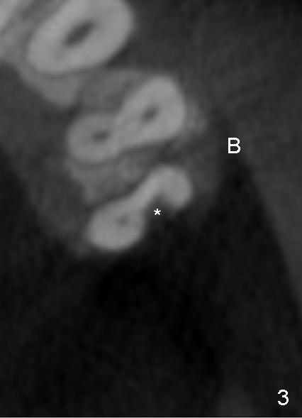
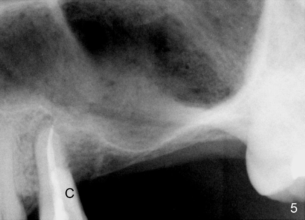
A 44-year-old man has severe periodontitis. The tooth #13 has a distal cervical carious lesion with spontaneous pain (Fig.1 (CT sagittal section) *). The caries is continuous with the buccal canal (Fig.2 (coronal), 3 (axial section) *).
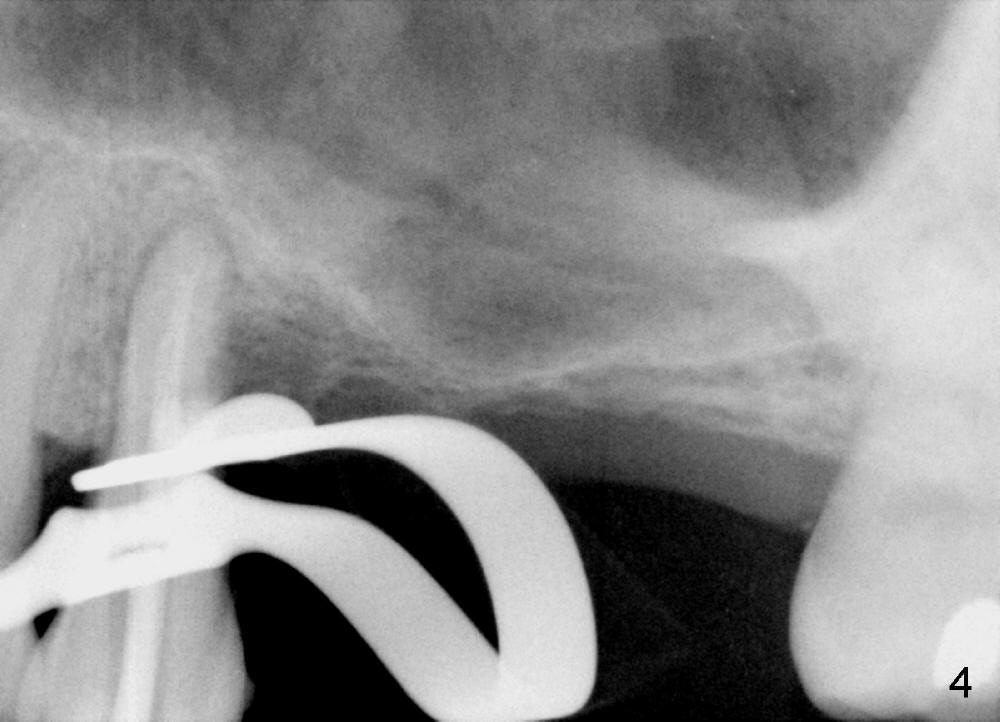
After local anesthesia and rubber dam isolation, caries is removed from the distal cervical region. Access is provided occlusally. Initial debridement is done with hand files until #20 with copious irrigation with 3% sodium hypochlorite. The shortcoming is bleach leakage. While #15 and 20 files are inserted in the buccal and lingual canals, the distal cavity is filled with Cavit temporarily. There is no more leakage, while the canals remain patent. RCT is finished with ease (Fig.4). Then the distal cervical Cavit is removed, followed by routine composite build up (Fig.5 C). The patient remains asymptomatic for 2 months now.
Return to Professionals
Xin Wei, DDS, PhD, MS 1st edition 11/23/2014, last revision 11/23/2014