|
|
|
Why does RCT Fails in Abutment Tooth?
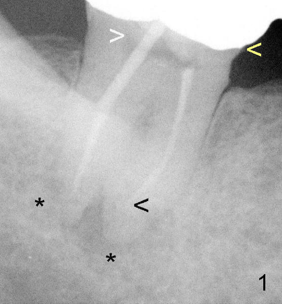
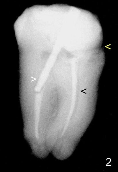
Fig. 1 and 2 show the abutment tooth #32 in vivo and in vitro, respectively. Yellow < in Fig.1,2 point to open margin in the mesiobuccal aspect of the crown/tooth. While the white > in Fig.1 points to unfilled pulpal chamber, the one in Fig.2 shows more clearly the void around the post in the distal canal. Oblique projection shows curved void associated with the mesial canal (Fig.2). RCT failure is complex, both endodontic and restorative (coronal leakage). Return to main text
Xin Wei, DDS, PhD, MS 1st edition 09/20/2011, last revision 09/21/2011