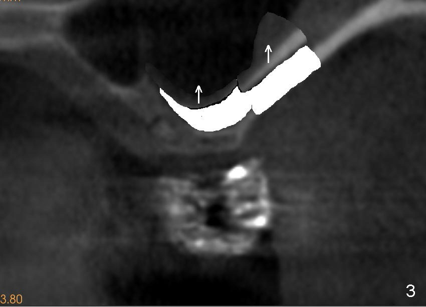
Fig.3 (illustration): The lateral wall of the maxillary sinus and sinus membrane have been lifted (arrows).
Return to Design
Xin Wei, DDS, PhD, MS 1st edition 04/18/2014, last revision 04/20/2014