|
|
|
|
|
||
 |
 |
 |
 |
||
 |
 |
 |
 |
||
 |
 |
 |
 |
||
 |
 |
 |
 |
 |
 |
 |
 |
 |
 |
||
 |
 |
 |
 |
||
 |
|||||
Trust and Royalty
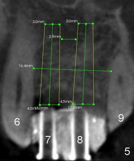
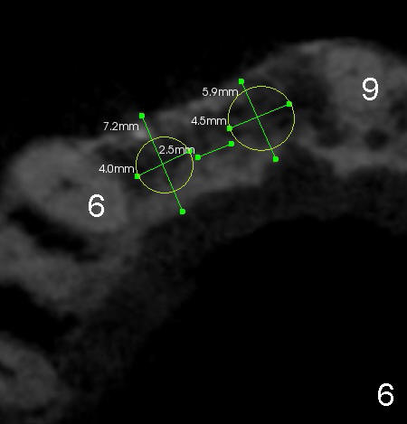
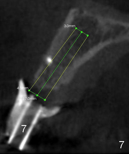
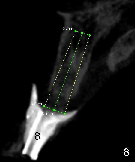
Sergio is from South America. He is seen by a Chinese physician. He also wants a Chinese dentist. That is how he ends up in my office. Every time he visits us, he shows appreciation with hugging and handshaking. We feel like that he is our family member. He has two dreams about his teeth: to have two fixed teeth for top ones and fix bottom crooked teeth (Fig.1). It appears that implants are the best solution to his first dream. But the bone and gums over the missing tooth areas shrink (Fig.1,2 *). Wax up is done on the model to see how the final teeth look like (Fig.3,4: #7,8). Surgical stent (#7,8 in Fig.5-8) is made for CT scan and treatment planning of implant placement.
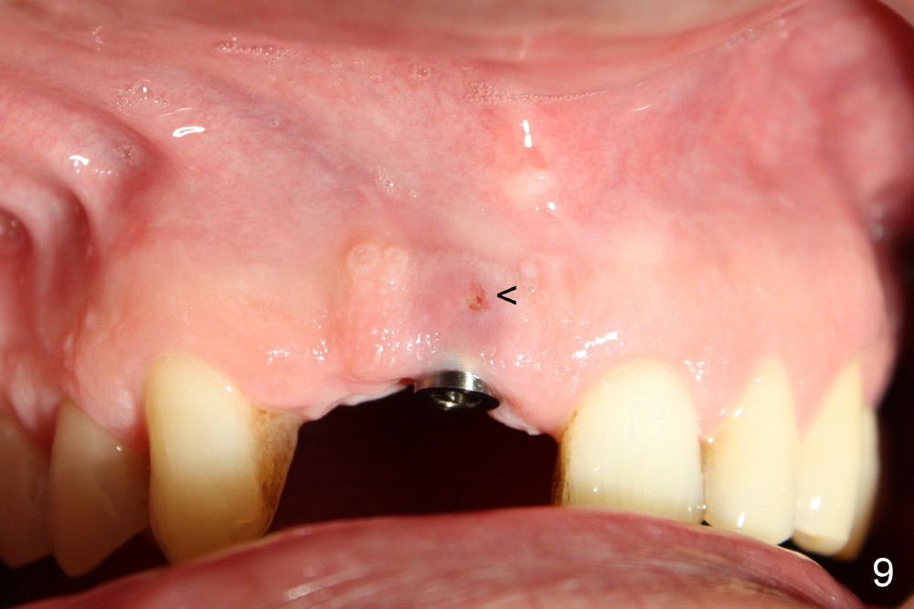
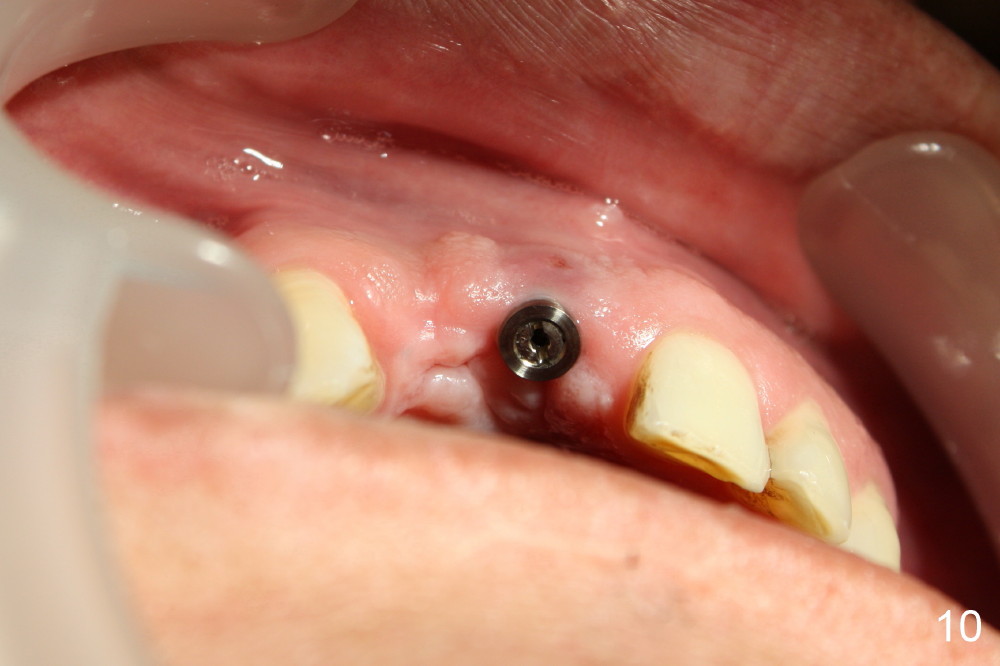
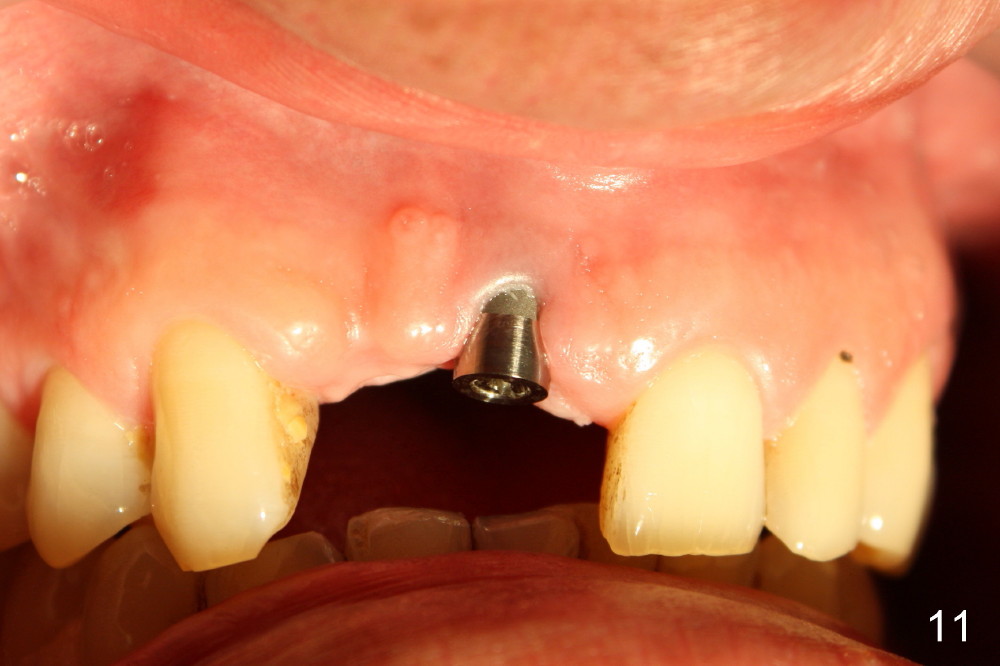
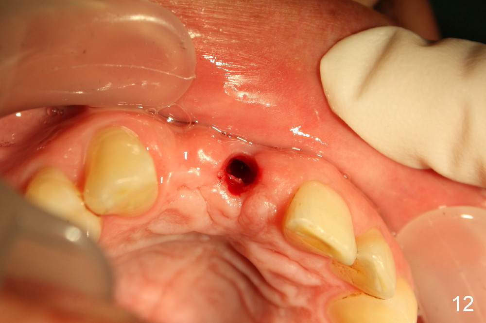
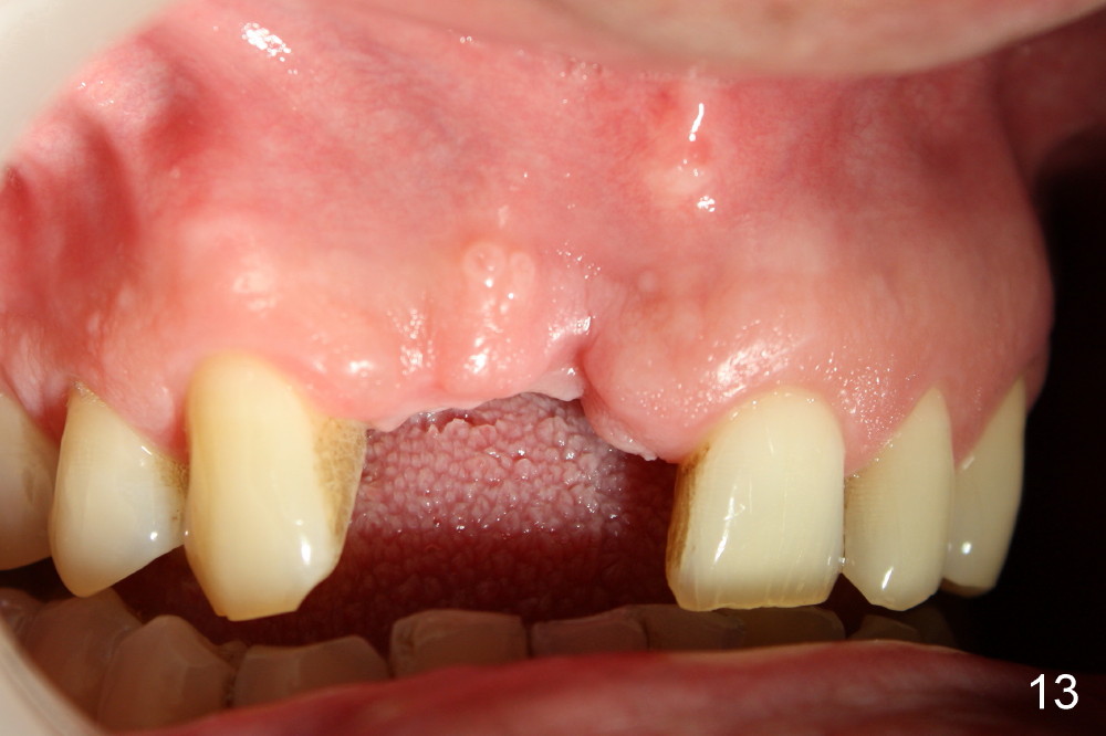
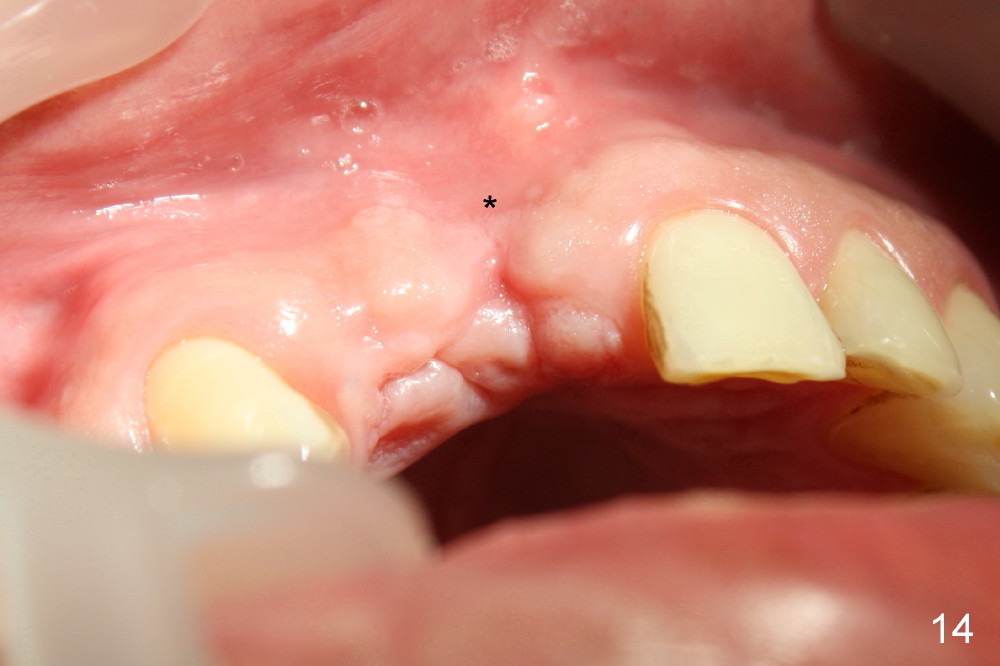
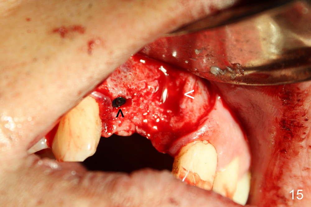
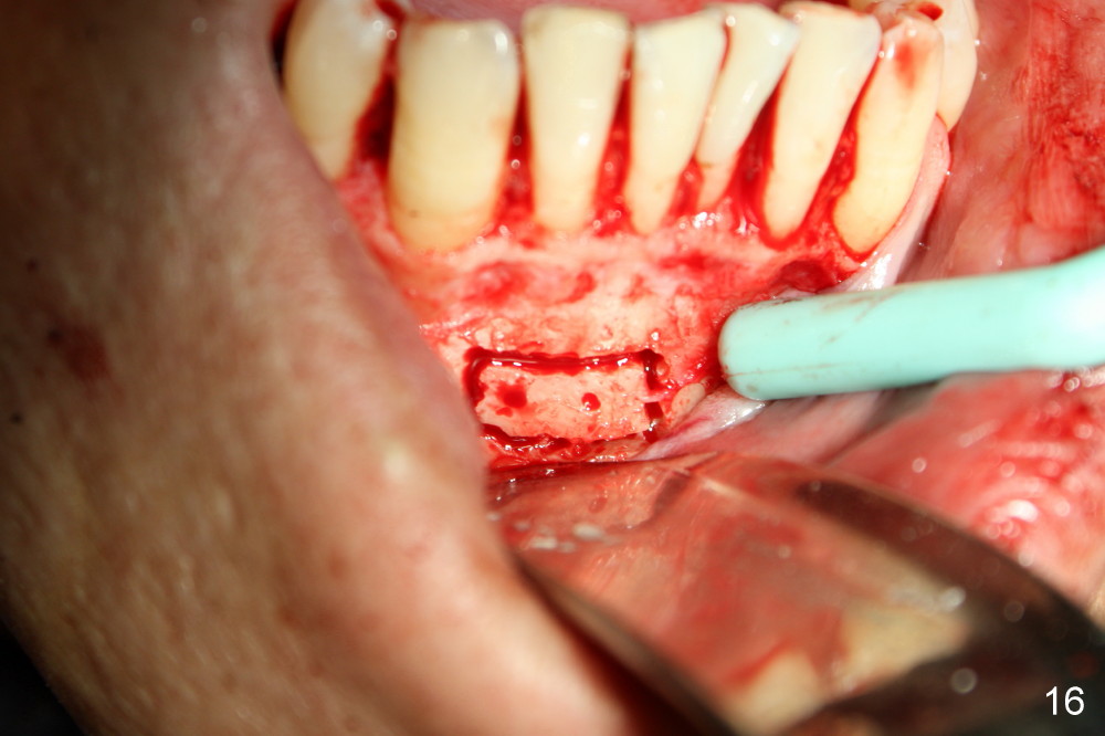
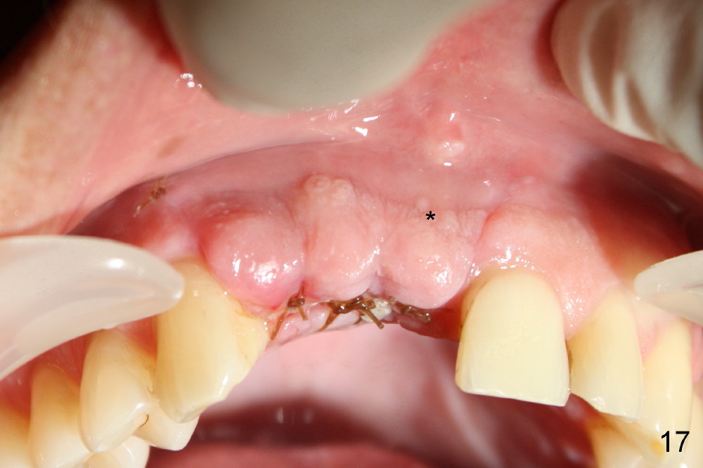
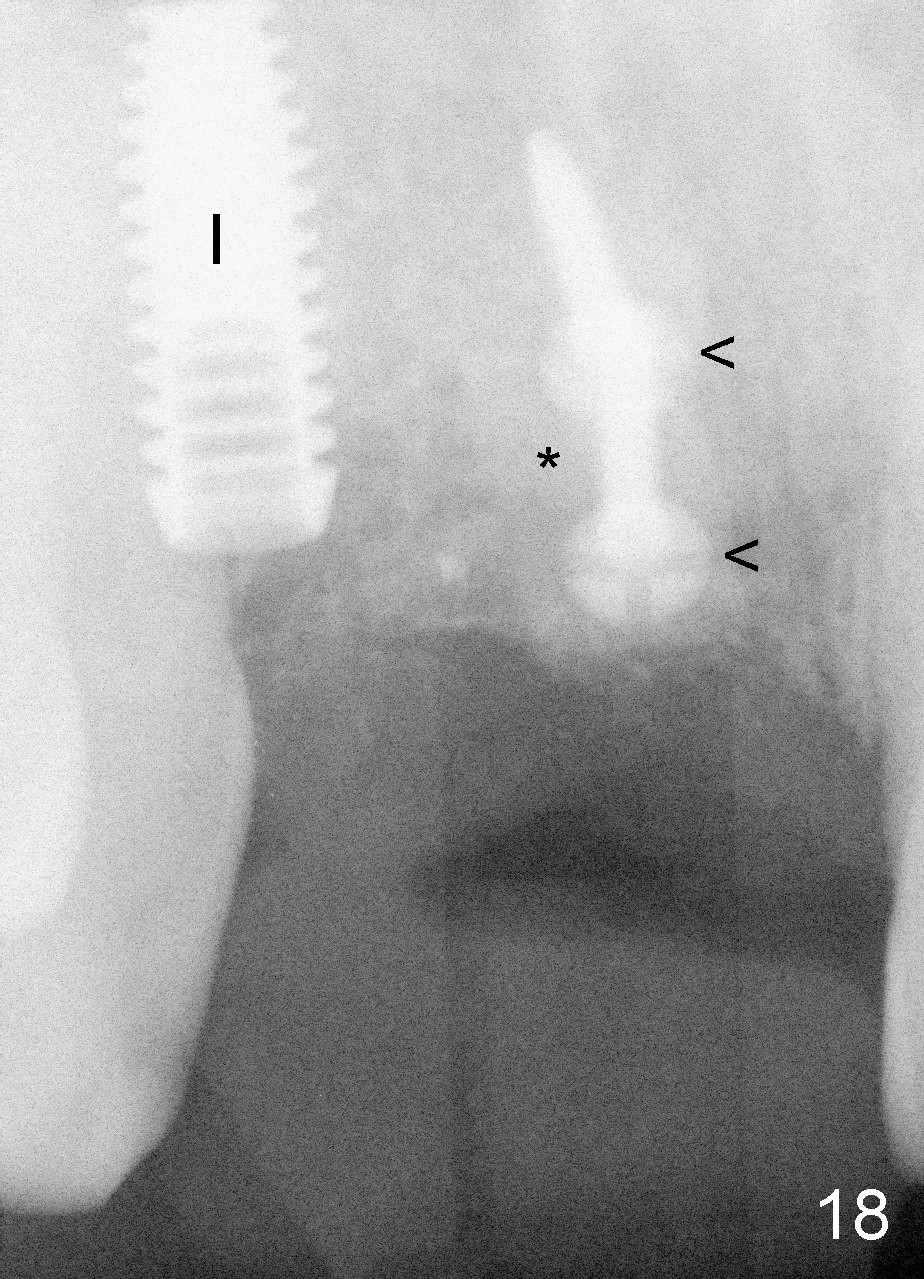
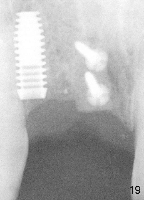
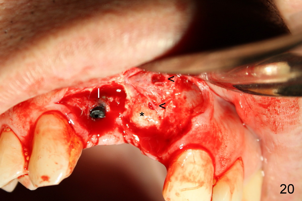
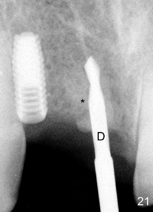
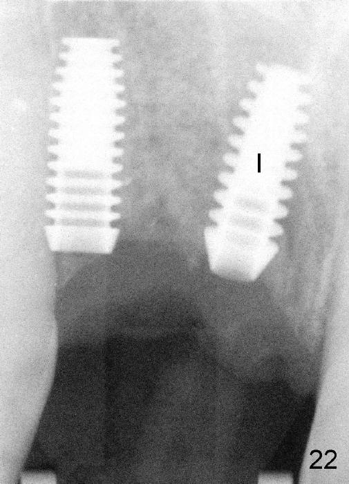
An implant is placed at the site of the tooth #8. It does not heal well (Fig.9 <, 10), gets worse (Fig.11) and has to be taken out (Fig.12). Everything needs to be done over again, but Sergio does not complains. Fortunately the wound heals, but the bone and gums shrinks more (Fig.13,14*). While an implant is placed at the site of #7 (Fig.15 black ^), the bony defect at the site of #8 (white <) is going to receive bone graft from the chin (Fig.16). One week later, the defect does not look so bad anymore (Fig.17*). X-ray shows the implant at #7 (Fig.18 I) and bone graft (*) and 2 fixation screws (<) at #8. Both the implant and the bone graft heals well a few months later (Fig.19). Fig.20 shows that the fixation screws (<) are just removed and that both the implant (I) and bone (*) heal. A drill is being used to create a hole underneath the bone graft (*) for future implant at #8 (Fig.21 D). Finally the implant is placed (Fig.22 I).
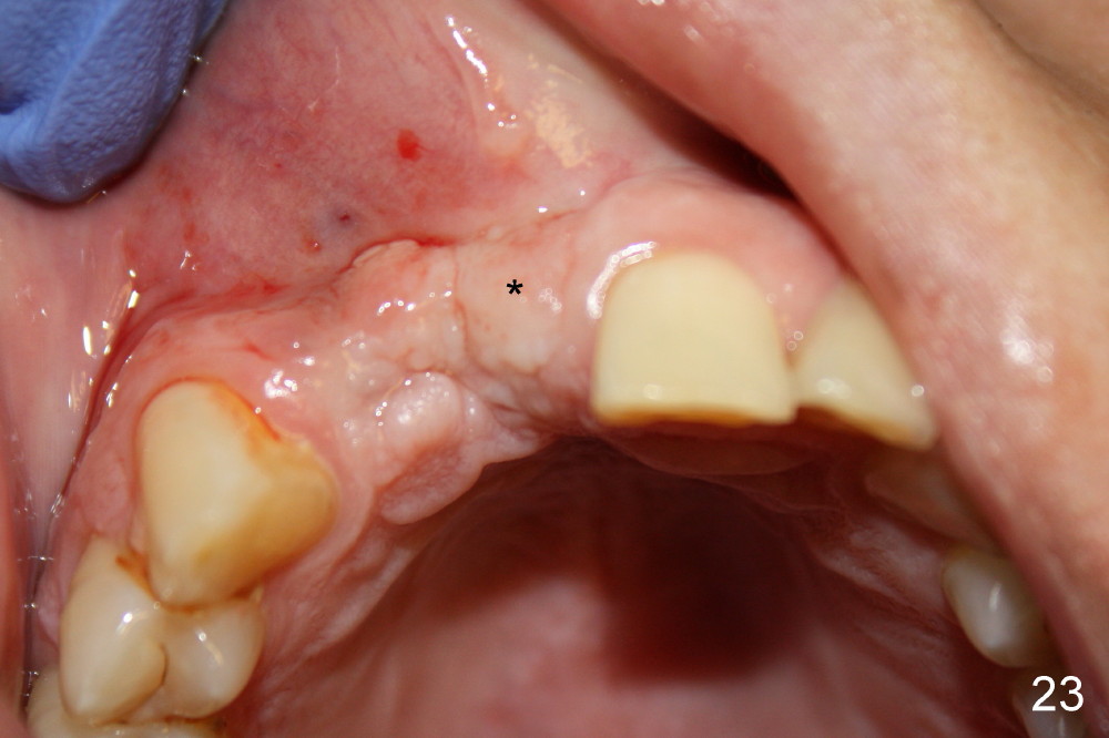
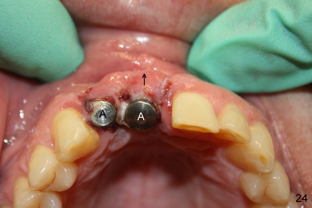
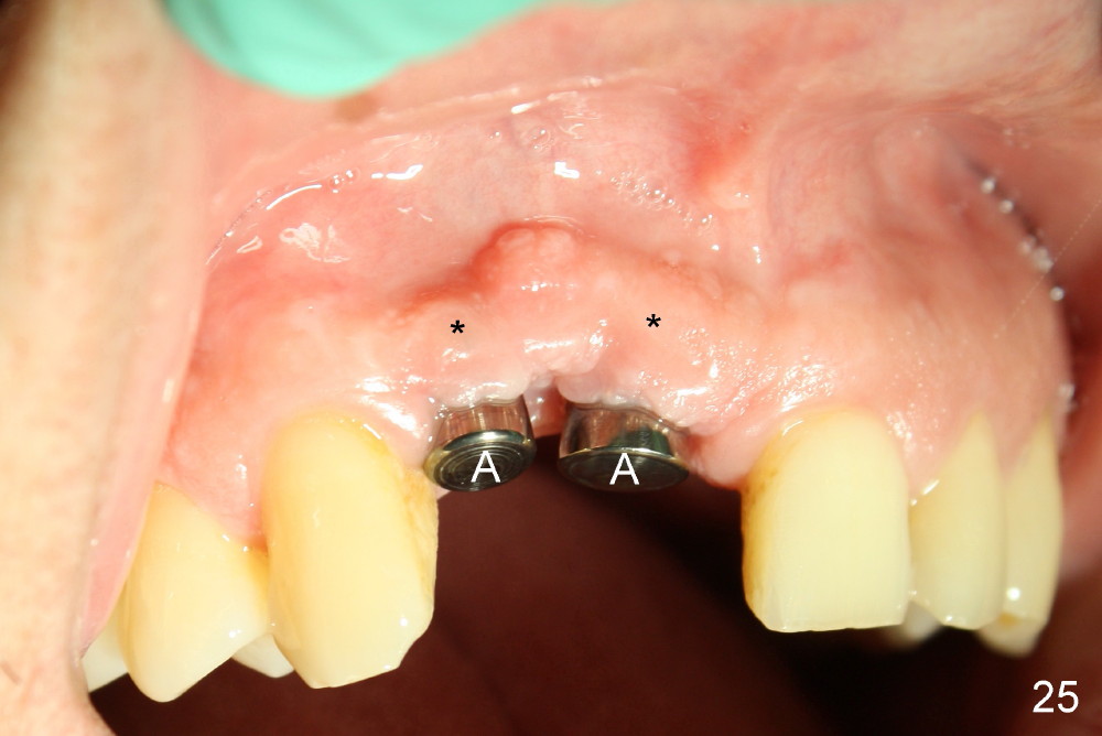
Time passes quickly particularly with the royal patient. We cannot wait to see him again. The wound heals wonderfully without sign of indentation on the gums (Fig.23 *). After small surgery, impression is taken and two temporary abutments (Fig.24 A) are installed to push the gums outward (arrow). Fig.24 is taken one day after the surgery. It looks again bad, but the patient feels great. In one month the gums (Fig.25 *) heal well around the temporary abutments (A).
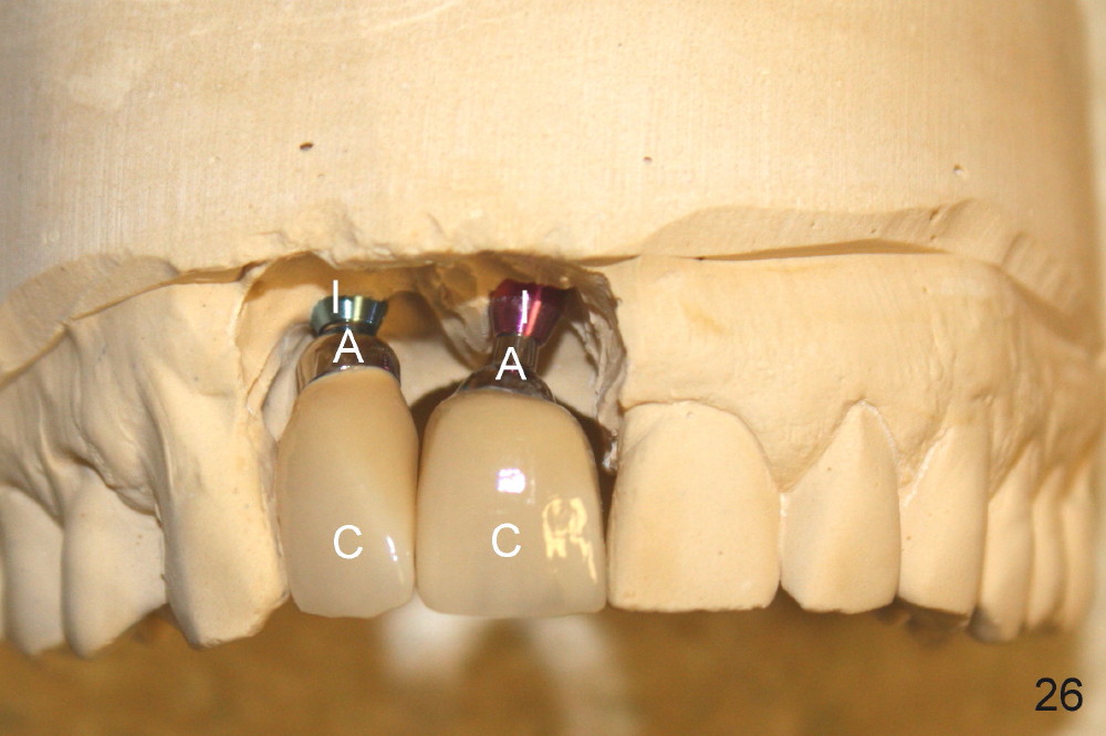
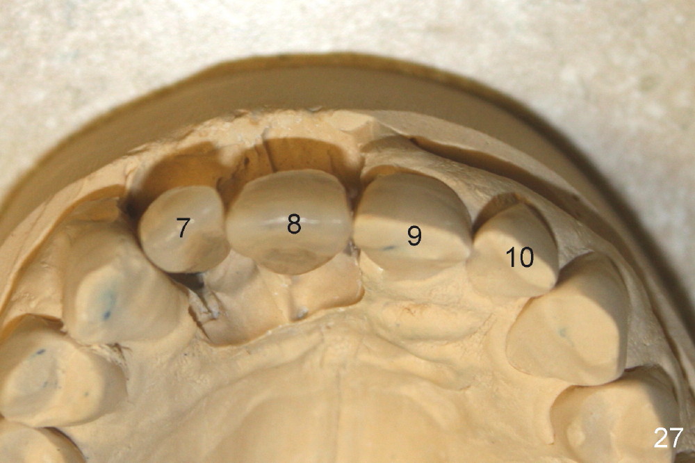
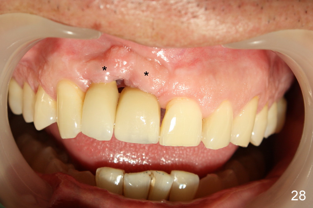
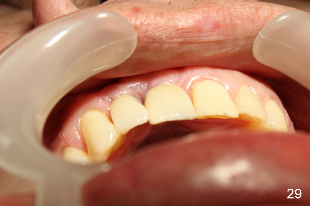
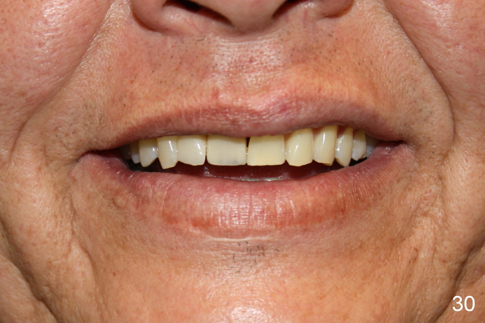
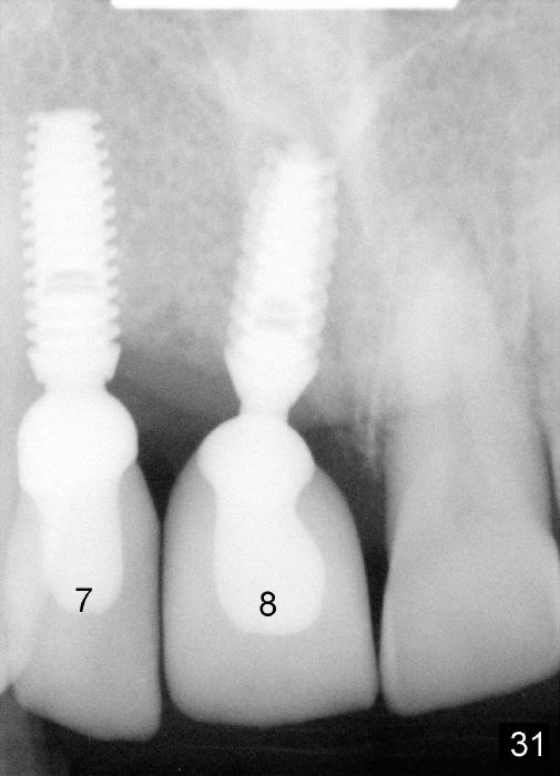
Fig.26 shows two crowns (C), permanent abutments (A), and implant analogs (I) in a dental model. The alignment of the four front teeth are unique (Fig.23 #7-10). After removal of the temporary abutments, the new crowns with the permanent abutments are inserted to the implants. The two new teeth look natural in his mouth (Fig.28, 29). The overlying gums are bulging (Fig.28*). The patient is as happy as ever (Fig.30). Fig.31 is X-ray taken after crown/abutment seating.
The hardest thing in dentistry is to handle complications associated with treatment. Patients with trust and royalty stick around to finish treatment. The result is good, as demonstrated in this case. In fact, there is no other better alternative.
Xin Wei, DDS, PhD, MS 1st edition 09/08/2013, last revision 09/08/2013