|
|
|
|
|
|
|
|
|
|
|
|
|
|
|
|
|
|
|
||
|
|
|
||||
How to Correct Discoloration after Luxation
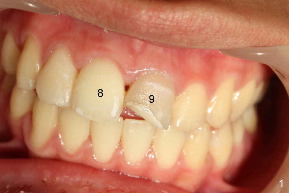
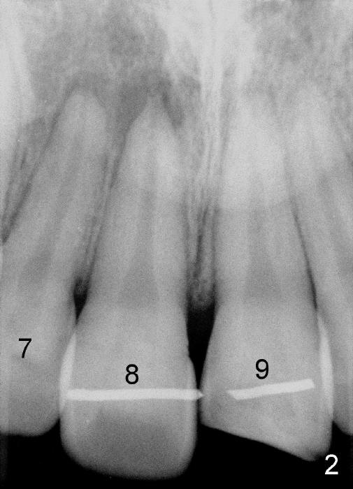
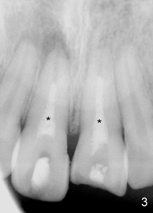
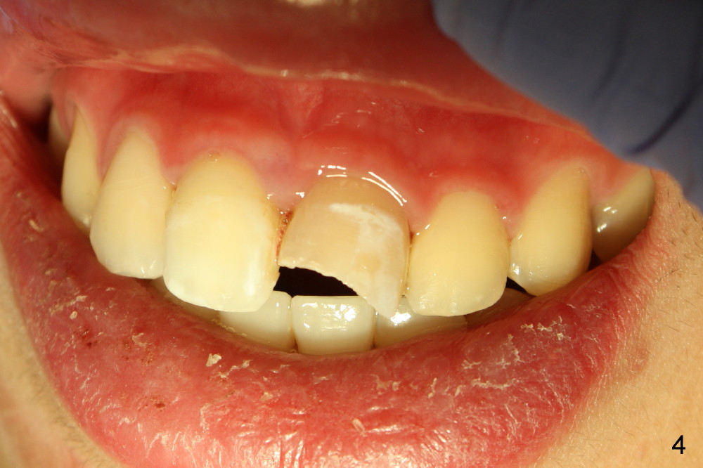
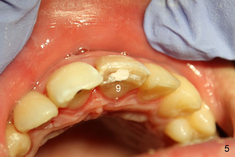
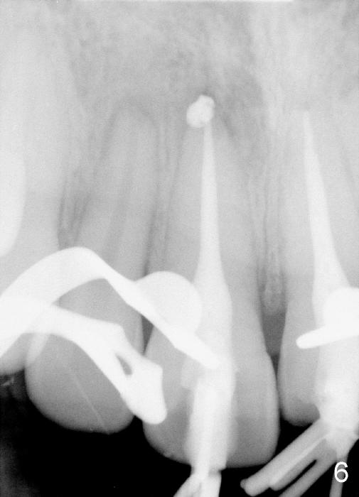
A 16-year-old boy had bicycle accident. The teeth #8 and 9 were luxated. They were placed back in the sockets in a timely manner and splinted. One month later, the tooth #8 had percussion, while #9 discolored (Fig.1). Although there is periapical radiolucency associated with #7 (Fig.2), RCT was initiated for #8,9 (Fig.3 *: CaOH paste). The shade of #9 improved post initial RCT (Fig.4,5).
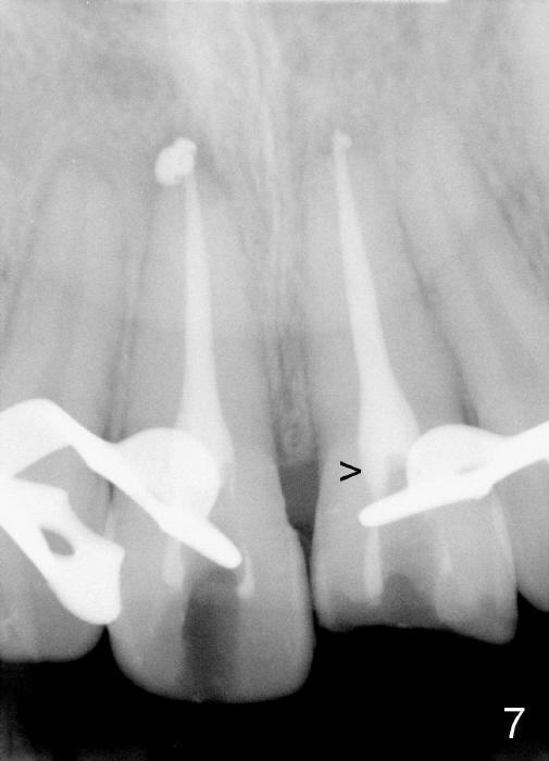
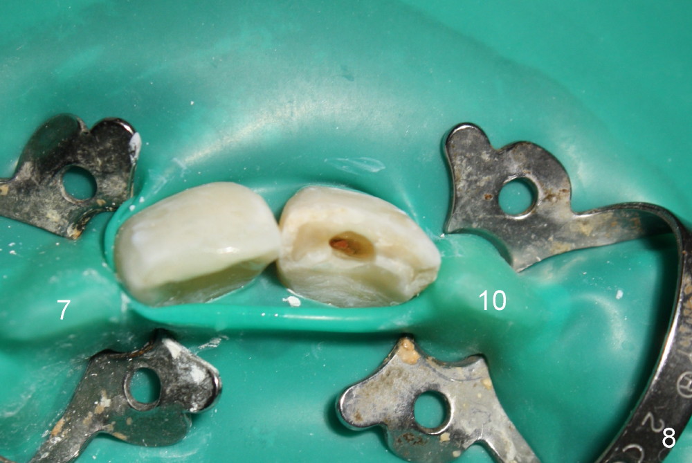
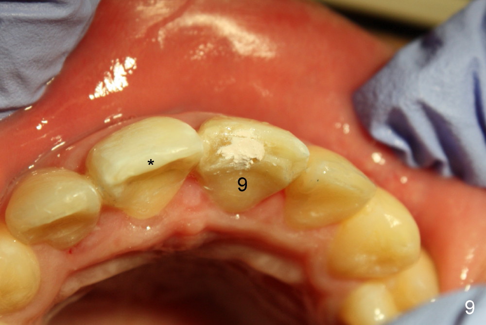
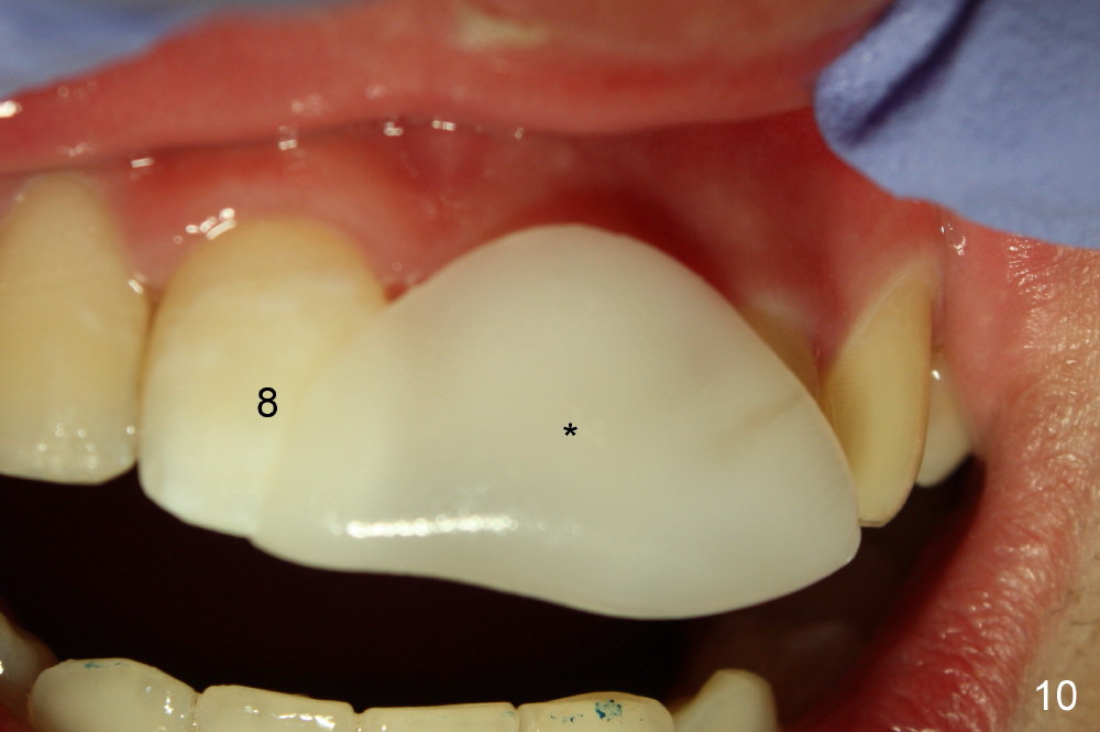
The patient returned to finish RCT 1.5 months later (Fig.6). Gutta percha was removed as much (high) as possible (Fig.7 >). In order to prevent butterfly rubber dam clamp interference in access and GP removal, smaller clamps should be placed in the neighboring teeth (Fig.8). When RCT was done, the tooth #9 was still the darkest (Fig.9). Thermoplastic tab was used to make local external bleaching tray (Fig.10 *). The patient and his mother were instructed to do external and internal bleaching daily.
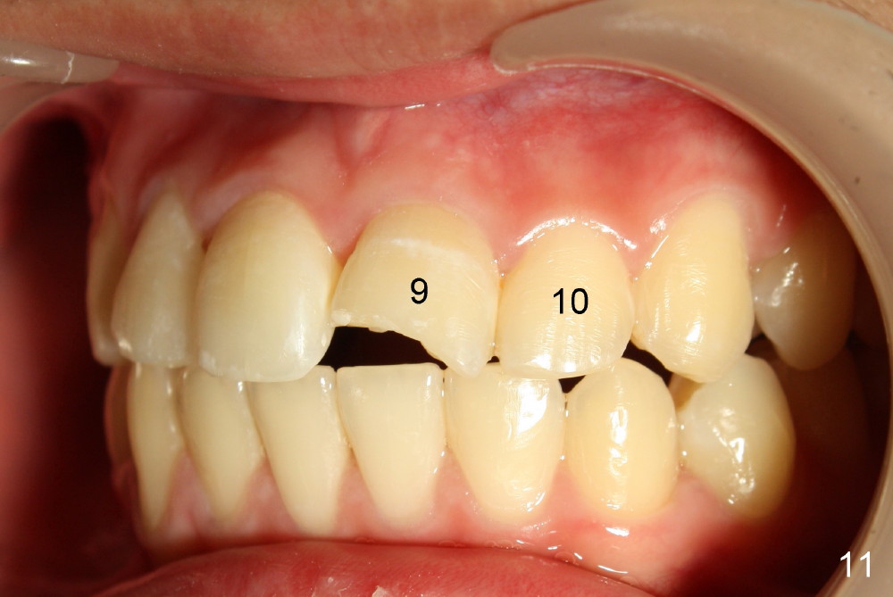
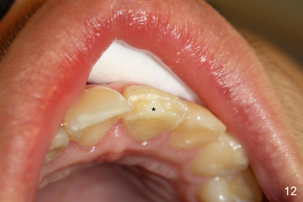
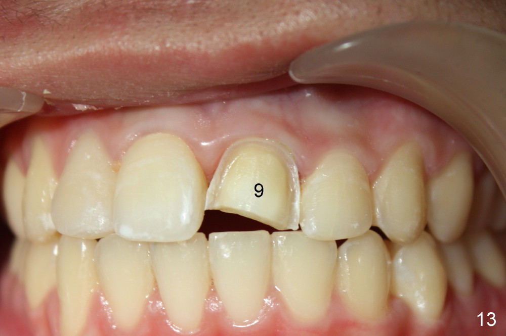
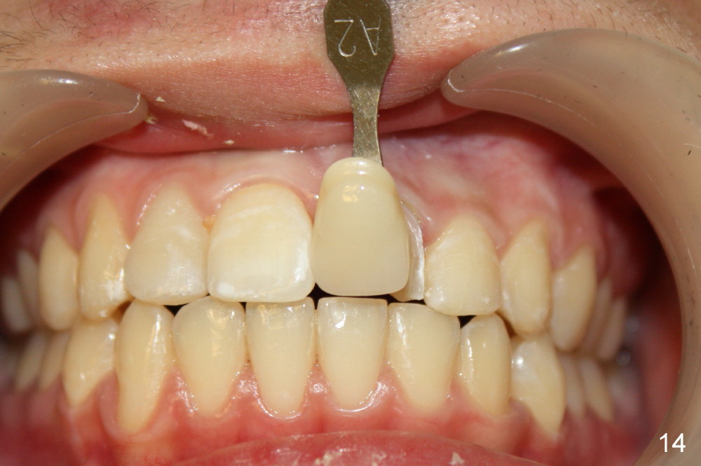
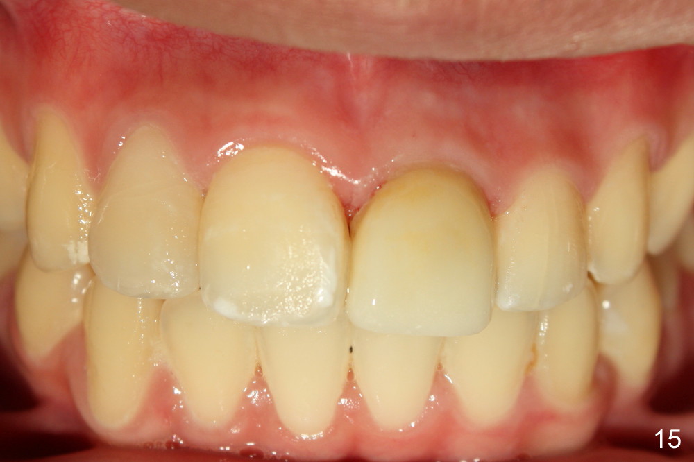
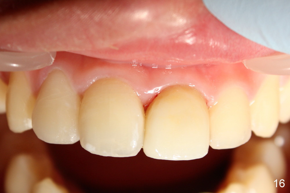
Two weeks later, the shade of #9 was similar to that of #10 (Fig.11). Composite with the lightest shades were used to finish build up (Fig.12 *). The tooth #9 was prepared for veneer; the dentine looks pretty light (Fig.13). Proper shade was chosen (Fig.14). Veneer at the site of #9 is bonded (Fig.15). The patient returns for excess cement removal 1 week post cementation (Fig.16).
Return to
Assistants
Xin Wei, DDS, PhD, MS 1st edition 10/29/2014, last revision 11/12/2014