,%20trimmed,%20bone%20graft.jpg)
,%20trimmed,%20bone%20graft.jpg)
|
|
|
|
|
|
|
|
|
|
|
|
|
|
|
|
|
|
|||
|
|
|
||||
Use of Osteotomy Plug for Sinus Lift
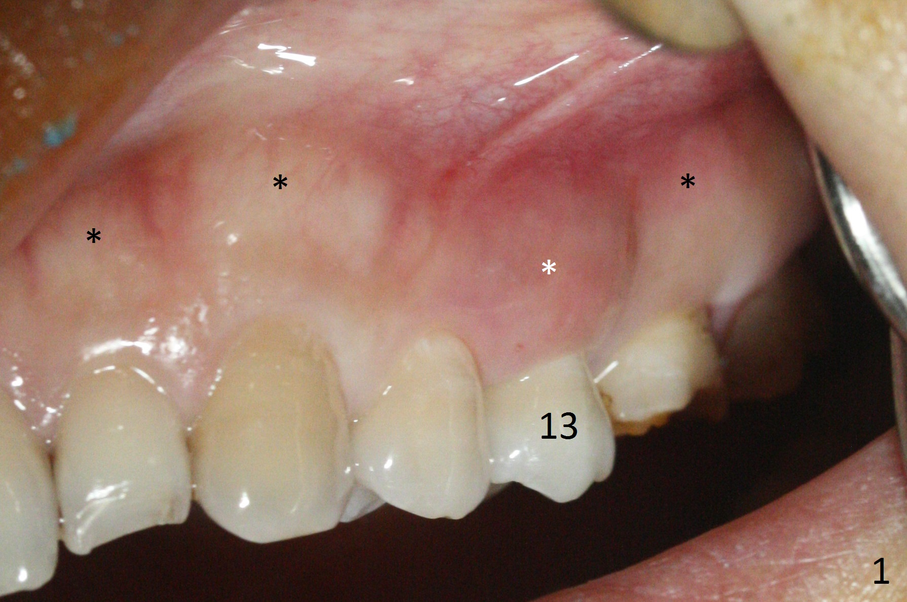
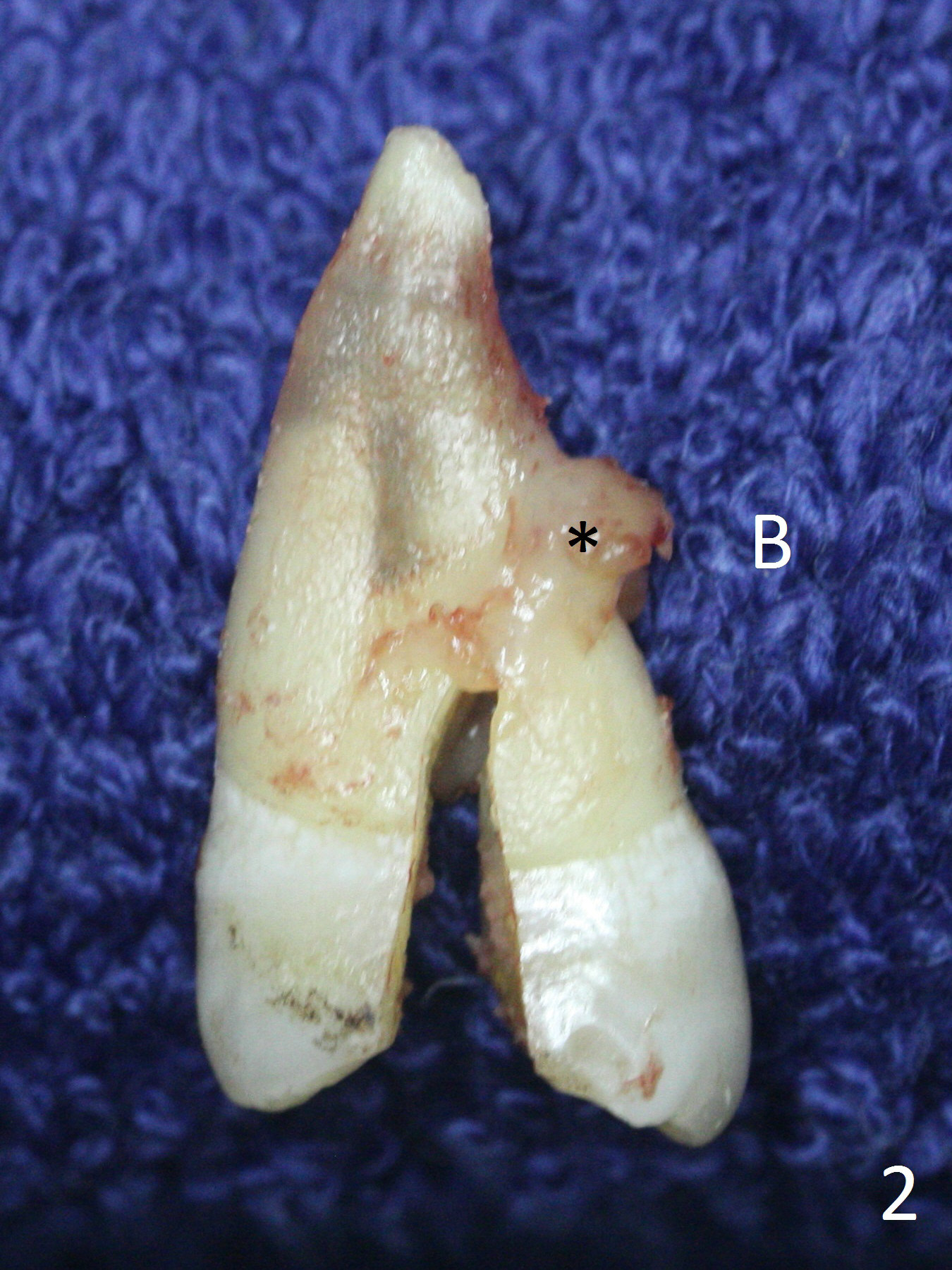
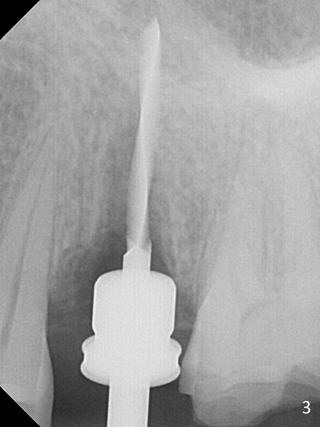
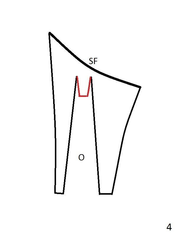
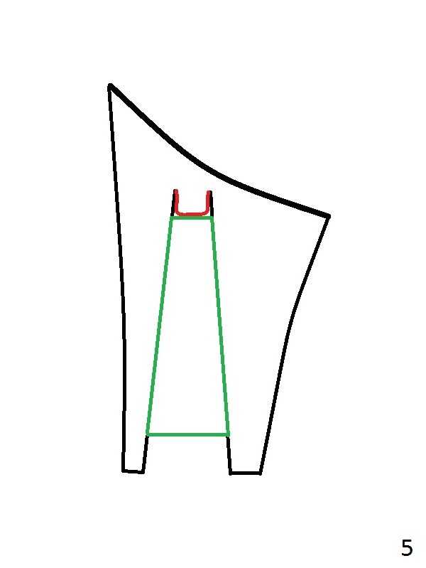
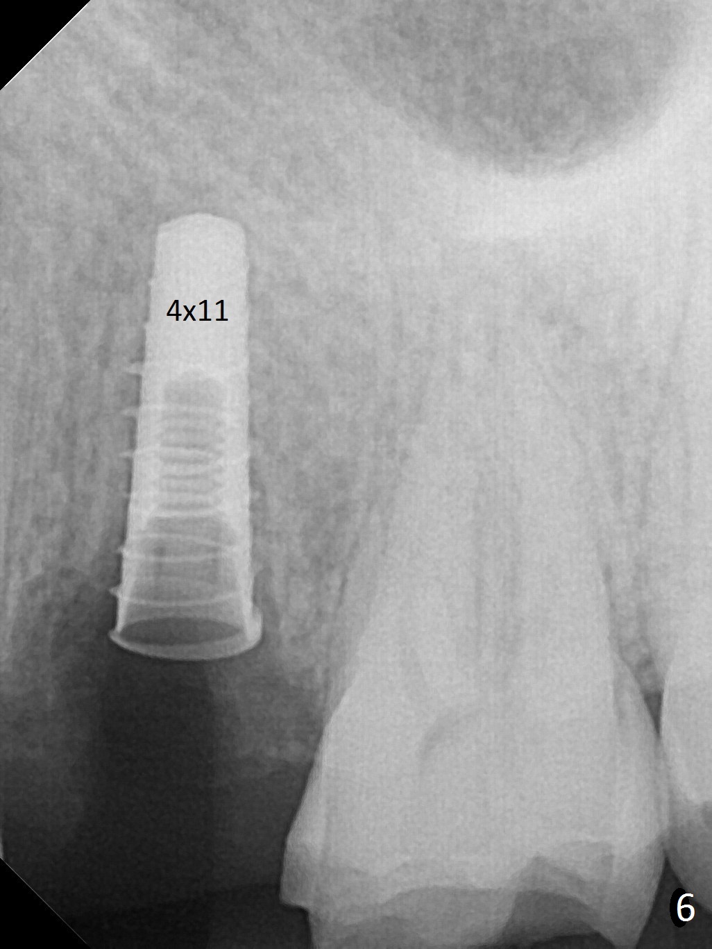
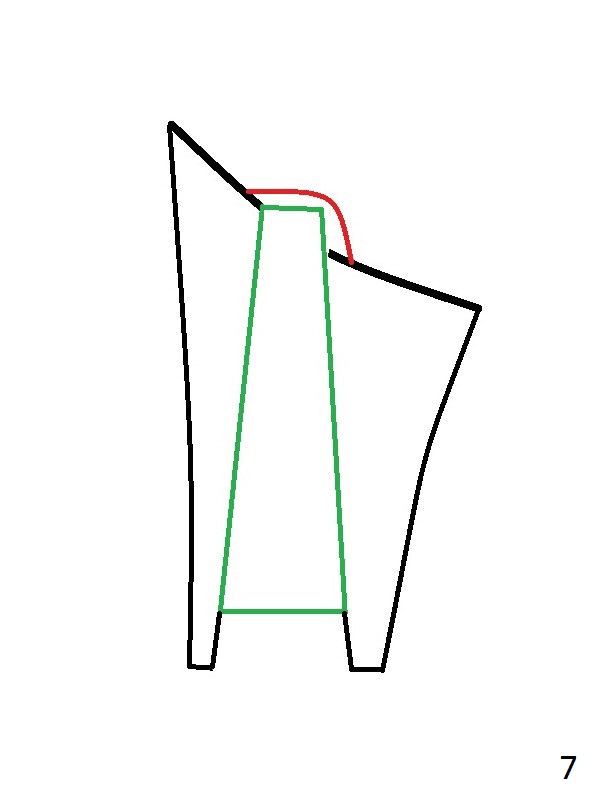
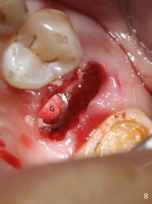
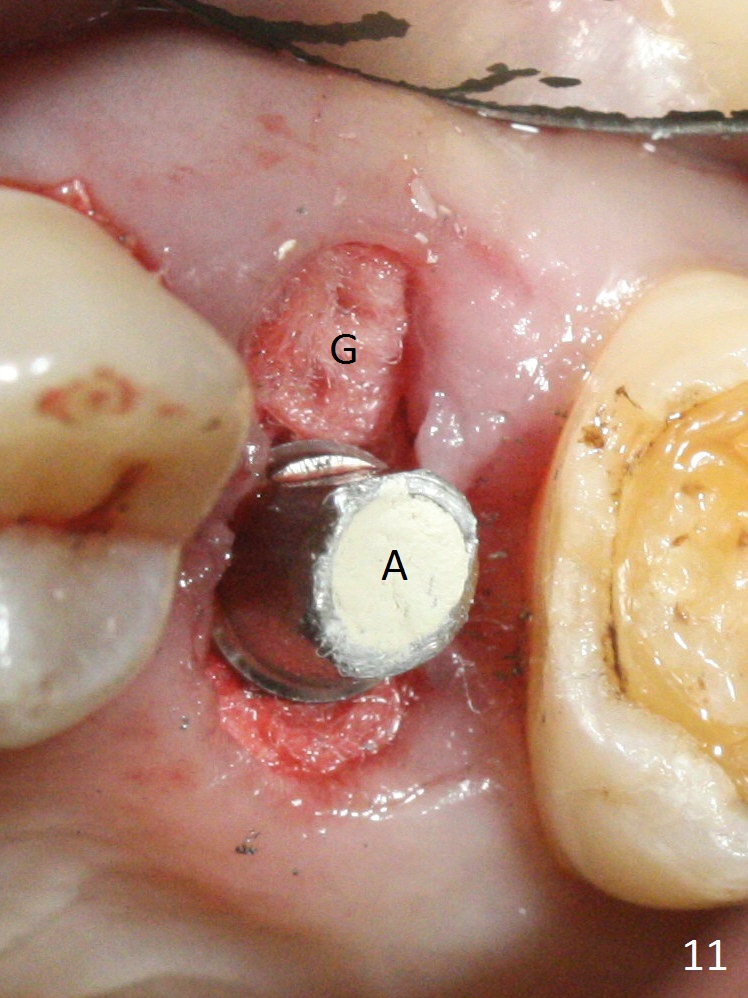
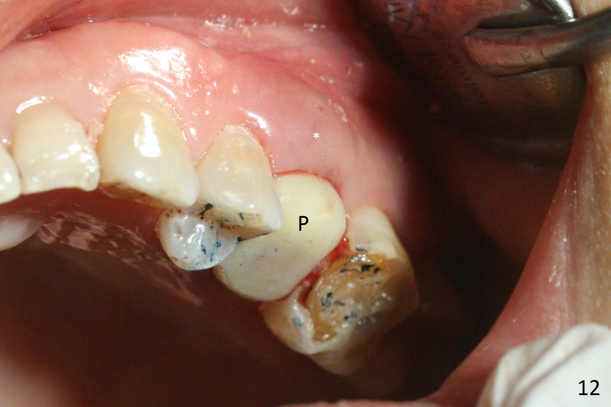
The buccal gingiva over the tooth #13 (Fig.1 white *) with crack (Fig.2 (mesial view of the extracted tooth)) is erythematous with deep buccal (B) pockets, which do not appear to extend the level of exostosis of the neighboring teeth (Fig.1 black *), i.e., coronal to the apical end of the crack with granulation tissue (Fig.2 *). Osteotomy is initiated with a 1.6 mm drill in the palatal aspect of the socket (Fig.3) so that an implant will be placed palatal (Fig.8,11) and there is enough buccal gap for bone graft (Fig.8 *). After withdrawal of 3.3 mm Magic Drill (trephine bur), the osteotomy (Fig.4 O) plug (red outline) is intentionally left in situ. When a 4x11 mm dummy implant is placed (Fig.5 (green outline), 6), the plug is compressed (Fig.5). With placement of a definitive implant (4.5x13 mm, Fig.7 (green), 9), the plug as well as the sinus floor (Fig.4 SF) is lifted (Fig.7 red curved line), 9 (arrowheads)). In brief the autogenous bone is used for sinus lift. There is no intra- or post-op nasal hemorrhage. With a small piece of gauze (Fig.8 G) in the implant well, allograft is placed in the buccal (mainly) and palatal gaps until the level of the implant plateau. Then a 4.5x4(3) mm abutment is placed (Fig.9-10 A). Next another piece of gauze is placed in the space corresponding to the abutment cuff (Fig.11 G) for fabrication of an immediate provisional (Fig.12 P). More bone graft is placed in the soft tissue zone (dual zone technique) after gauze removal and before provisional seating. With dual zone bone graft technique and provisional support, the soft tissue atrophy should be expected to be minimal (Fig.12 *). The zone of exostosis (more coronal) should be much less, since the bone density in the zone is high.
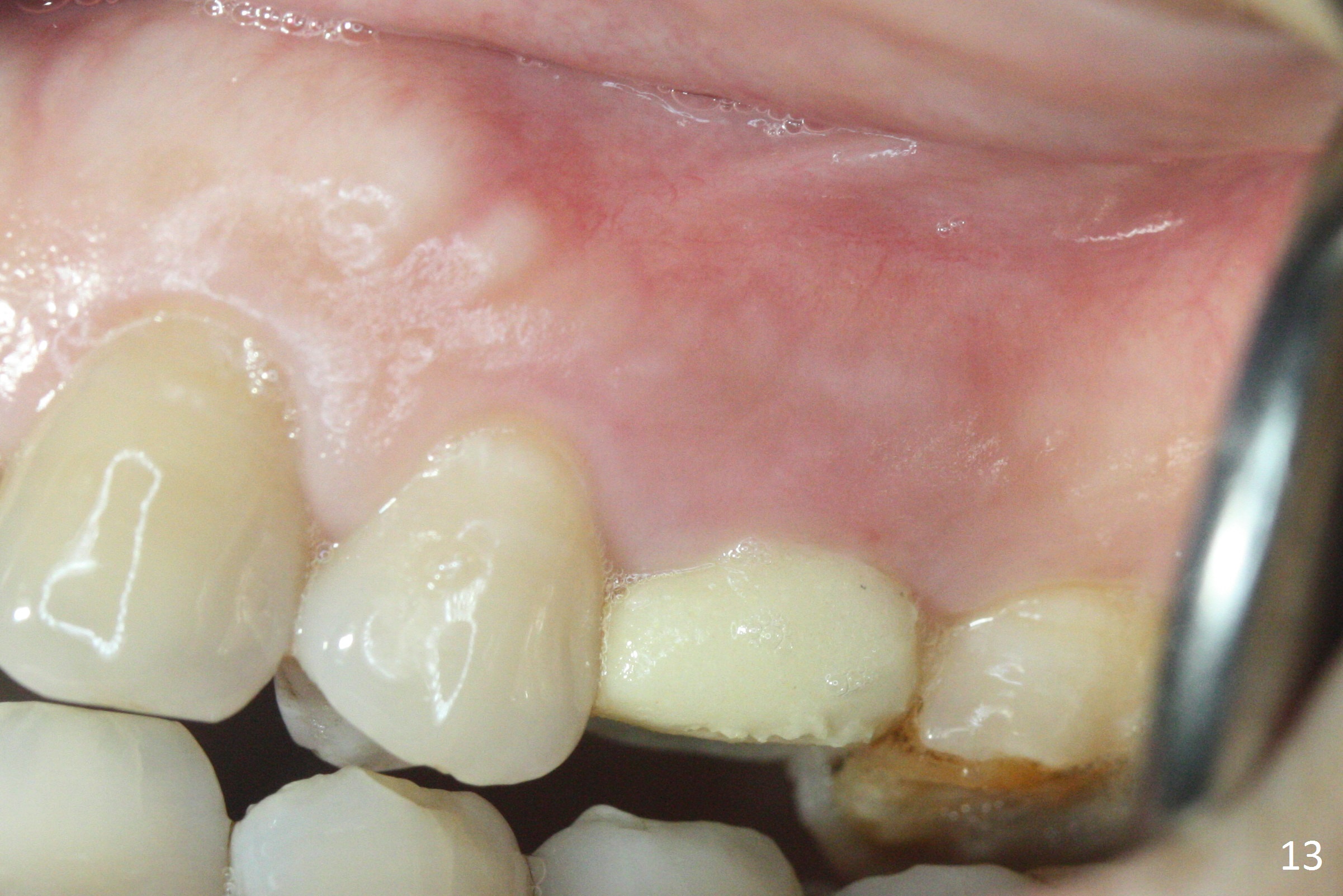
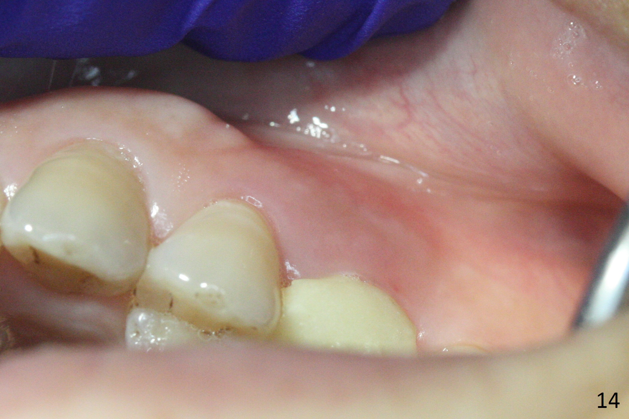
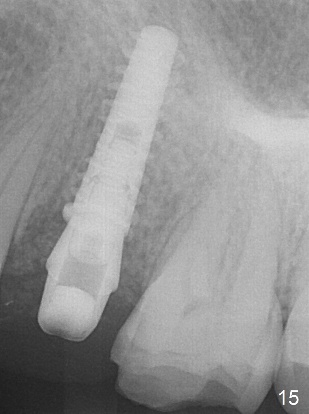
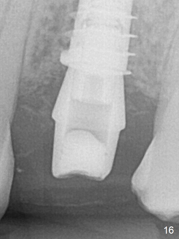
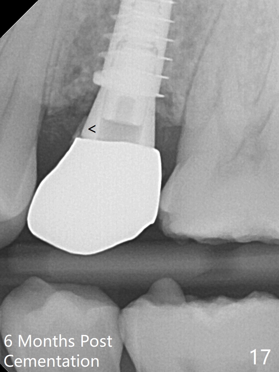
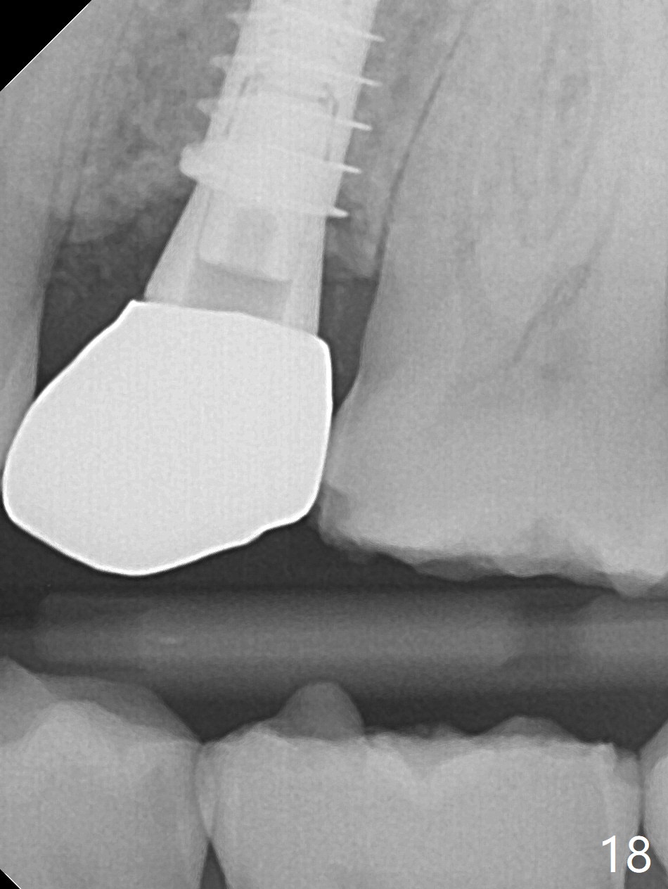
The buccal gingival inflammation subsides 1 week postop (Fig.13,14). There is no bone loss 4 months postop (Fig.15,16). The crown is recemented 6 months post cementation (probably due to short abutment); there is a residual cement (Fig.17 <), which is removed (Fig.18).
Return to
Upper Premolar Immediate Implant,
IBS,
1st
Year, Sinus Lift with
Implant,
Course
2
3
4
Xin Wei, DDS, PhD, MS 1st edition 05/16/2017, last revision 04/02/2018