|
|
|
|
|
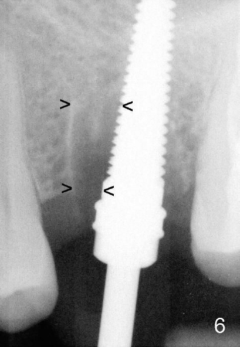
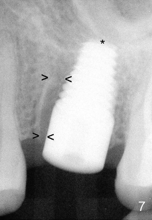
Fig.6 shows 4.5 mm tap in place with stability. It appears too high. When it is removed, the sinus membrane is found to have been perforated. The next taps (5,6 and 7 mm) are placed shorter by 3 mm with stability. A piece of collagen dressing and a small amount of bone graft (Fig.7 *) are inserted to the osteotomy before placement of a 7x14 mm implant. Arrowheads: the remaining mesial socket.
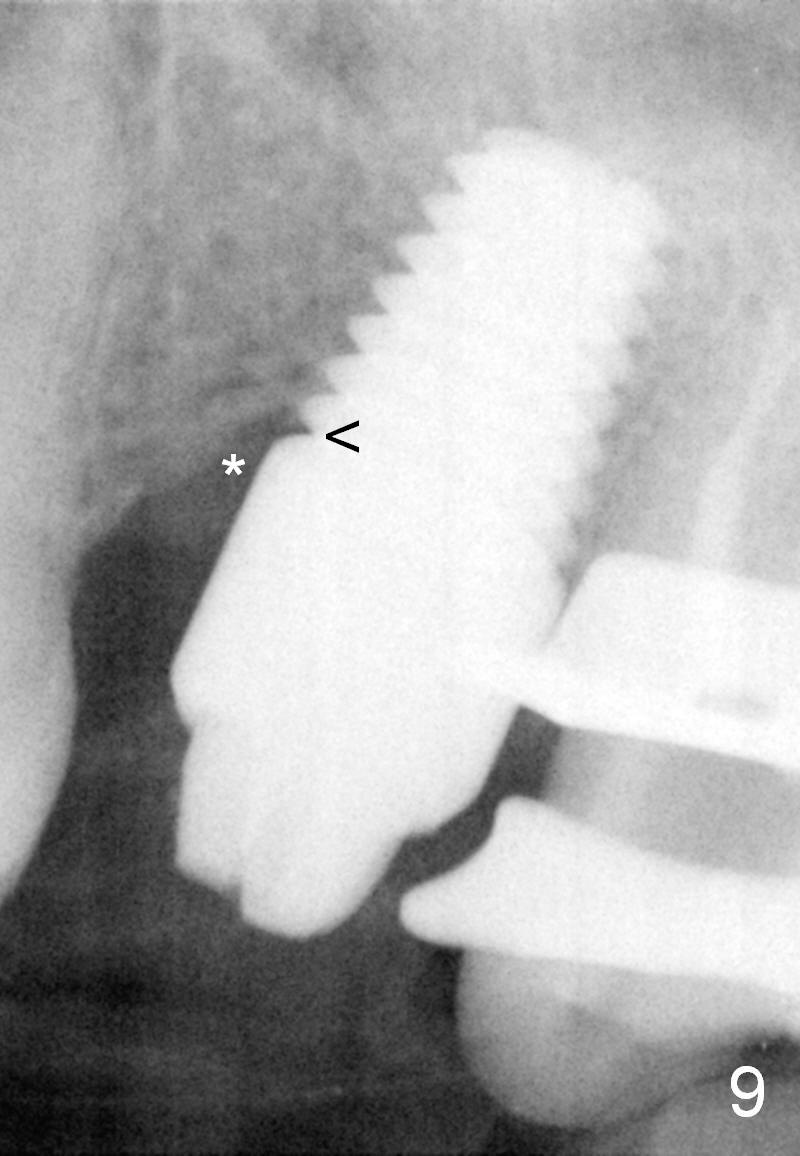
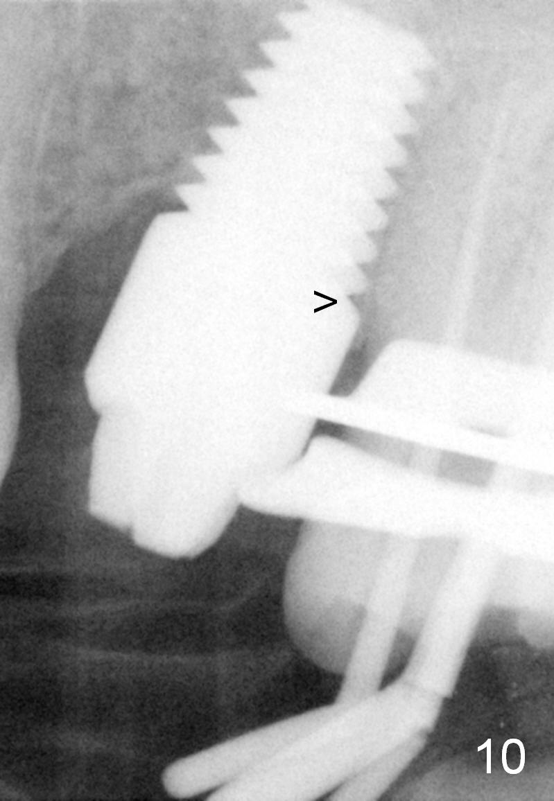
Twenty-one months postop, the patient returns for #15 RCT. PAs show that although there is a narrow space in the original mesial socket (Fig.9 *), the 1st thread space is partially obliterated with the bone (<). In contrast, the distal 1st thread space is completely obliterated (Fig.10 >). After the RCT, impression is taken for #14 and 15.
Immediate Provisional for Upper 1st Molar Last Next
Xin Wei, DDS, PhD, MS 1st edition 12/15/2014, last revision 08/15/2021