|
|
|
|
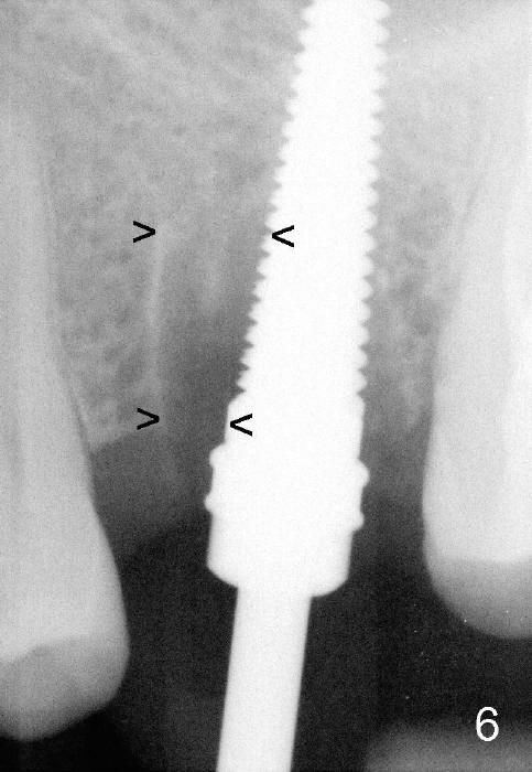
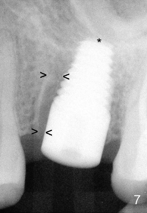
Fig.6 shows 4.5 mm tap in place with stability. It appears too high. When it is removed, the sinus membrane is found to have been perforated. The next taps (5,6 and 7 mm) are placed shorter by 3 mm with stability. A piece of collagen dressing and a small amount of bone graft (Fig.7 *) are inserted to the osteotomy before placement of a 7x14 mm implant. Arrowheads: the remaining mesial socket.
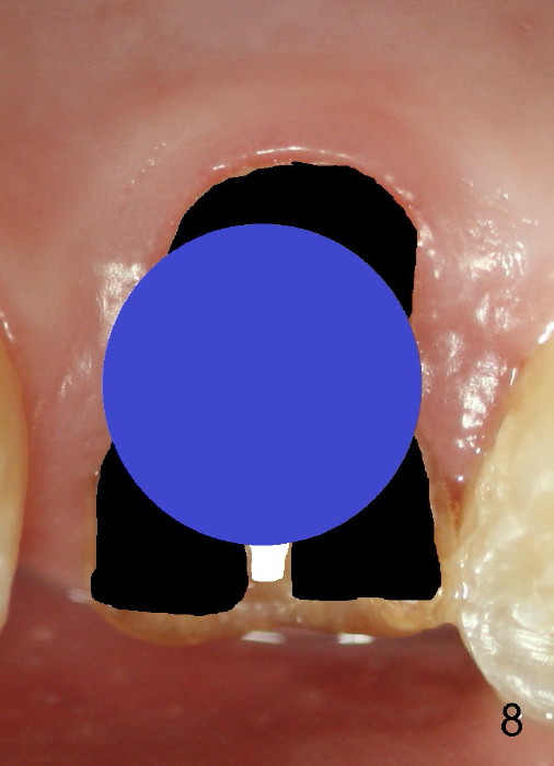
Fig.8 illustrates the implant (blue) placed in the middle of the socket. The remaining buccal and lingual gaps (black) are then closed by graft and membrane. Placing the graft in the deeper portion of the mesiobuccal socket is not so easy because the large implant blocks the entrance (Fig.7 between lower arrowheads). If more graft has been dispensed and the earlier PA shows larger upper space exists (Fig.6 between upper arrowheads), the graft should be placed to that region prior to implant placement.
Immediate Provisional for Upper 1st Molar Last Next
Xin Wei, DDS, PhD, MS 1st edition 12/15/2014, last revision 08/15/2021