|
|
|
|
|
|
|
|
Granulation Tissue in Septal Undercut
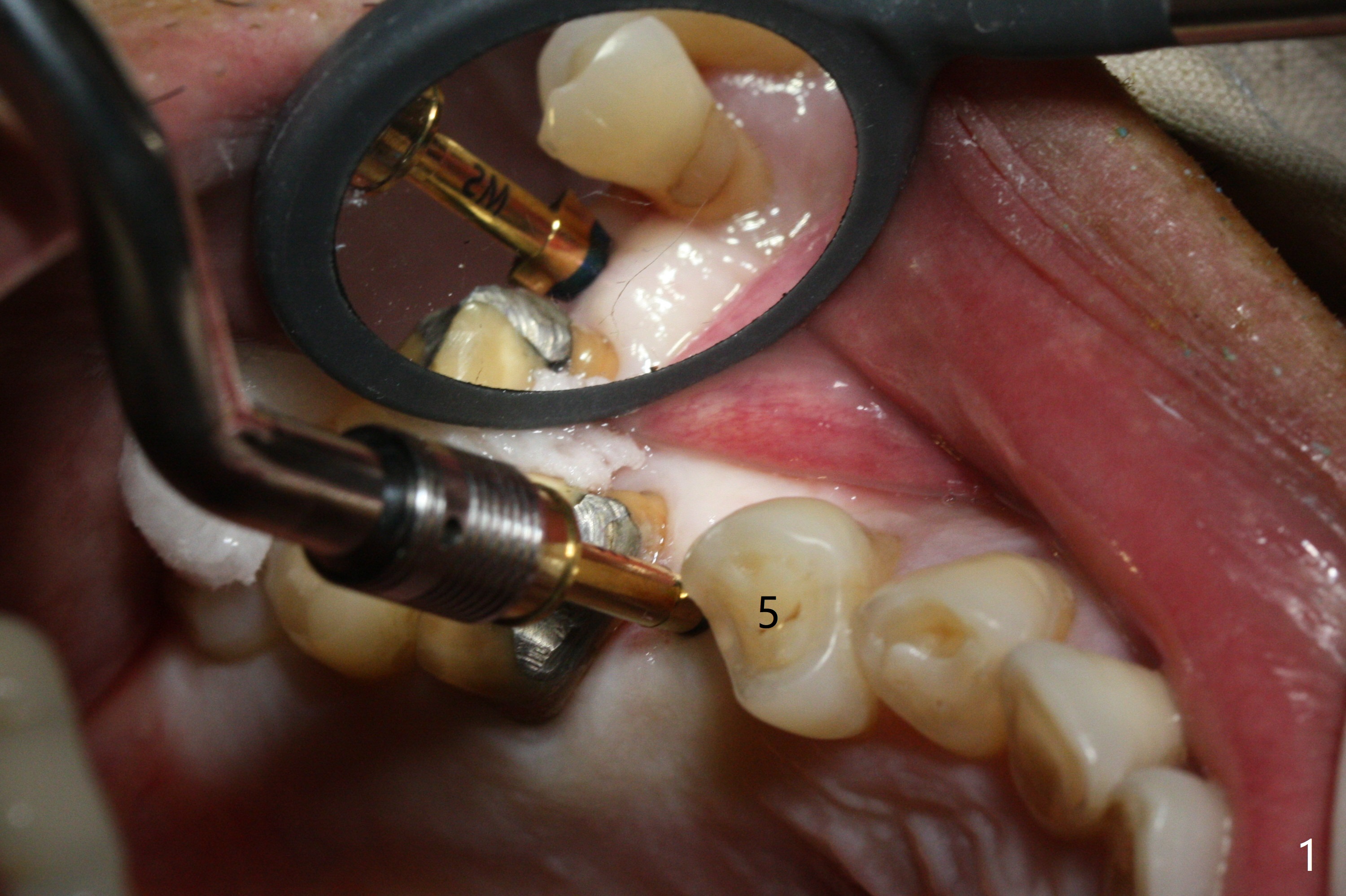
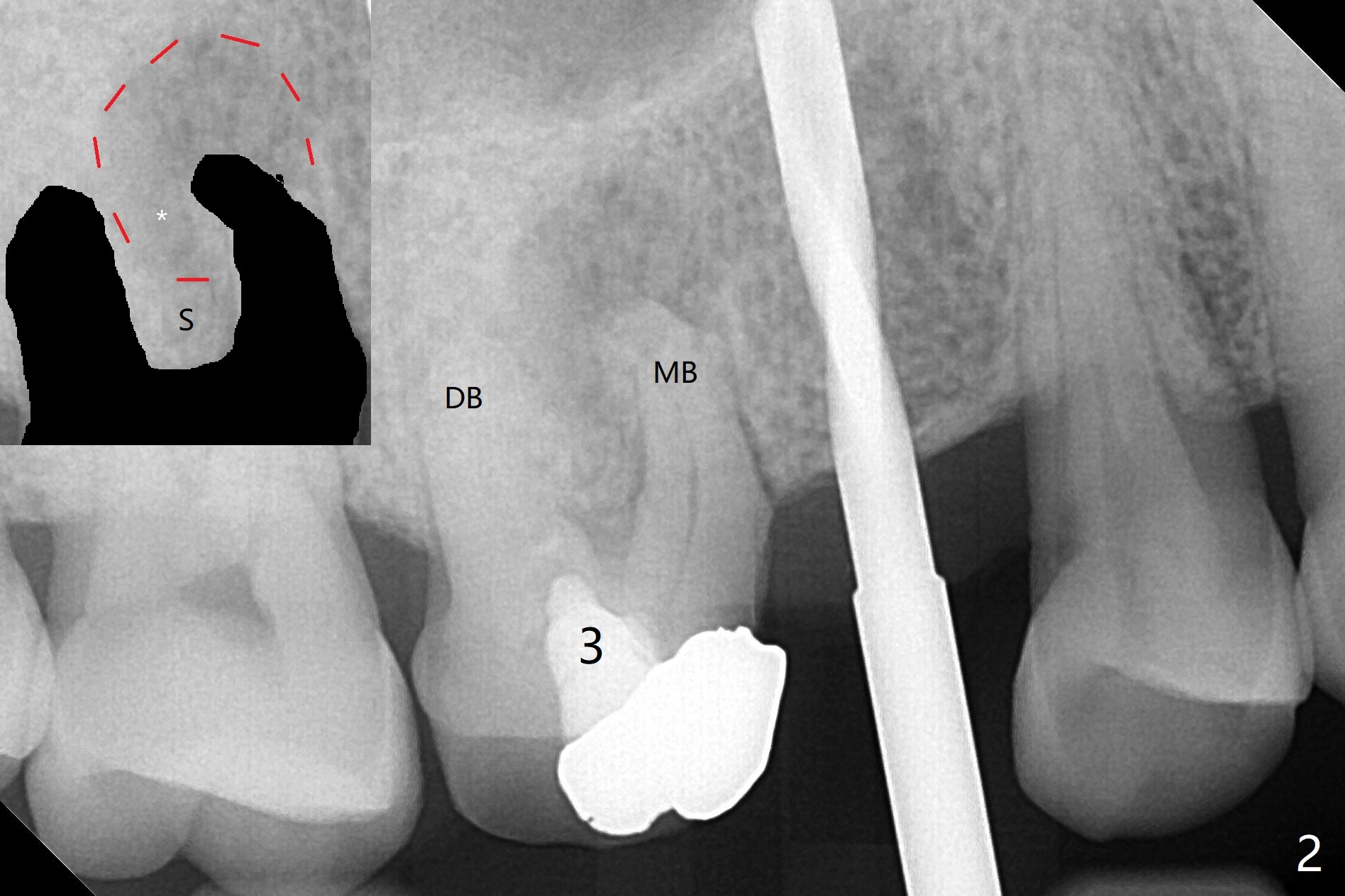
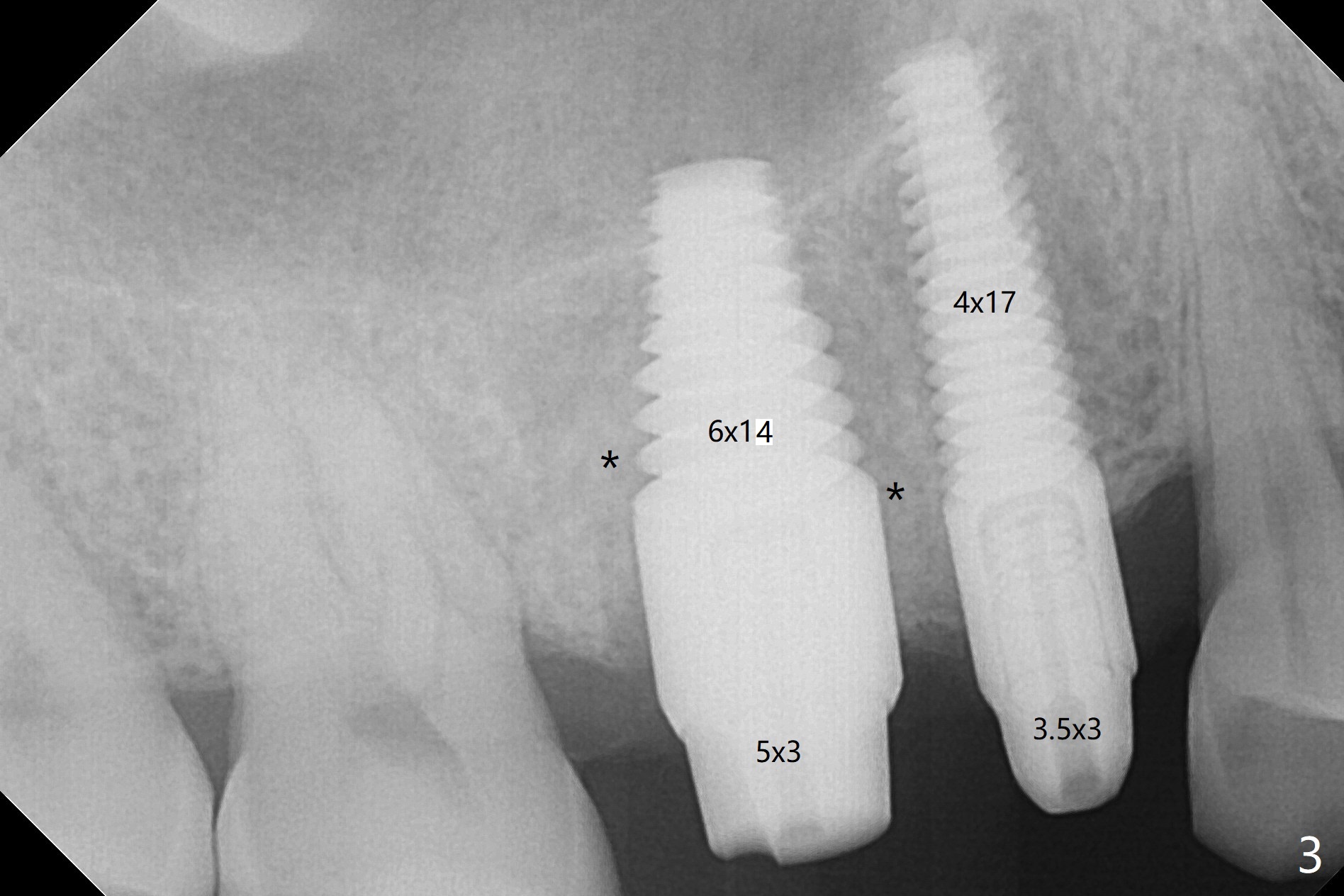
As planned, osteotomy starts at #4 prior to #3 extraction (Fig.1,2). In fact the bone density at the edentulous area is high. Drills are used to finish placing a 4x17 mm tissue-level implant (Fig.3).
A challenge associated with #3 extraction is large amount of granulation tissue apical to the mesiobuccal root (MB, Fig.2 (inset: red dashed line)). To remove the granulation tissue (*) apical to the septum (S), the septum between the buccal roots has to be chiseled. The remaining septum for osteotomy becomes less.
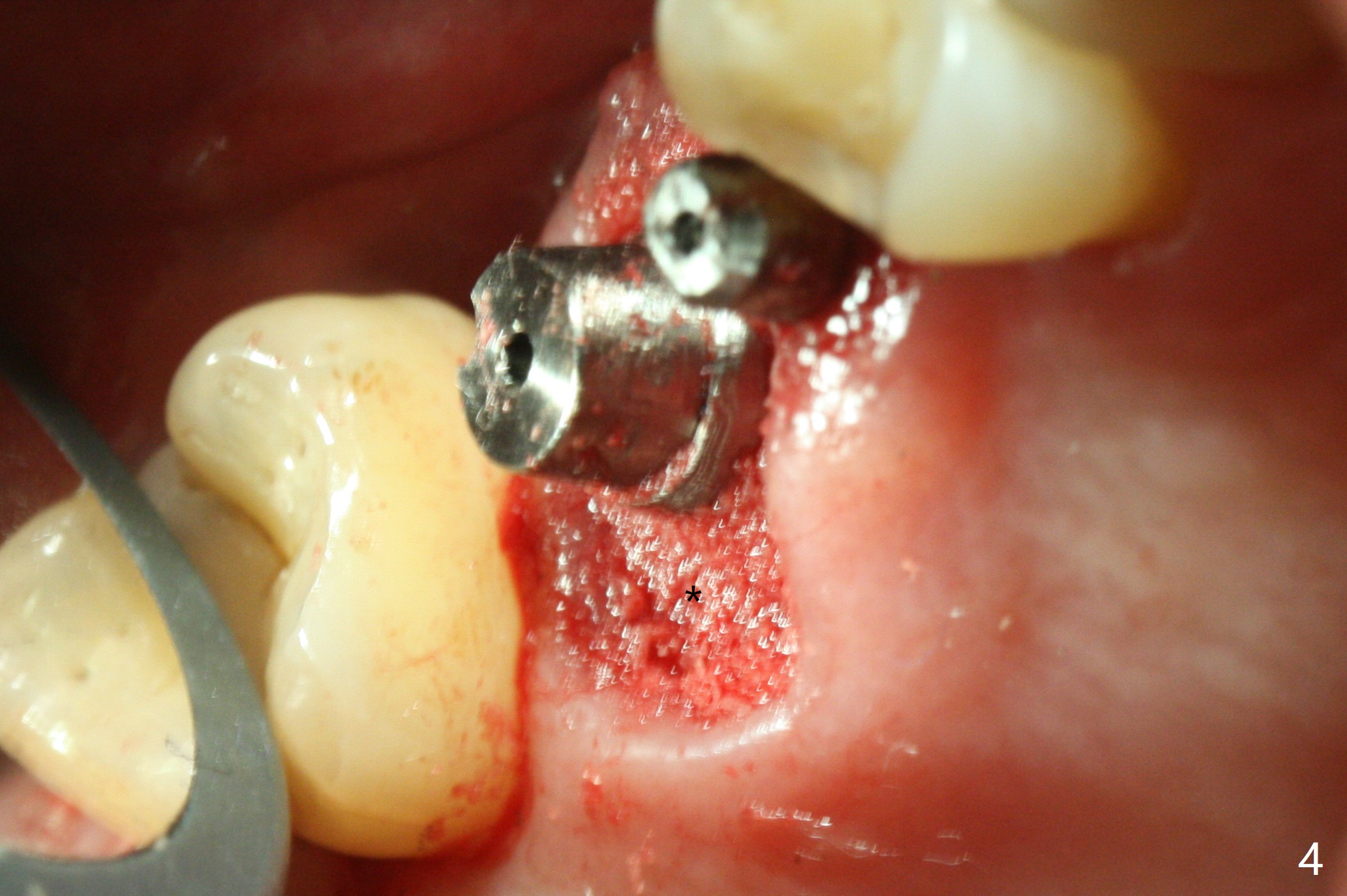
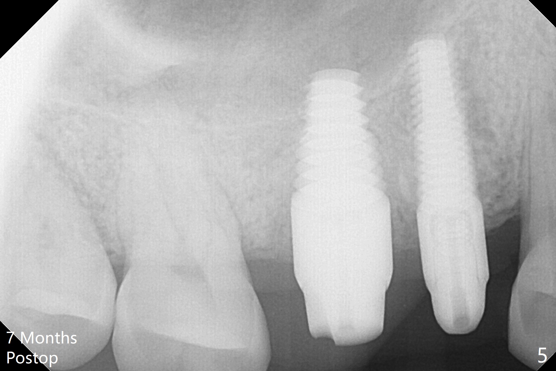
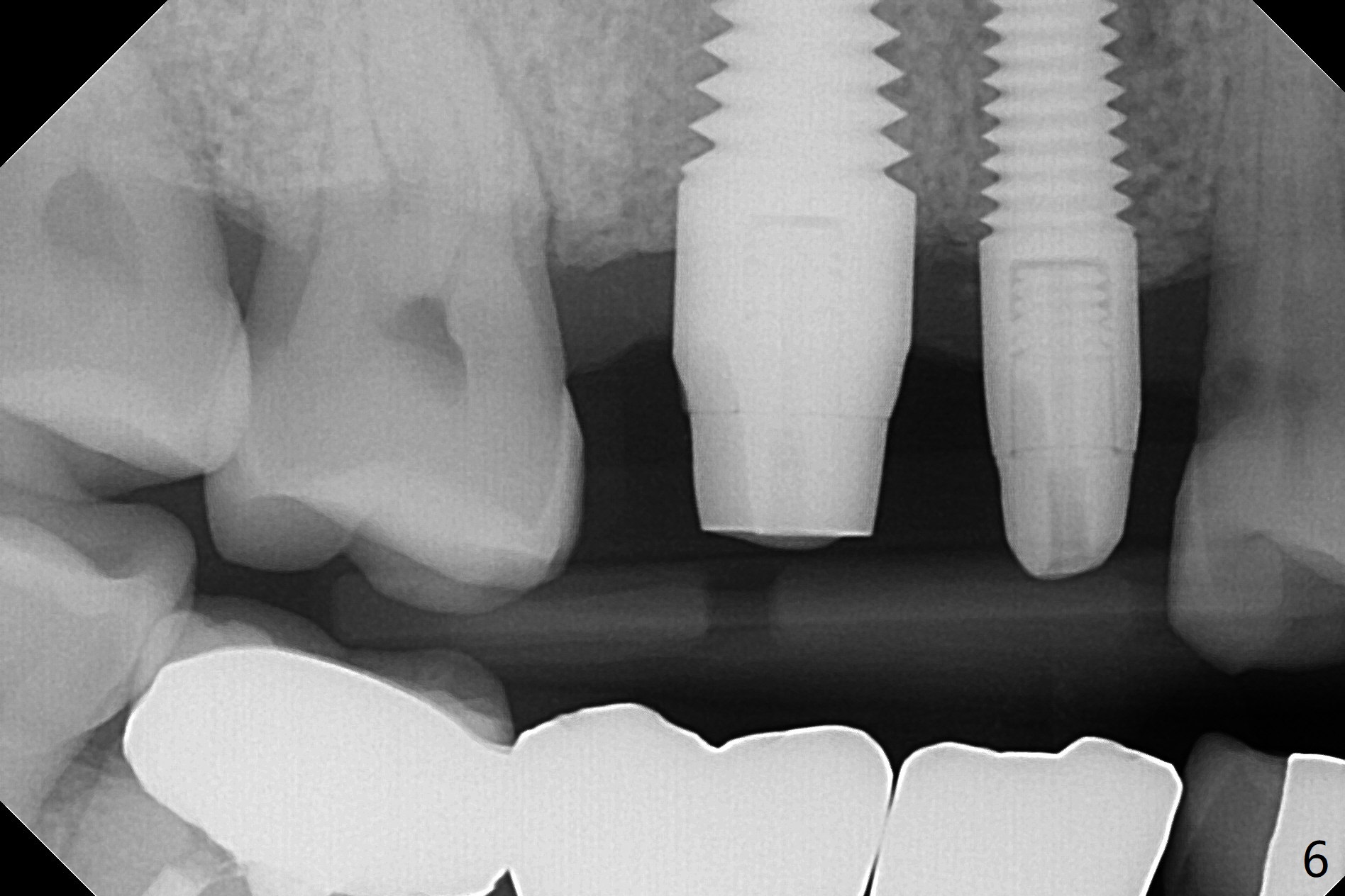
With proper manipulation, a 6x14 mm implant is placed with primary stability (Fig.3). The buccal and palatal socket opening is closed with Vera Graft (allograft, Fig.4 *), which is in turn covered by an immediate splinted provisional. No gap exists 7 months postop (Fig.5,6). Return to Upper Molar Immediate Implant, Prevent Molar Periimplantitis (Protocols, Table), IBS Xin Wei, DDS, PhD, MS 1st edition 09/26/2017, last revision 04/24/2018