,%20then%20autogenous,%20Vanilla,%20mesial%20bone%20less.jpg)
|
|
|
|
|
|
|
|
|
|
Bone Graft Should Be Placed Prior to Abutment
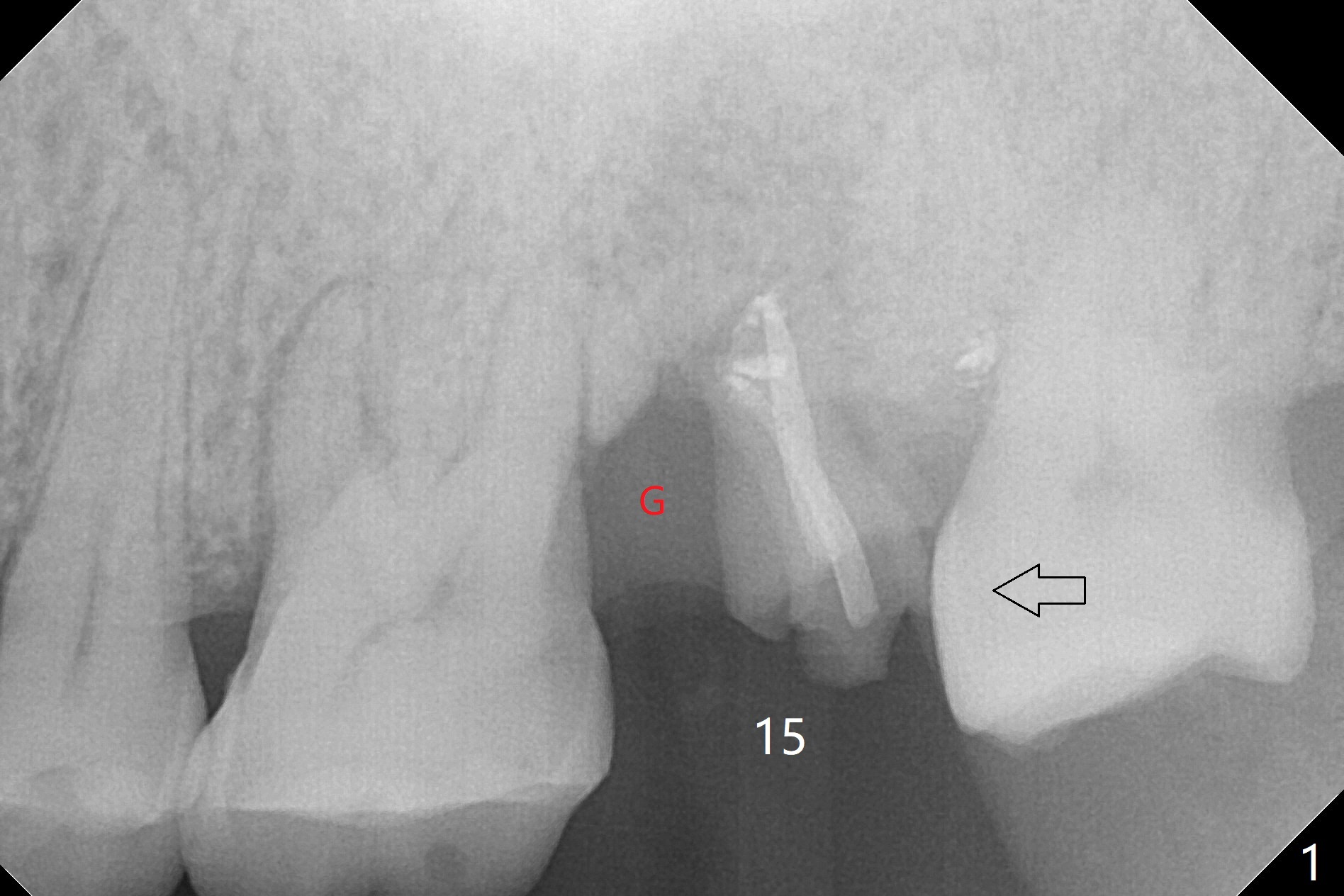
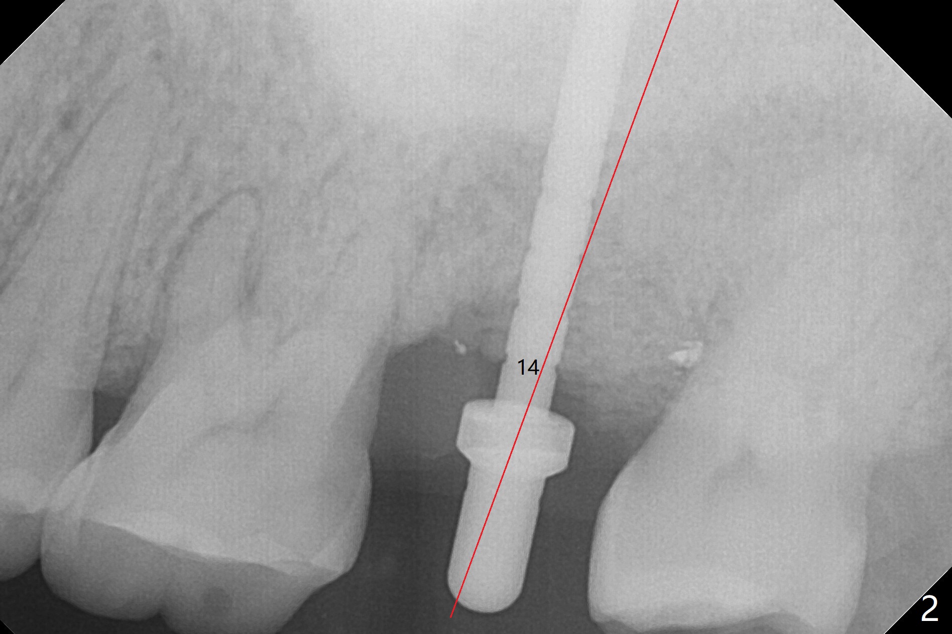
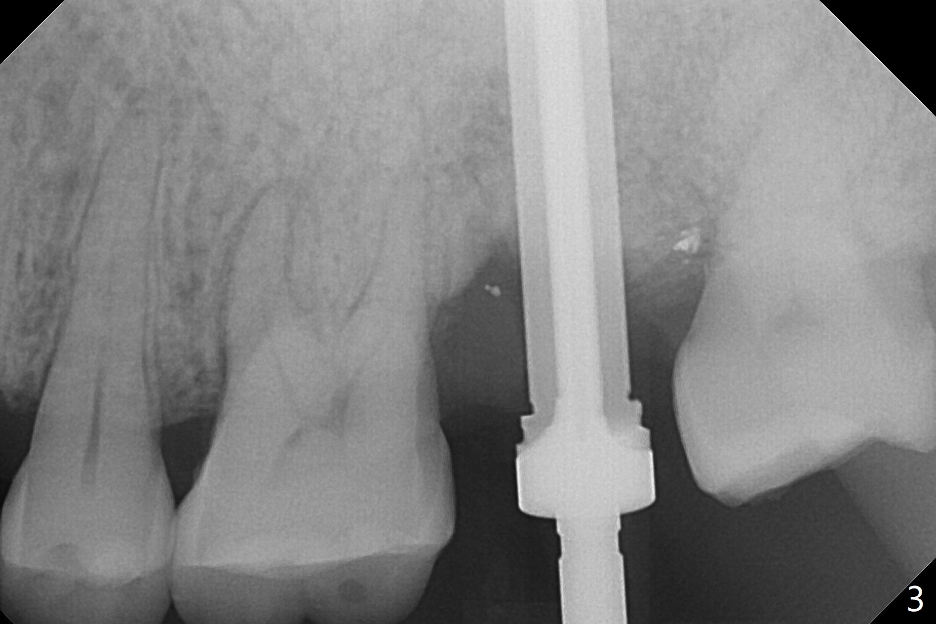
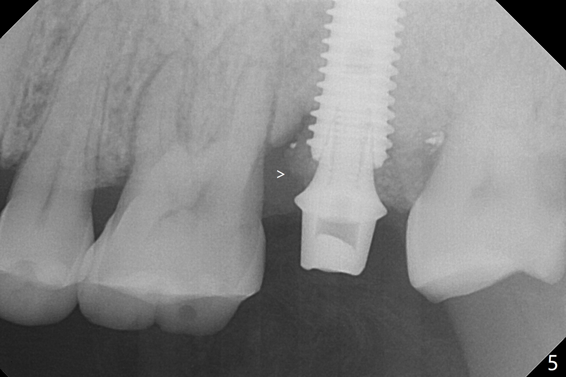
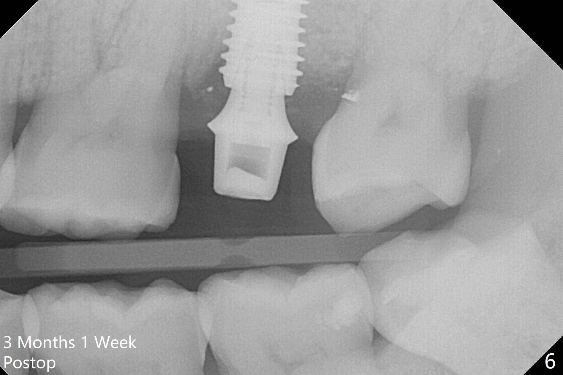
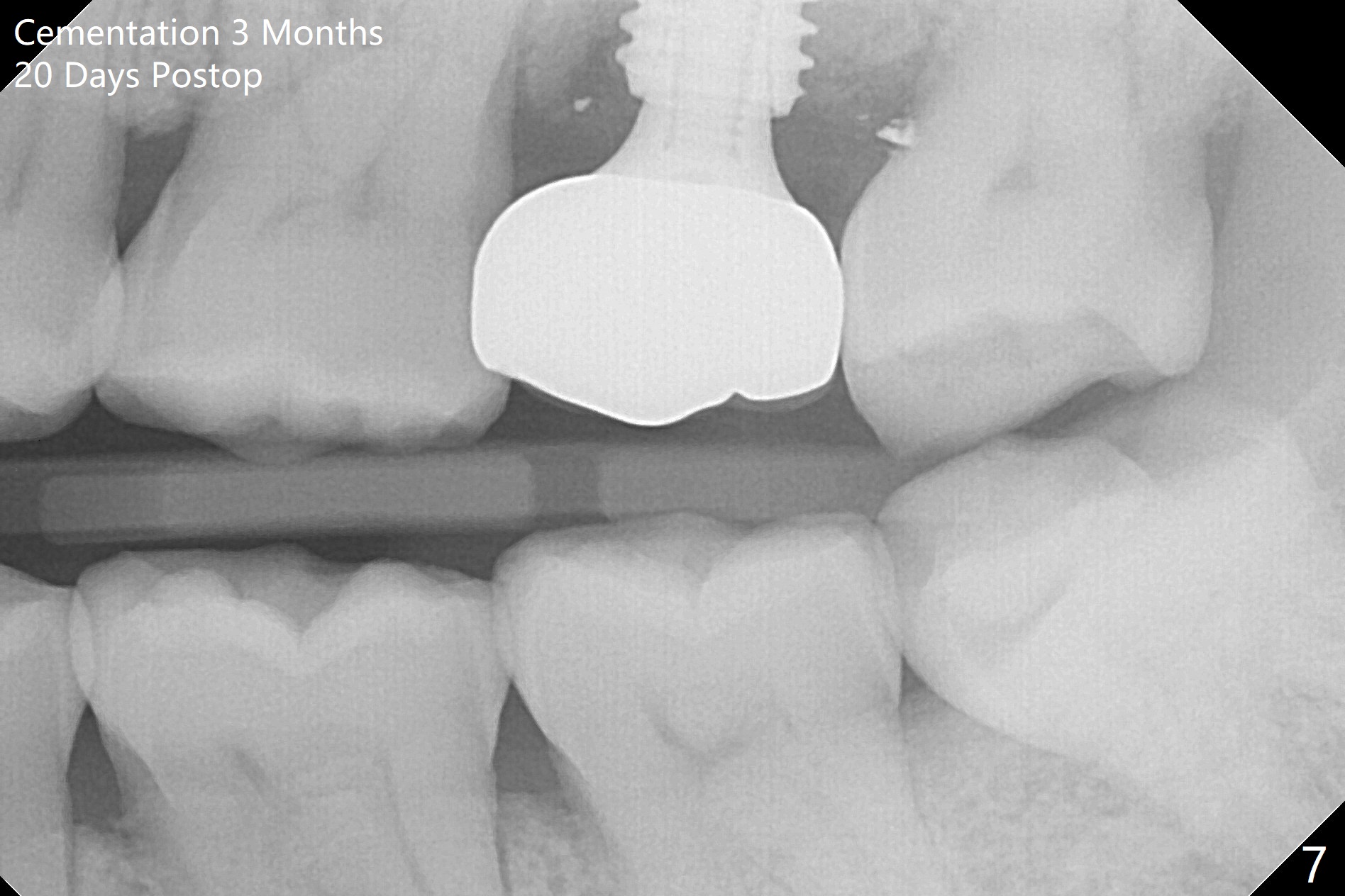
When the patient returns for the tooth #15 extraction and implant, the mesiobuccal residual root has been expelled, while the distobuccal and palatal roots seem to have extruded (Fig.1). The mesial portion of the gingiva (G) is intact and thick. The trajectory of the initial osteotomy is to be changed as shown by red line in Fig.2. The 3.8 mm drill appears to be distal (Fig.3). With mesial bone removal with Lindamann bur, the position of the final implant (5x13 mm) is within normal limit (Fig.4 (50 Ncm)). Because of the thick mesial gingiva (Fig.4 G) and placement of the 5.5x4(2) mm abutment, insertion of mixture of autogenous and Vanilla Graft (*) into the mesial aspect of the implant is difficult (Fig.4 >). Further pushing of the bone graft from the buccal and palatal socket gaps results in more ideal packing (Fig.5 >). If the bone graft were placed first, packing would have been easier. An immediate provisional is fabricated to prevent further mesial shifting of the 3rd molar (Fig.1 arrow). The implant remains stable, while the provisional and abutment are loose 3 months 1 week postop (Fig.6). Impression is taken after abutment cleaning and retightening. Because of the long implant (13 mm), a permanent crown can be cemented early (3 months 20 days postop, Fig.7).
Return to Upper Molar Immediate Implant, Prevent Molar Periimplantitis (Protocols, Table), IBS, #31
Xin Wei, DDS, PhD, MS 1st edition 10/25/2017, last revision 03/04/2018