.jpg)
|
|
|
||
|
|
|
|
|
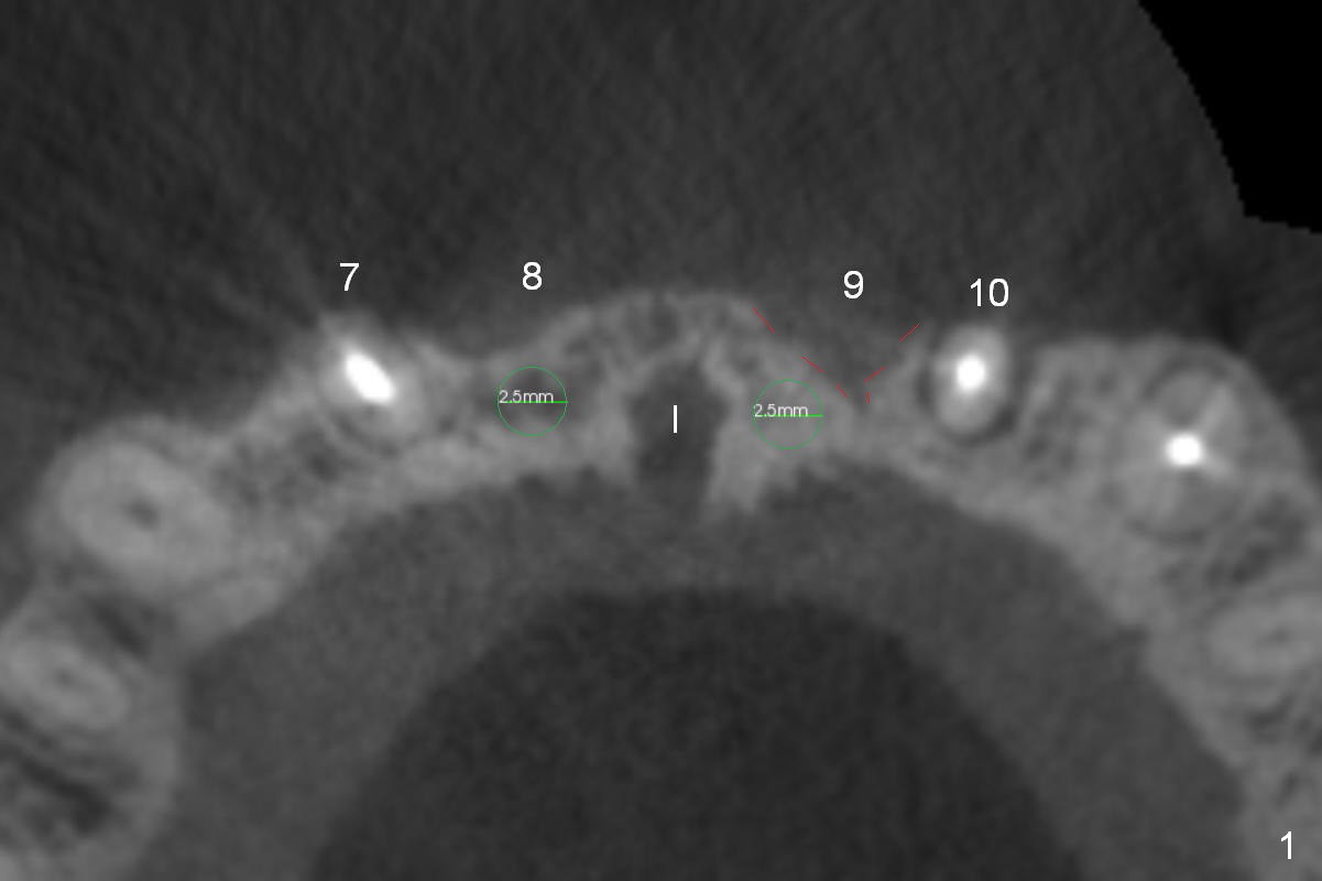
2.5 mm 1-Piece Implants in Anterior Atrophic Ridge
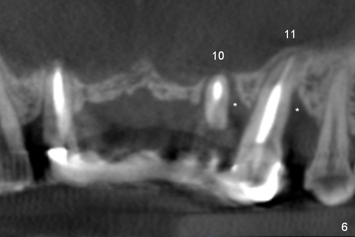
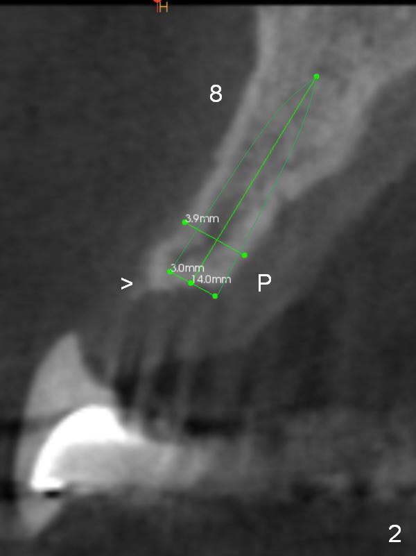
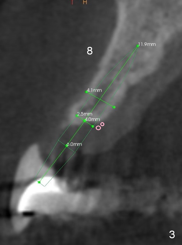
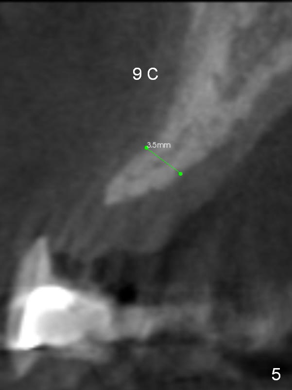
A 53-year-old man chips porcelain of the upper anterior bridge after implant placement at #14 and 15. Since he will return to home country for visit in the next few months, he is eager to restore the anterior restoration. Two of the abutments (#10, 11) appear to have severe bone loos (Fig.1,2 *). It does not seem to be a ideal treatment plan to redo the bridge. The edentulous ridge is atrophic at #8 and 9. It appears that 2.5 mm 1-piece implants (Fig.3,4) fit better than 3.0 mm one (Fig.2). There is a buccal concavity at #9 (Fig.1 red dashed line). The 2.5 mm implant at #9 should be placed between the Incisive Canal (Fig.1 I) and the buccal concavity (Fig.4). The ridge at the buccal concavity is not suitable for implant placement (Fig.5). The palatal plate (Fig.2 P) is usually denser and thicker than the buccal one. The osteotomy for the implant is initiated palatally; the implant is placed below the buccal crest (Fig.2 >). The exposed lingual thread will be covered by bone graft (Fig.3 pink circles) and collagen dressing. Since the ridge at #9 appears more atrophic, a 2.0 mm 1-piece implant may be indicated. The bridge will be sectioned between 7 and 8 and 9 and 10.
Return to
Full Arch Reconstruction,
IBS,
Atrophic Ridge
Xin Wei, DDS, PhD, MS 1st edition 01/21/2017, last revision 05/24/2019