|
|
|
|
|
|
|
|
|
|
|
|
||
|
|
|
|
|
||
One Piece Implant in a Narrow Space
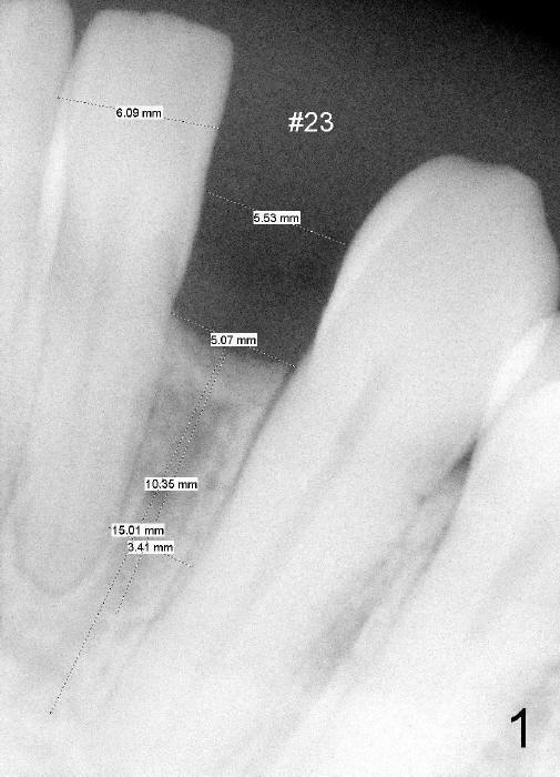
A 44-year-old phobic black lady requests implant restoration for #23, a congenital missing incisor (Fig.1). The space between the roots of the neighboring teeth is 3.41 mm. The smallest permanent implant is 3 mm in diameter. She had ortho probably 20 years ago. At first, diastemata among the lower anteriors were closed. They relapsed. Finally, a single edentulous space was created in hope to place an implant. A retainer has been worn ever since. Her oral hygiene is poor, partially due to long term wearing of the retainer. It appears that she cannot go back to the orthodontic office to redo ortho, most likely due to finance.
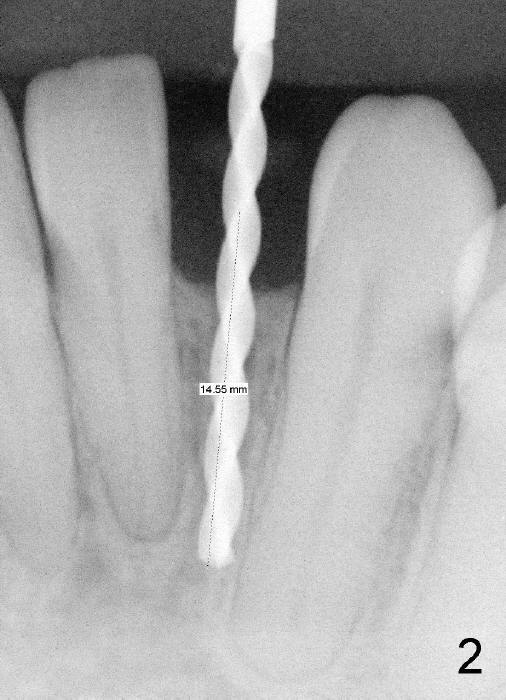
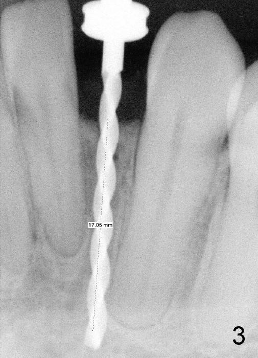
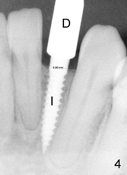
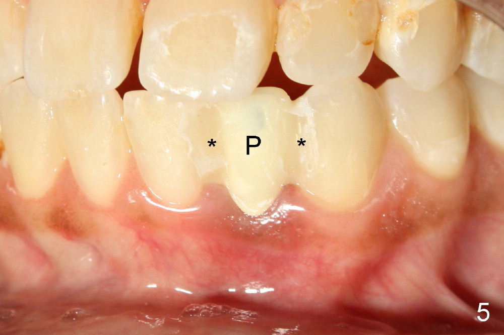
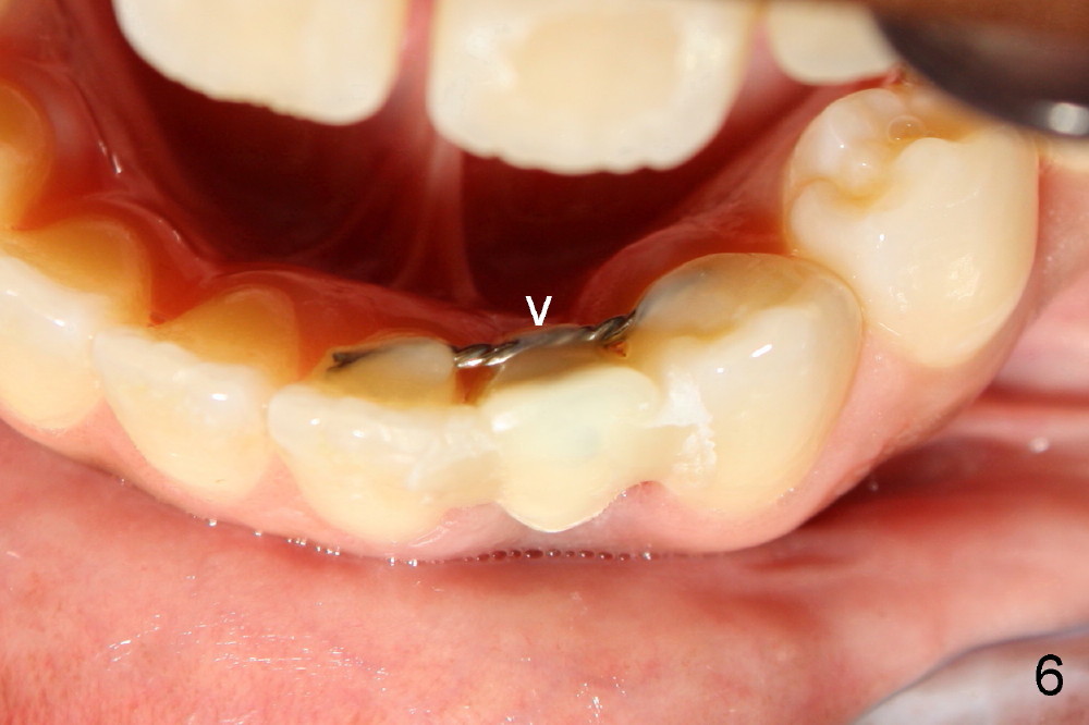
Informed consent is obtained with emphasis of potential damage to the neighboring roots. Two PAs have to be taken with the first pilot drill (1.5 mm) in place for determination of initial trajectory (Fig.2,3). Osteotomy is enlarged coronally with 2 mm pilot drill. Finally a 3x17 mm one piece implant is placed with primary stability, determined tactilely (Fig.4). Immediate provisional is fabricated. To avoid micromovement, the immediate provisional (Fig.5 P) is bonded to the neighboring teeth with composite (*); it is further fixed in place with a lingual retainer (Fig.6 arrowhead).
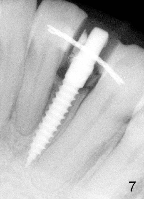
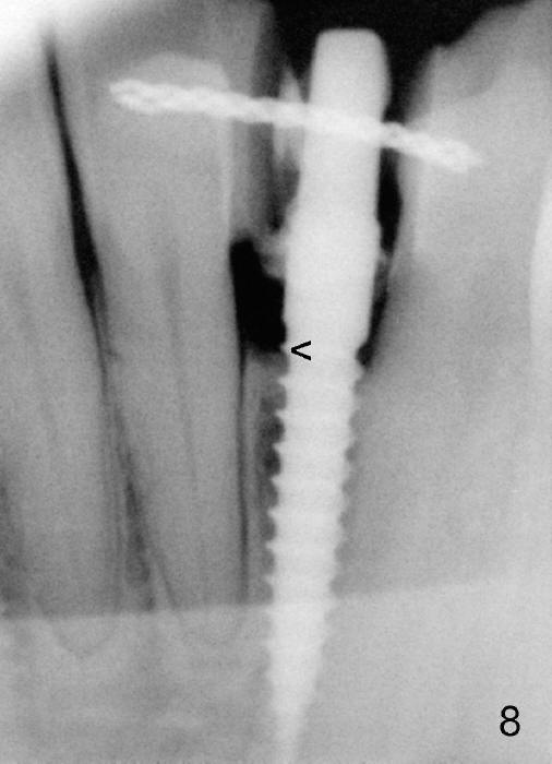
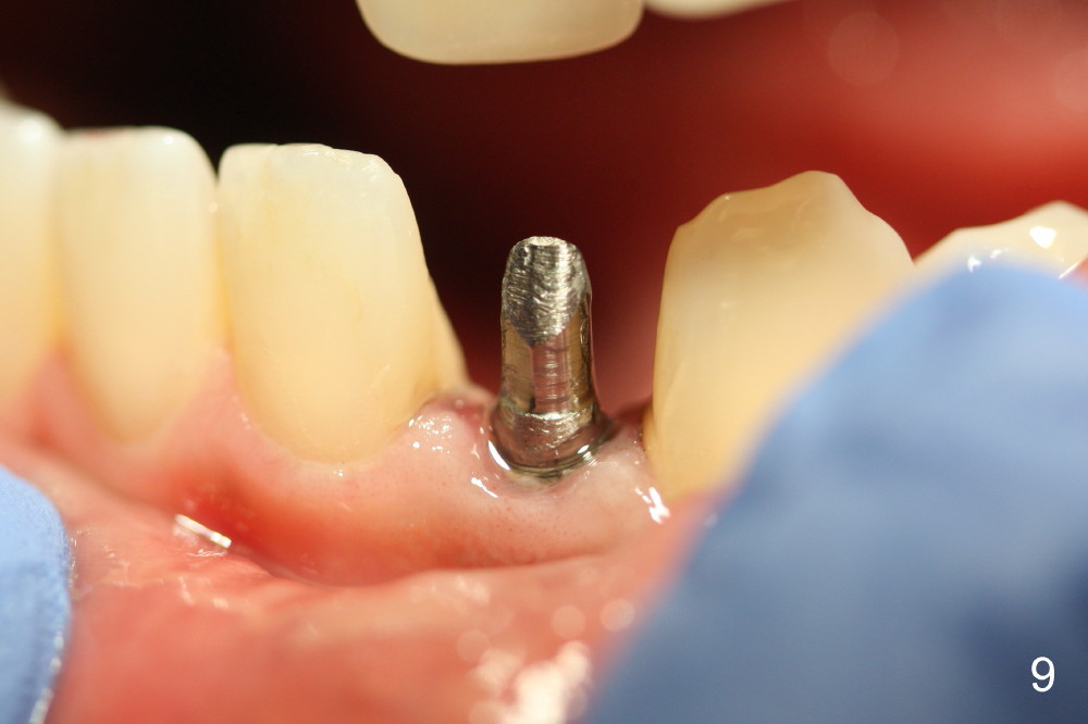
Within the first several days postop, the patient reports mild tenderness from one of the neighboring teeth. Otherwise she is doing fine. Again due to finance, she does not return for restoration for more than 1 year. PA taken 10 months postop shows that there is no abnormality (Fig.7). In fact bone resorption occurs gradually over 2 years 2 months postop, manifested as the 1st thread supracrestal (Fig.8 <). When the 1-piece implant is reprepped for impression 2 years 2 months postop, the micro threads are found apical to the prep margin (Fig.9). The nervous patient has had occasional tooth sensitivity 5 years postop. A narrower implant is more suitable for this case (2.0 or 2.5 mm).
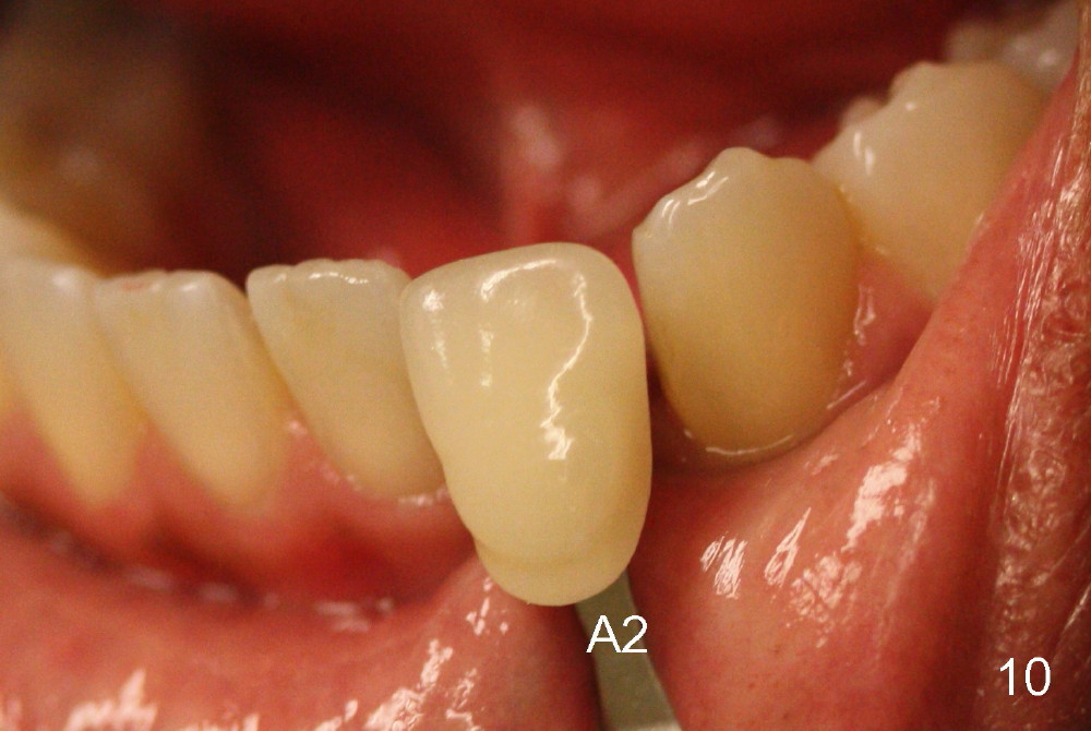
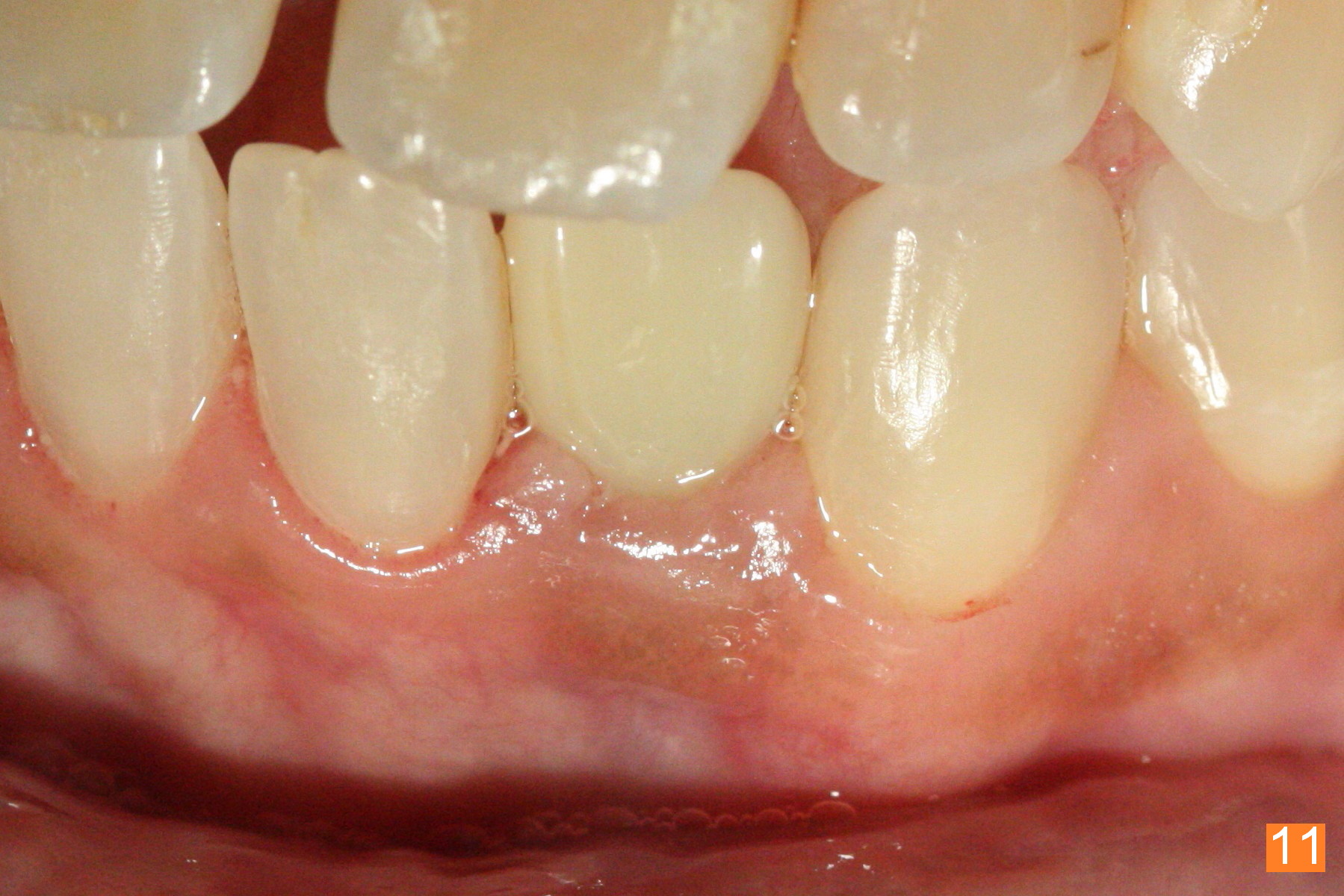
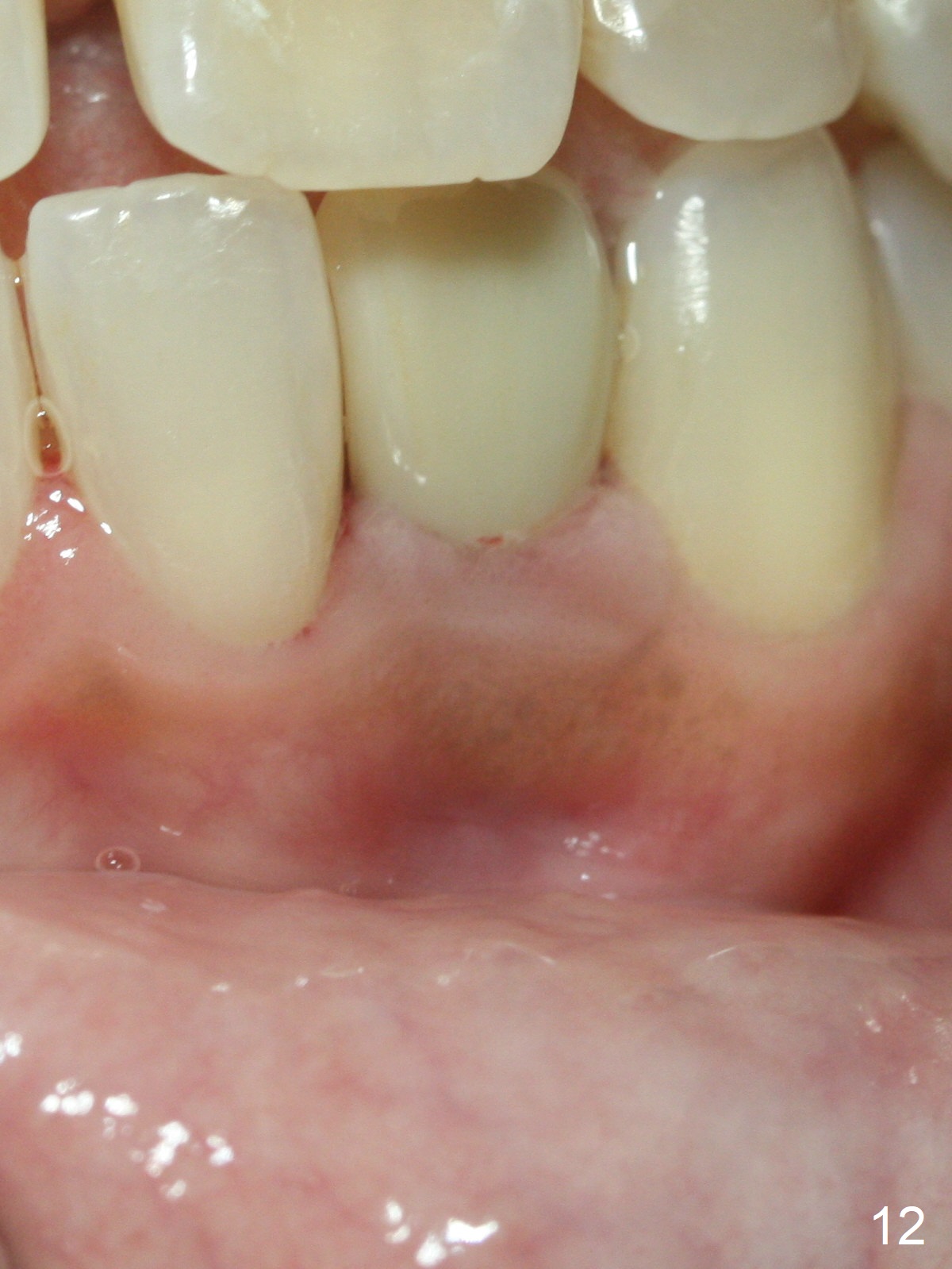
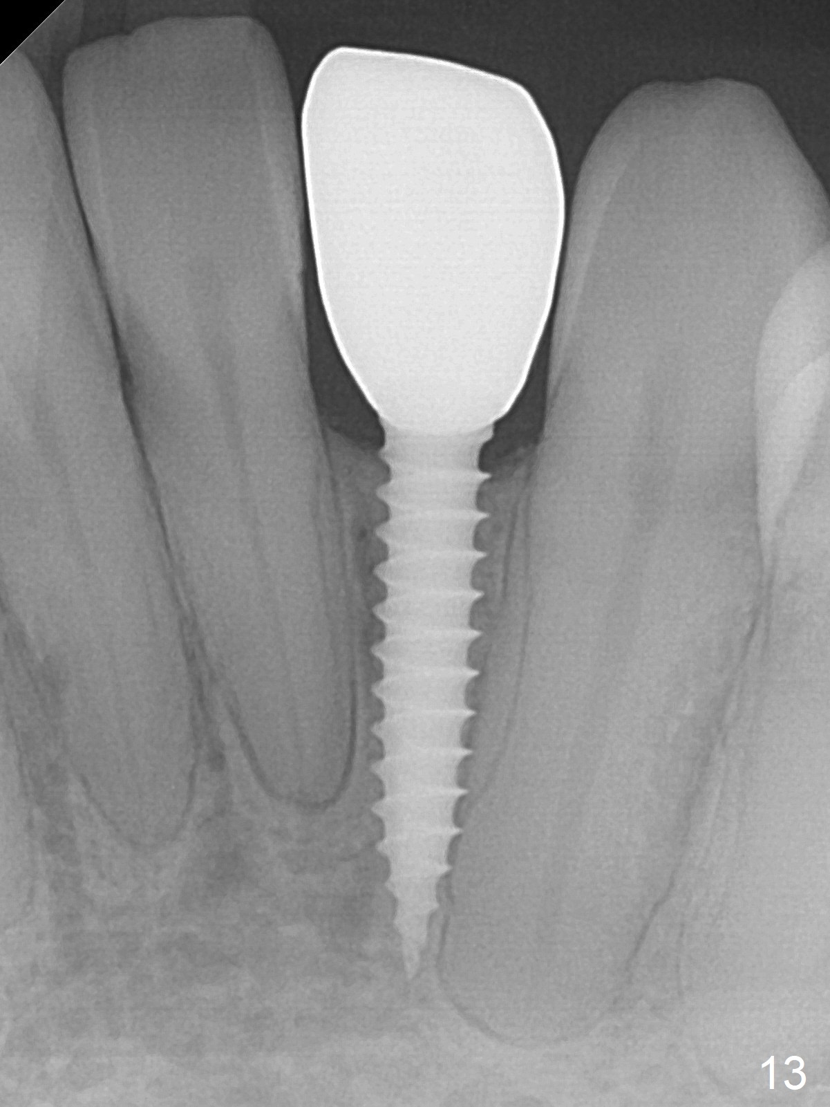
Fig.10: A2 shade guide. The patient requests A1 for the incisal edge and body and A2.5 for the cervical region. Fig.11 is taken 22 months post 2nd cementation (4 years postop). The implant crown remains normal 1 year 10 months post 2nd cementation (4 years 10 months postop, Fig.12,13).
Multiple units of immediate implants, immediate provisionals are to be introduced next.
Immediate Implant for Lower Incisors, Immediate Implant, 1-Piece
Xin Wei, DDS, PhD, MS 1st edition 10/02/2012, last revision 04/18/2020