,%20.5-1.5%20allograft.jpg)
|
|
|
|
|
|
|
|
|
|
|
|
|
|
|
|
|
|
|
|
Implant Placement Depth Control
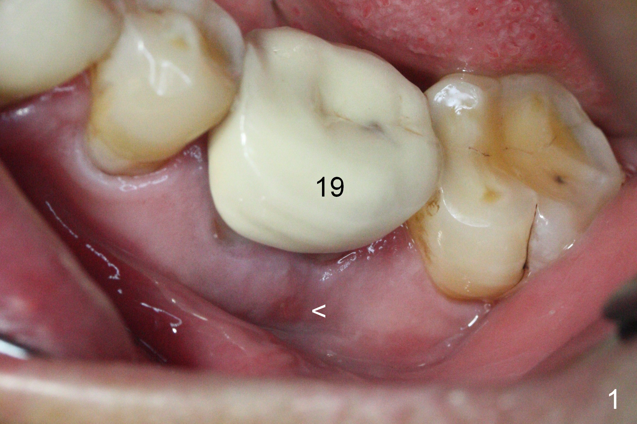
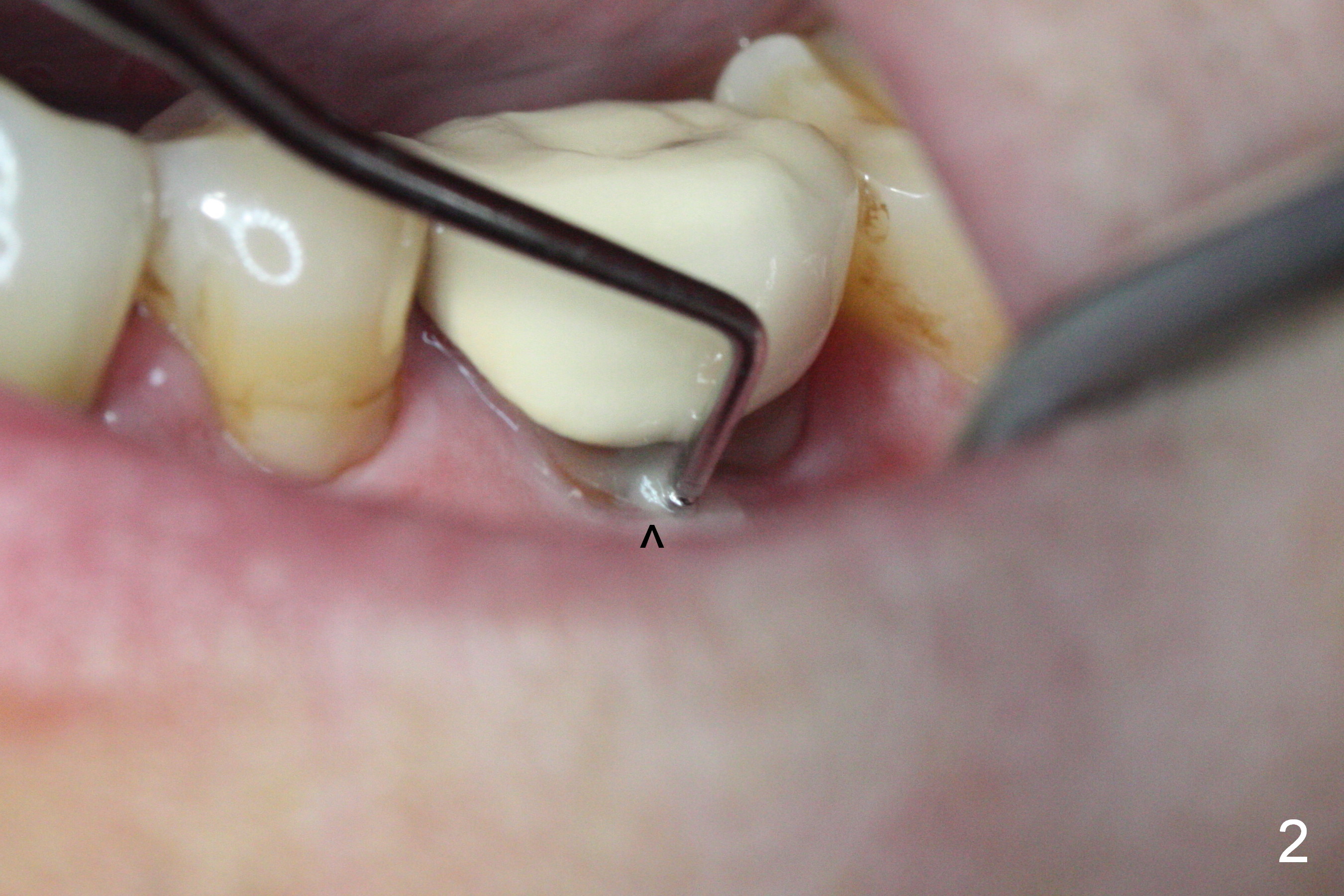
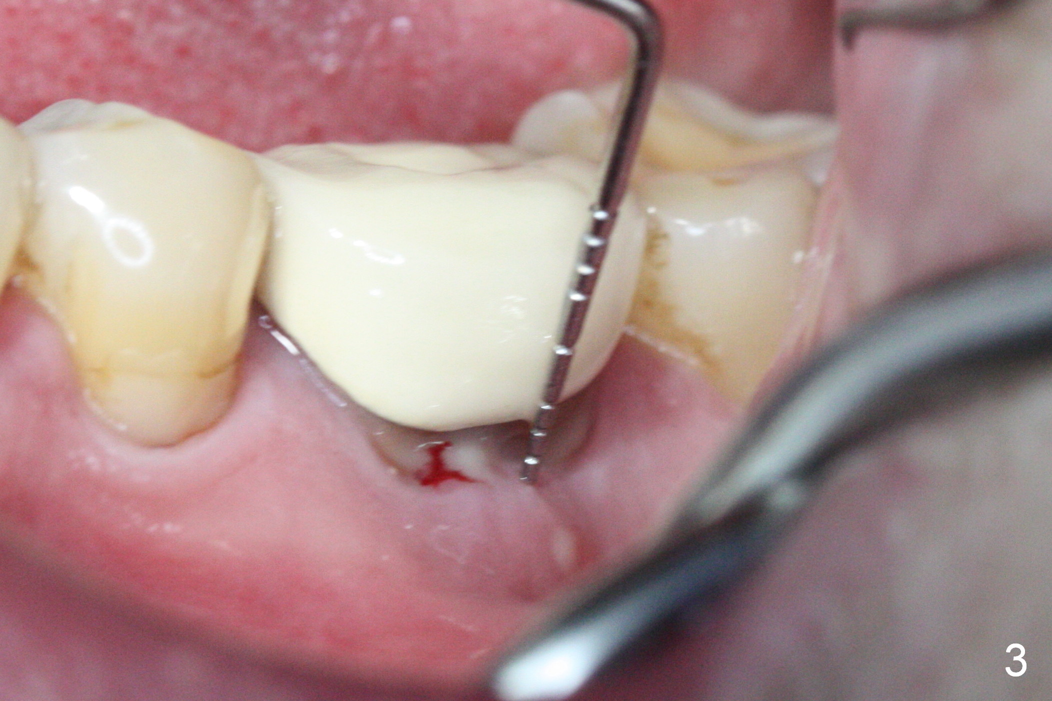
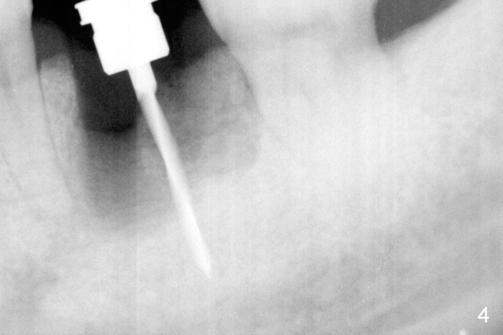
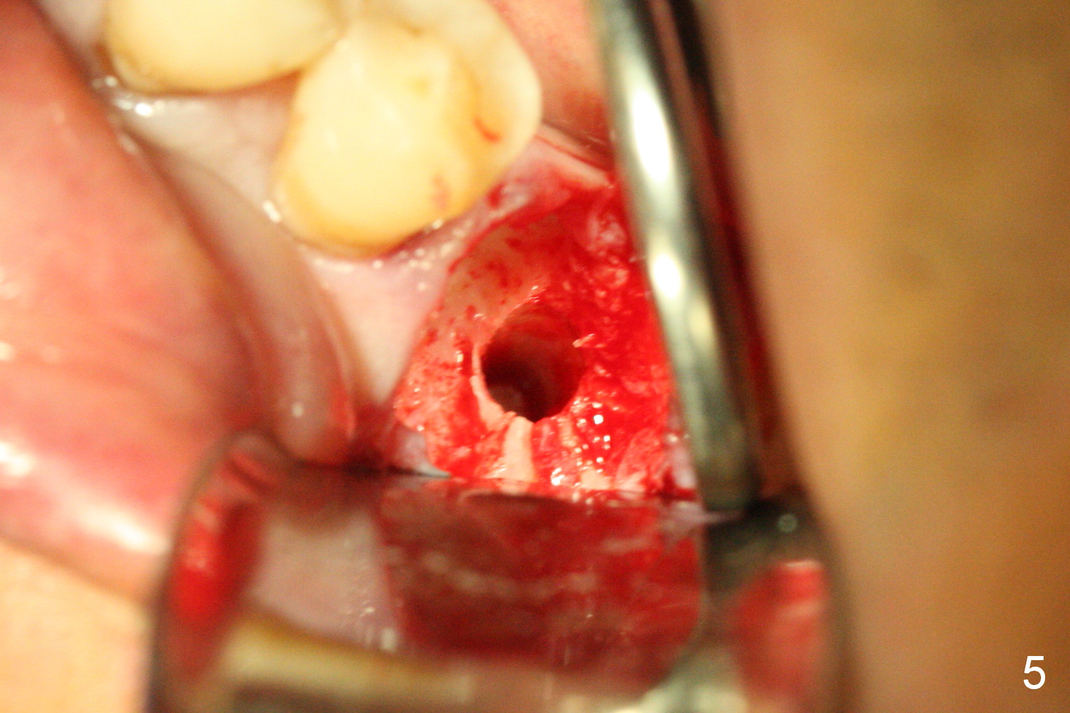
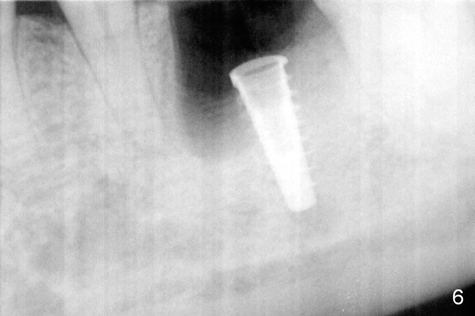
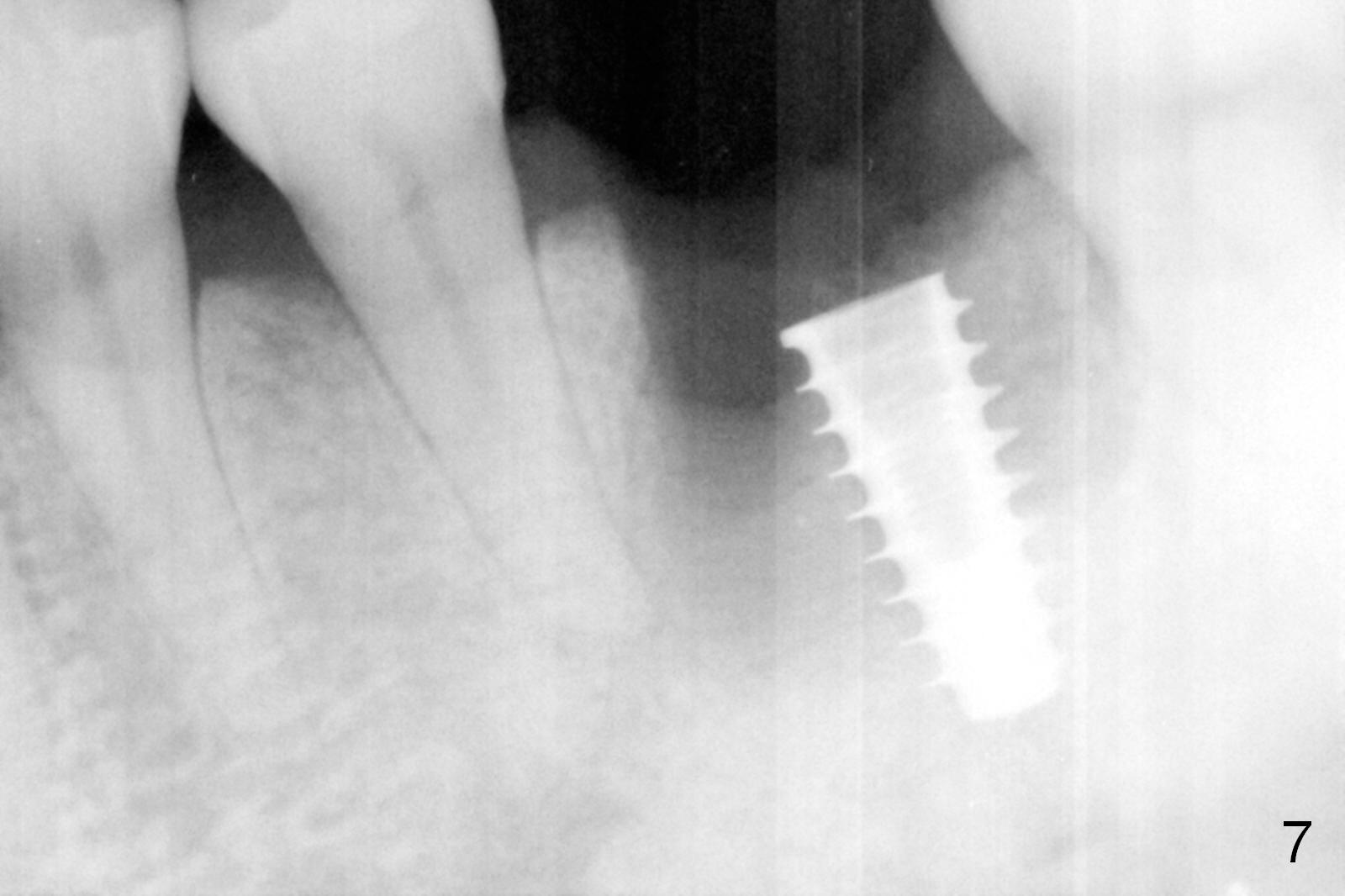
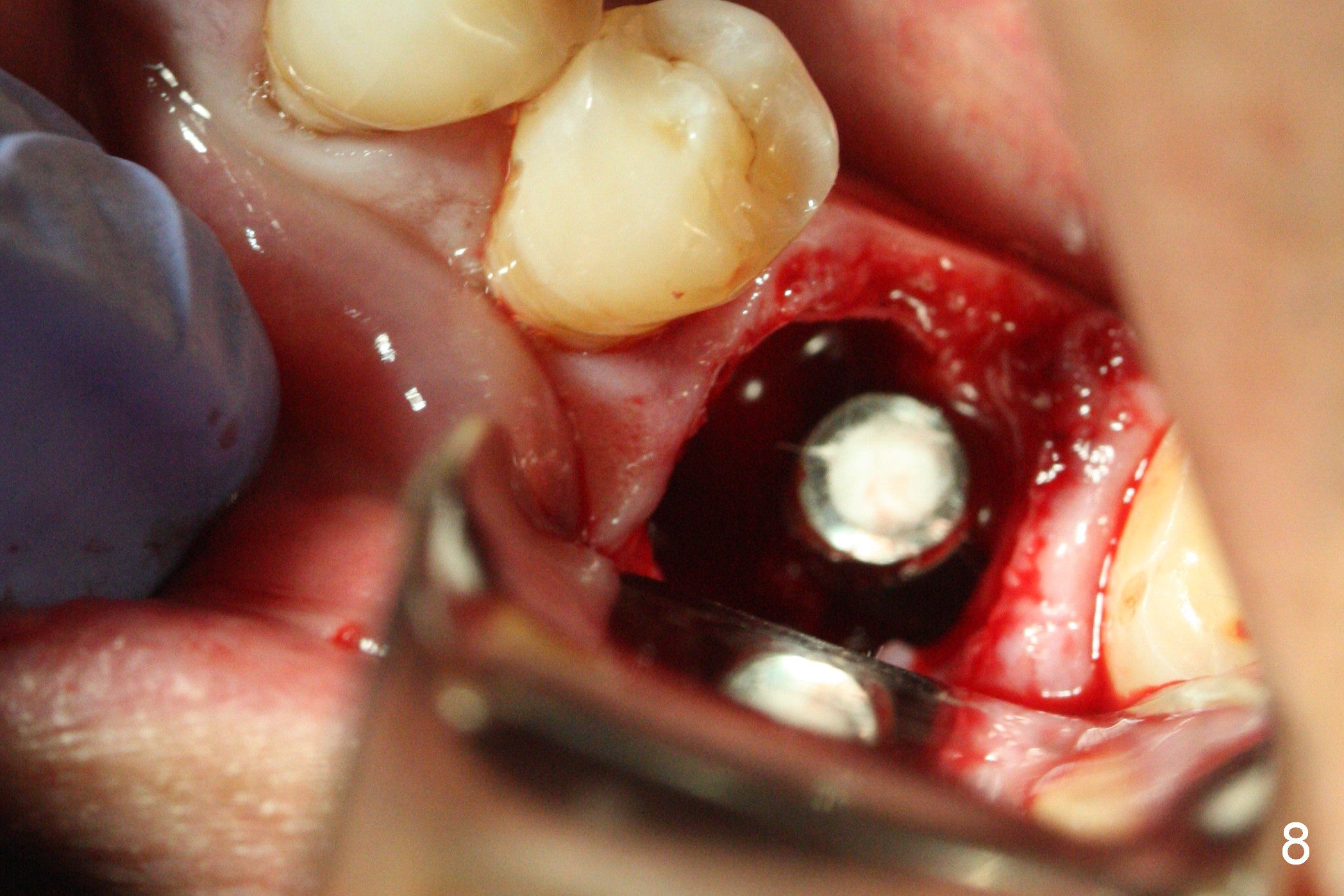
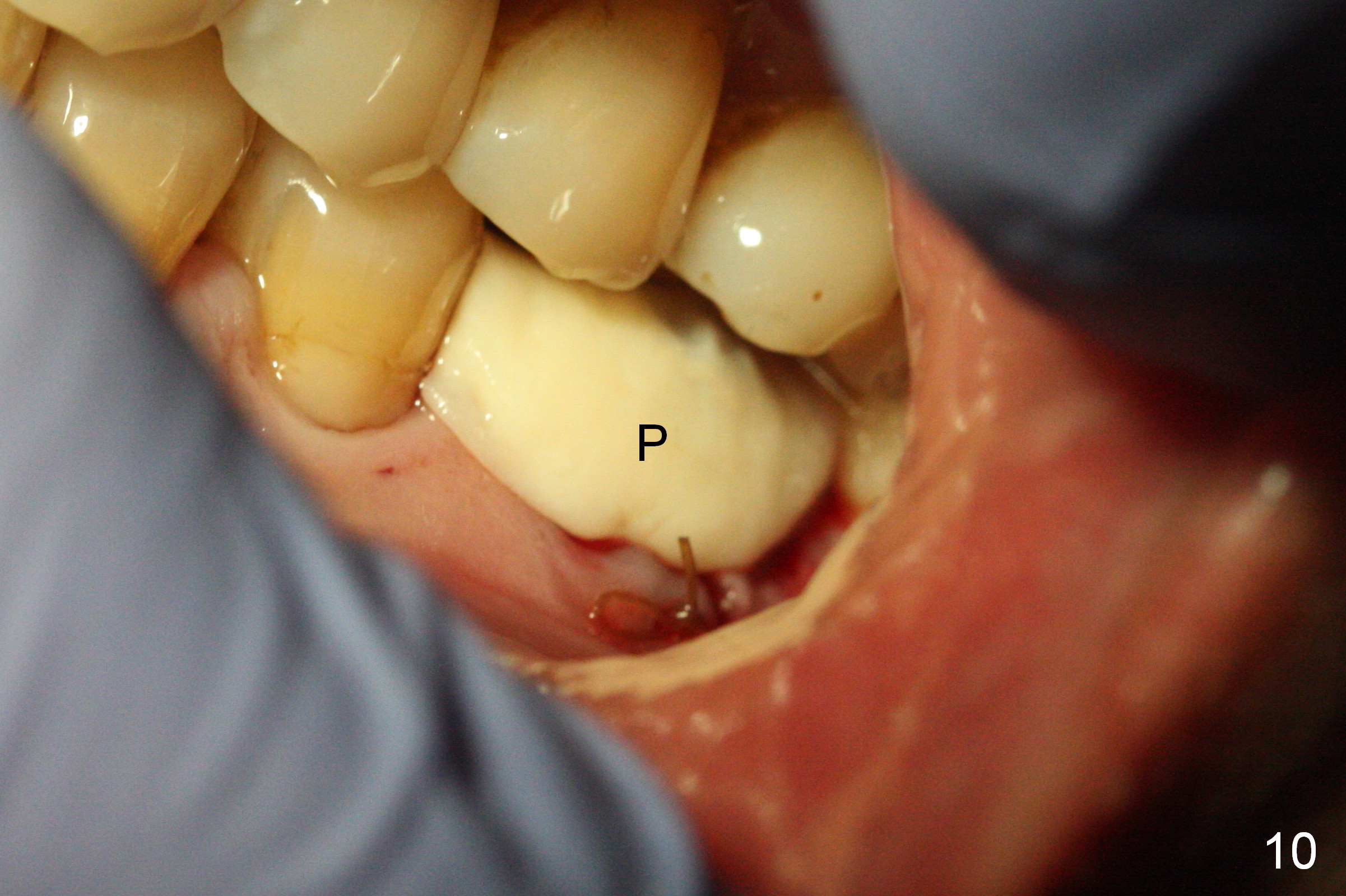
The asymptomatic tooth (#19) has a midbuccal fistula (Fig.1 <). The mesiobuccal pocket is 10 mm with purulent discharge from the sulcus (Fig.2,3). The mesiobuccal wall defect is confirmed when the tooth is extracted. Osteotomy is initiated lingually in the septum following septal crestoplasty (flattening) (Fig.4: using 1.6 mm drill for 9 mm). Since the lingual portion of the osteotomy is higher, it is difficult to use drill with stopper. For the narrow septum osteotomy, multiple drills are used sequentially (Fig.5 after 4.3 mm drill). A 4.5x11 mm dummy implant is placed (Fig.6) apparently too deep. When a 5x11 mm IBS implant is being placed, the depth is tightly controlled (Fig.7). The implant is apical to the lingual crest, whereas there is ~ 2 mm implant exposure buccally. That is, there is a large gap mesiobuccally (Fig.8), which is filled with .5-1.5 mm allograft (Fig.9 *). A 6.5x5.7(3) mm abutment (A) is placed and trimmed for an immediate provisional (Fig.10 P). The lacerated buccal gingiva is sutured as well as application of Perio Glue.
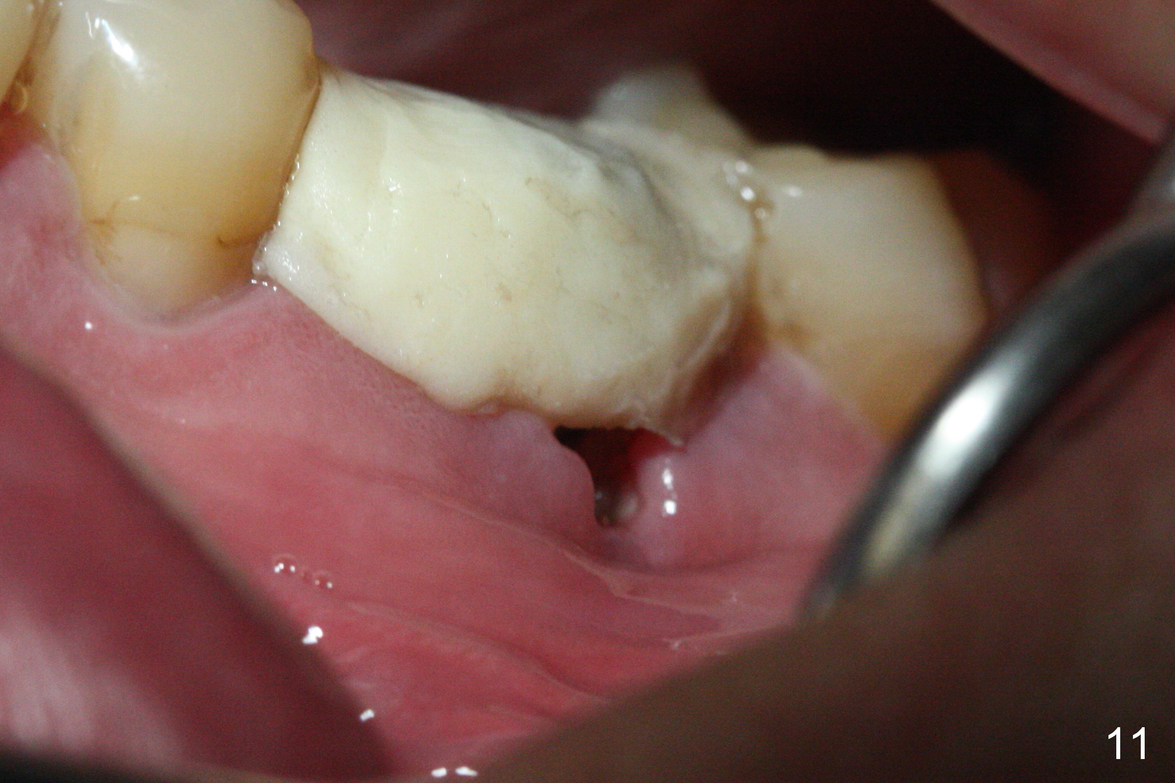
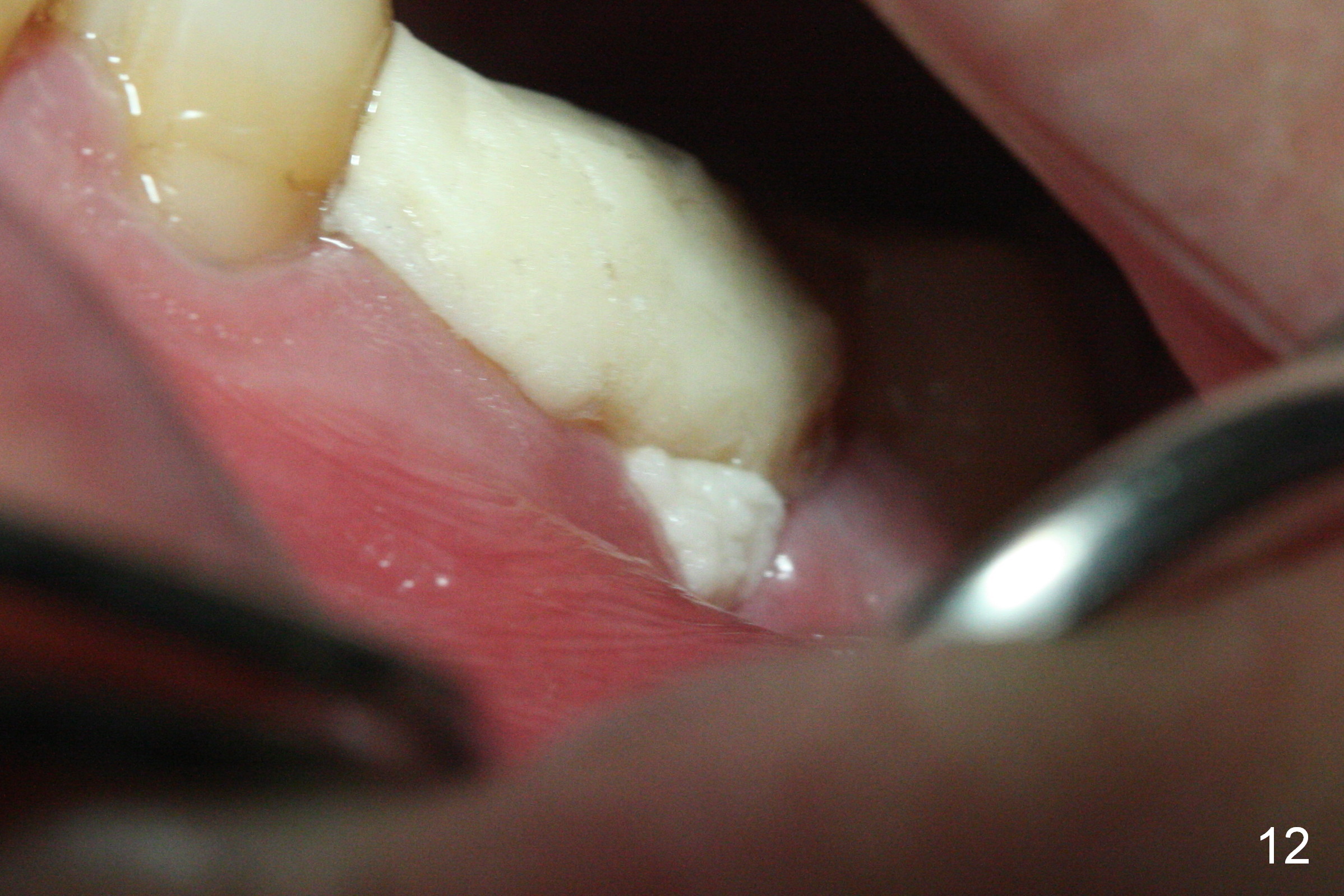
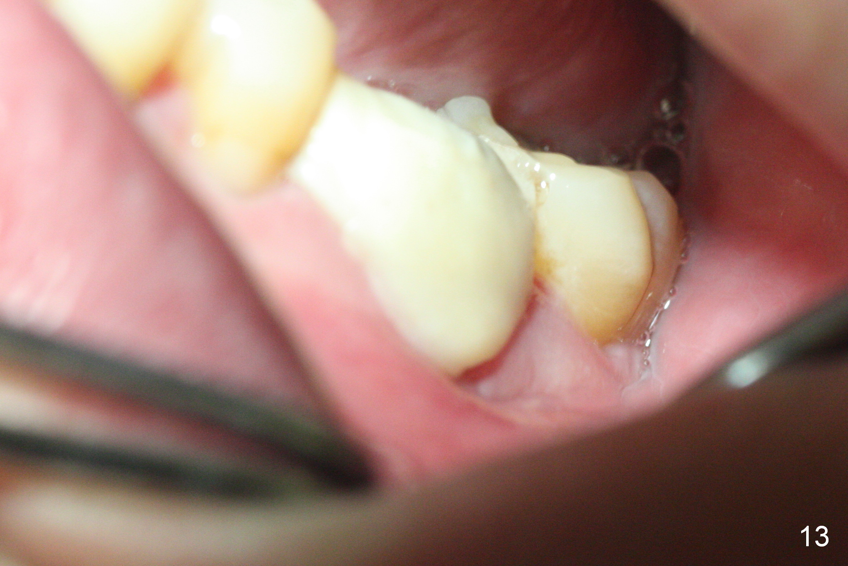
The mild postop pain lasts ~ 6 days with loss of bone graft for the 1st two days postop. Midbuccal dehiscence is found 7 days postop (Fig.11). After Chlorhexidine irrigation, a piece of Osteogen plug is inserted (Fig.12), followed by closure of the wound with a new layer of acrylic (Fig.13). It appears that placing a piece of collagen membrane against buccal plate before bone graft is not secure. Preferably, PRF membrane(s) should be used instead to facilitate wound healing internally, followed by externally acrylic coverage (to prevent bone graft from dislodgment if the membranes do not prevent dehiscence). Most importantly, do not hesitate to harvest blood for PRF if indicated. Once making it, keep it moist with the PRF box cover.
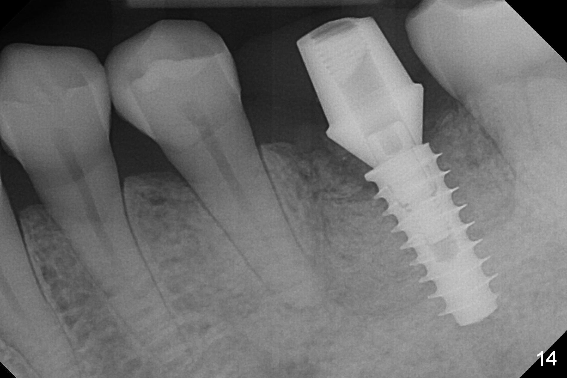
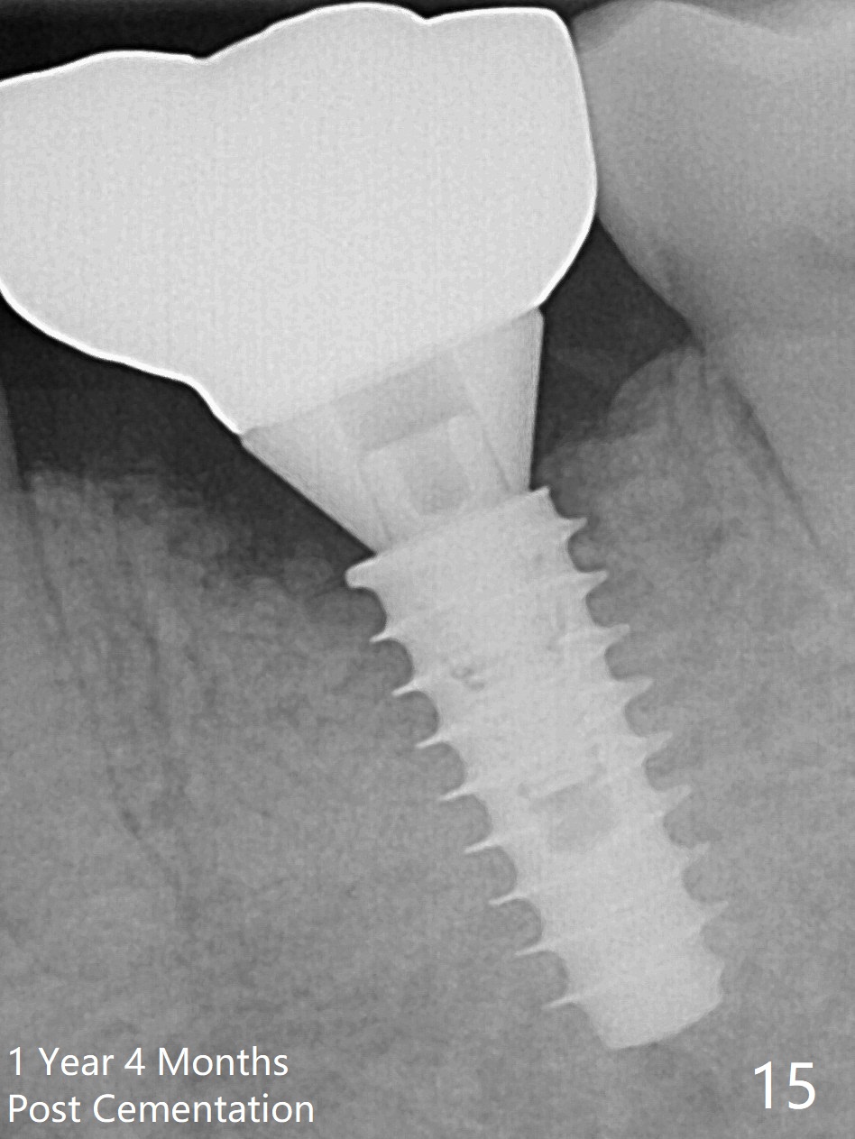
There is a new pattern of bony trabeculae around the implant 4.5 months postop (Fig.14). Bone density increases 1 year 4 months post cementation (Fig.15).
Return to Lower Molar Immediate Implant, Prevent Molar Periimplantitis (Protocols, Table) 1st Year, Course 1 2 GEM21S Cases Expect to Form Lamina Dura
Xin Wei, DDS, PhD, MS 1st edition 10/20/2016, last revision 03/31/2020