|
|
|
|
|
||
|
|
|
|
|||
|
|
|
|
|||
|
|
|
|
|
|
|
|
|
|||||
Large Defect, Large Implant I
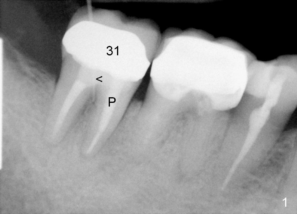
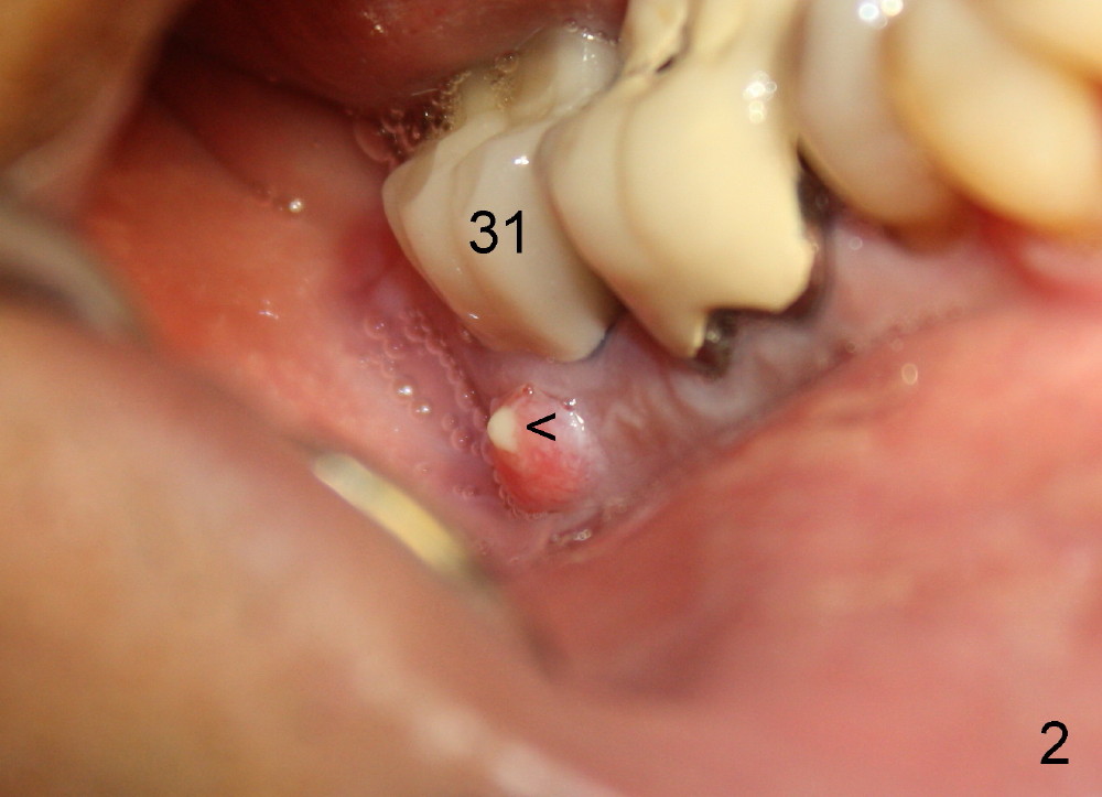
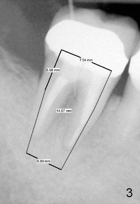
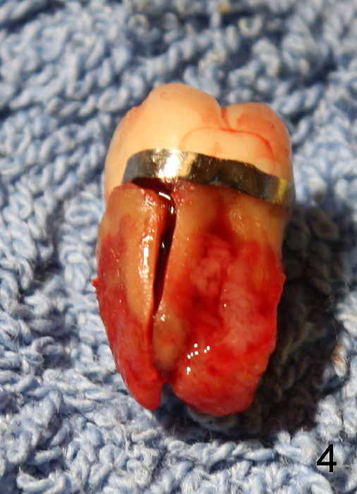
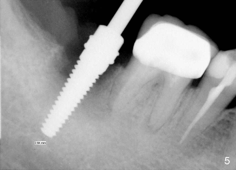
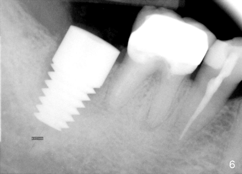
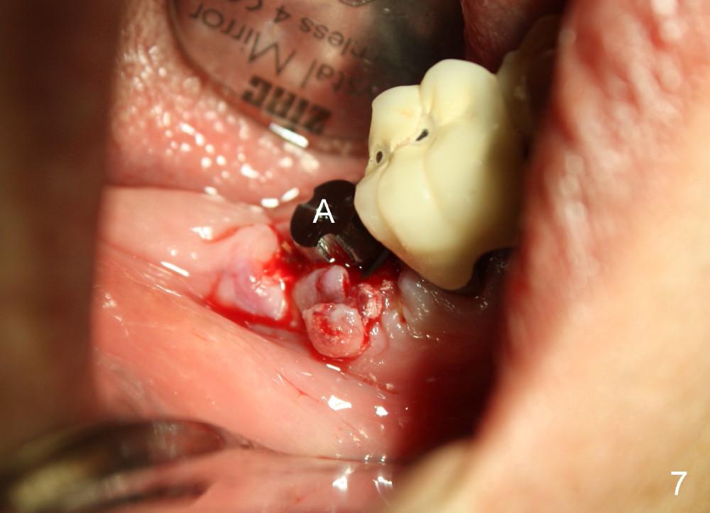
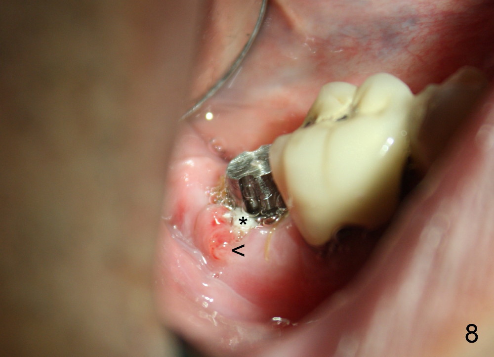
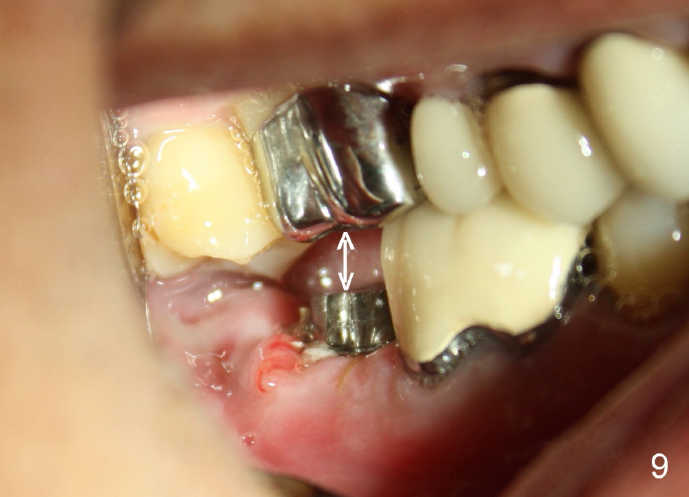
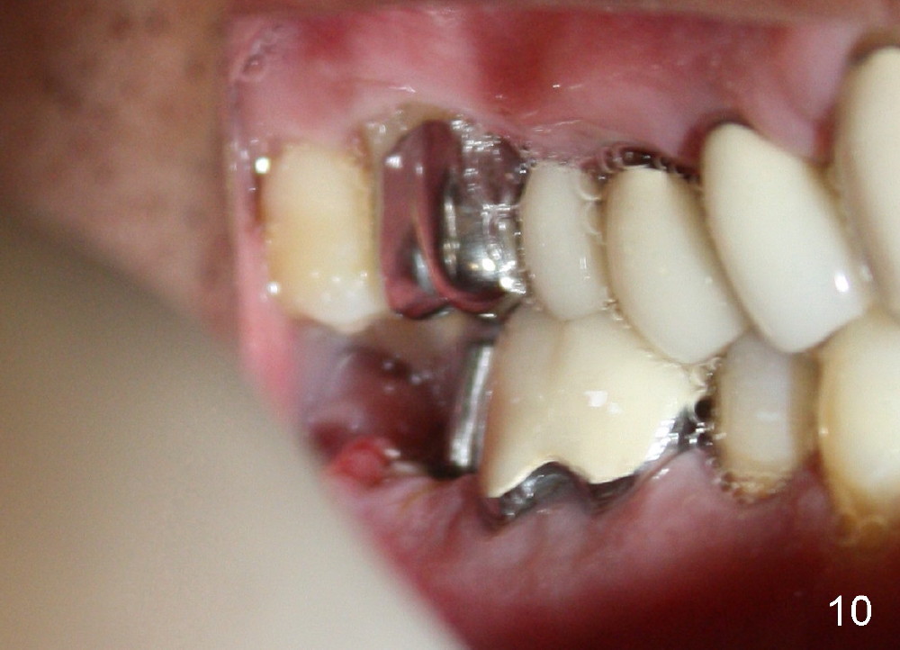
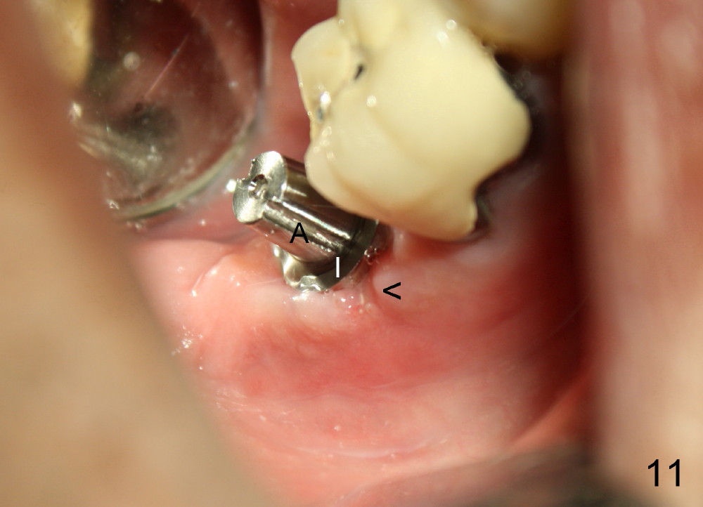
A 51-year-old man has experienced pain and swelling in the lower right 2nd molar for 7 months (Fig.1, P: post in the mesial root; <: gutta percha inserted into a buccal fistula). Fig.2 is taken immediately before extraction (<: purulent discharge from the mesiobuccal fistula) and immediate implant (Fig.3 implant design: 7x14 mm). A vertical root fracture is noted in the mesial root (Fig.4: lingual view). The septum is in fact absent, as compared to Fig.1. The mesiobuccal plate is low; the osteotomy starts lingual to the center of the socket. Fig.5 shows a 4.5x17 mm tap in place: approximately 3.5 mm in the new bone (~2.5 mm from the inferior alveolar canal). When a 7x17 mm tap (14 mm from the gingival margin) is placed in the socket, it looks relatively small. Fig.6 shows a 8x14 mm implant in place with a small gap distally. Mineralized cancellous allograft and Osteogen mixture is placed mainly buccally, followed by a thin strip of collagen dressing and sutures (Fig.7); an abutment (A) is placed to keep perio dressing in place. The perio dressing does not stay long. The buccal portion dislodges by itself 5 days postop. The lingual portion is removed in clinic. New dressing is going to be re-applied, because the buccal wound has not completely healed (Fig.8 <, albeit asymptomatic) with partial exposure of the bone graft (*). Why is the dressing lost so early? The abutment is not long enough; there is plenty of occlusal clearance (Fig.9 arrows). A longer abutment is used to increase mechanical retention for perio dressing (Fig.10). By the time the second perio dressing dislodges, the wound has healed (Fig.11, 13 days postop).
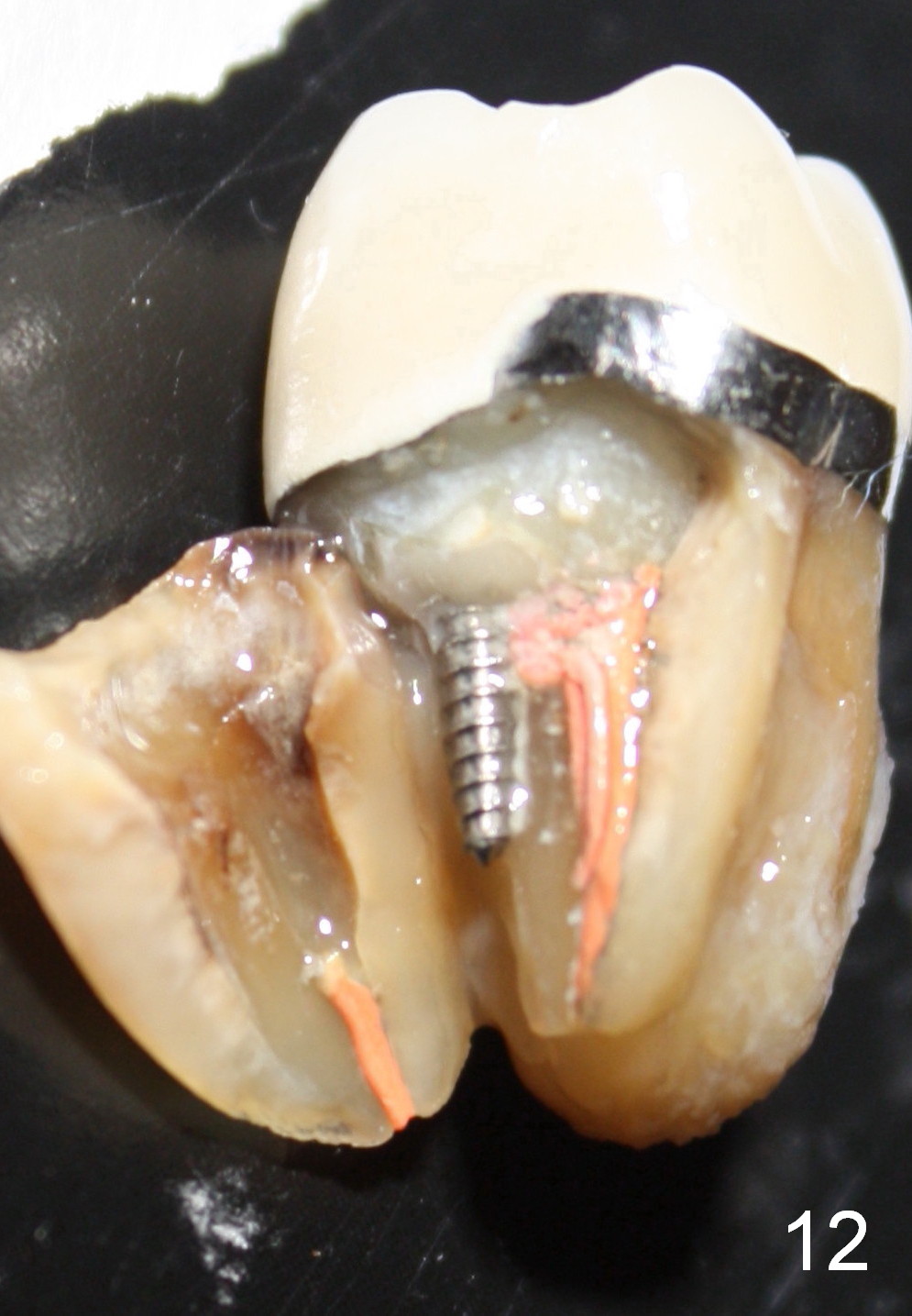
Fig.12 show root fracture associated with mesiobuccal post of the 2nd molar. In contrast the lower right 2nd premolar is intact without crown after root canal therapy. It appears that the post weakens the root.
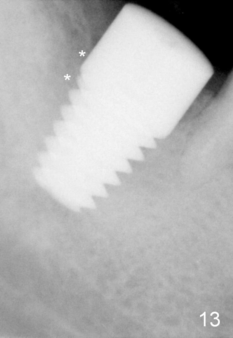
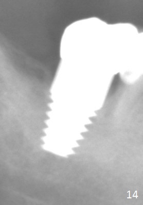
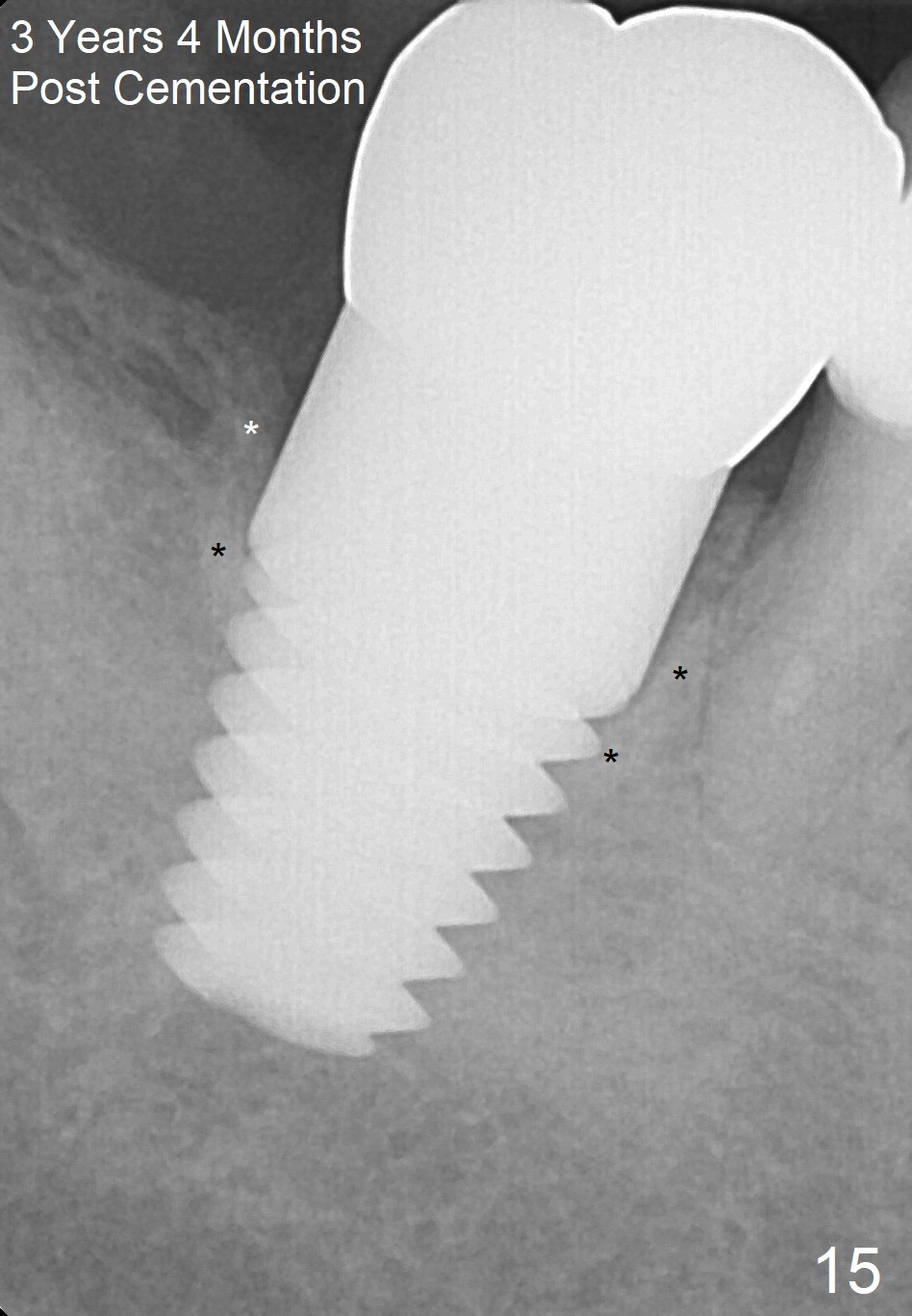
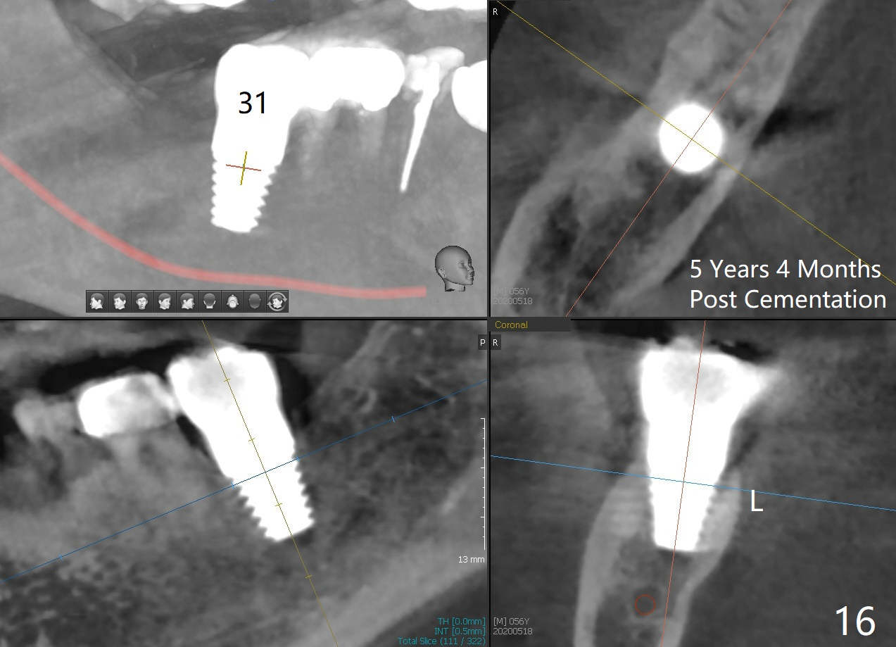
The patient returns for final restoration 5.5 months postop; it appears that the distal gap has disappeared (Fig.13 *). There is no bone loss 26 months post cementation (Fig.14). The lamina dura-like dense bone forms coronally 3 years 4 months post cementation (Fig.15 *). The 8 mm implant remains buried in the bone 5 years 4 months post cementation (Fig.16).
Return to
Lower Molar Immediate Implant
The patient's other implant cases (12,13,
3/4)
Failed Case
Xin Wei, DDS, PhD, MS 1st edition 07/24/2014, last revision 05/18/2020