,%204.5x17.jpg)
,%204.5x17.jpg)
|
|
|
|
|
|
|
|
|
|
|
|
|
|
|
|
||
|
|
|
|||
1- & 2-Piece Implants for Mandibular Anteriors
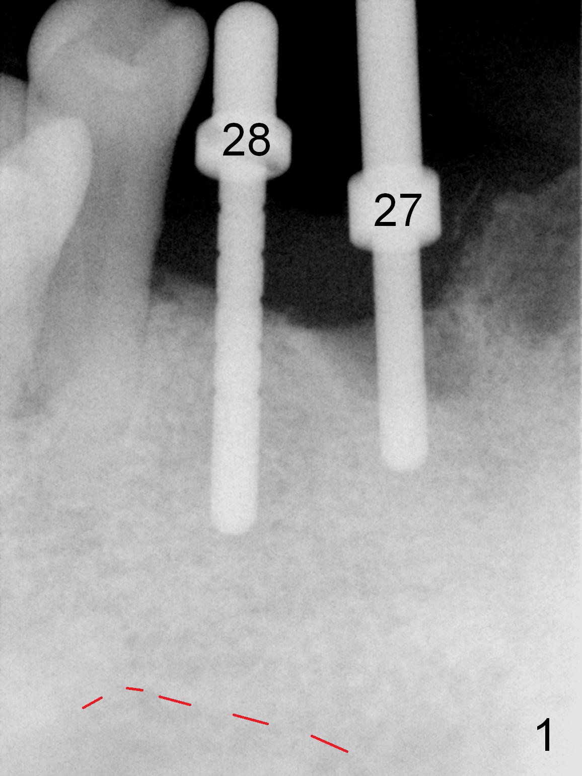
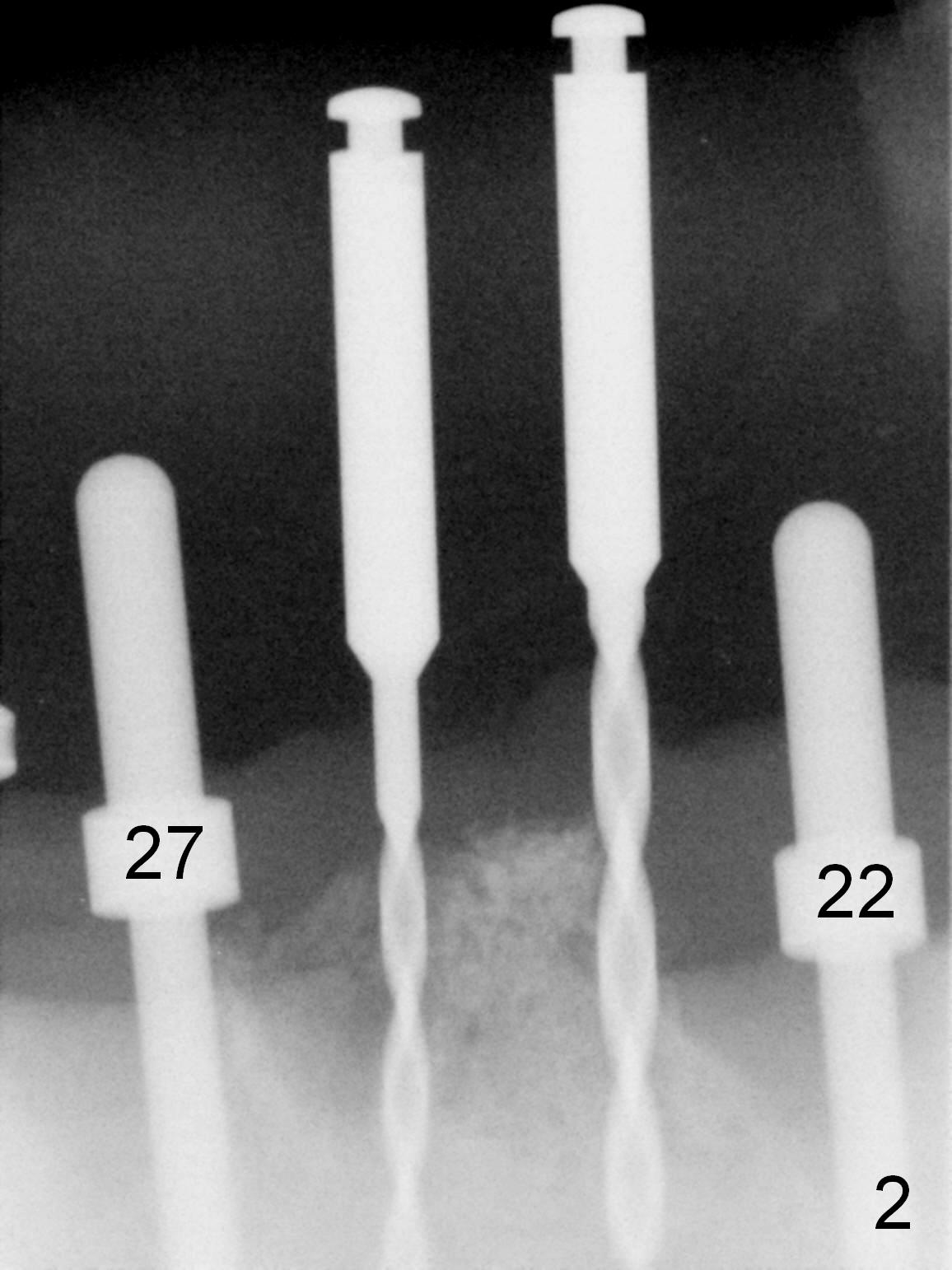
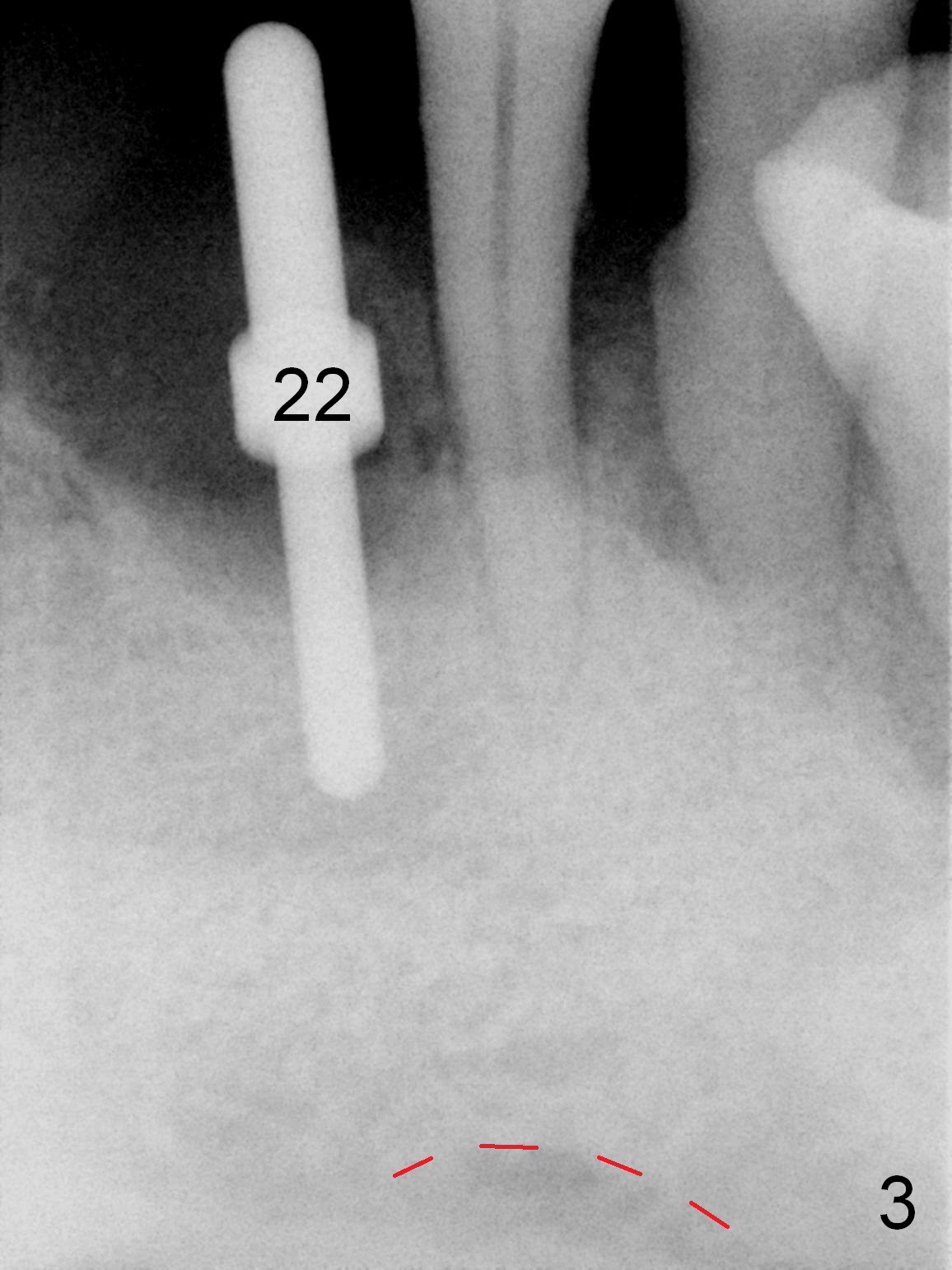
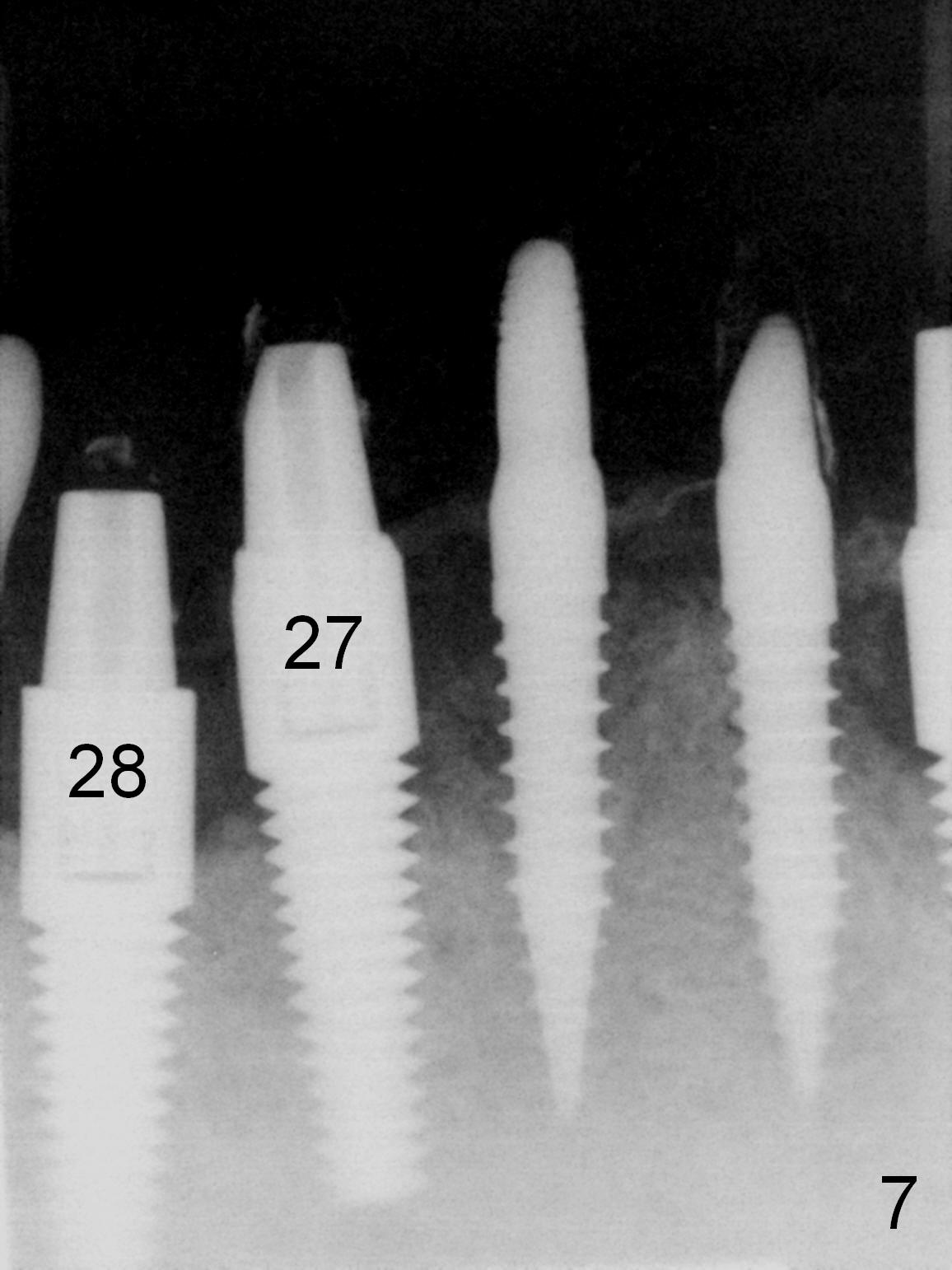
As expected, the bone density in the mandibular anterior region is found to be high during initial osteotomy (Fig.1-3). Five implants are placed at the sites between #22 and 28 (Fig.4,5): 3x14(2) mm 1-piece (bone-level) implants in the incisor region; 4.5x17 mm 2-piece (tissue-level) ones in the canine/premolar area. All of the implants are placed as lingual as possible. Bone graft is placed (*). Red dashed line: the superior border of the Inferior Alveolar Canal. Immediate splinted provisional bridge is fabricated. One week postop, periodontal dressing remains attached to the provisional and the gingiva.
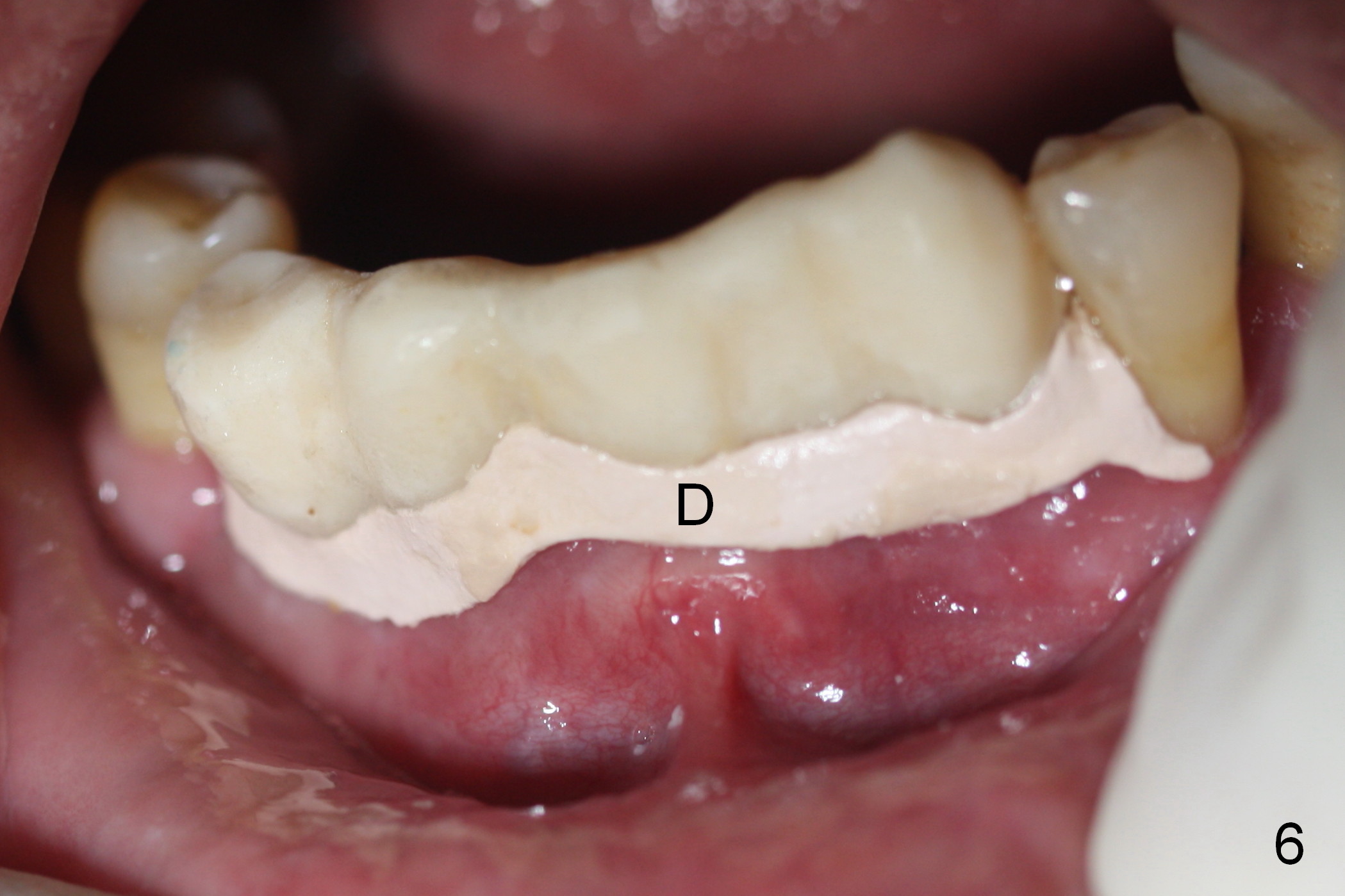
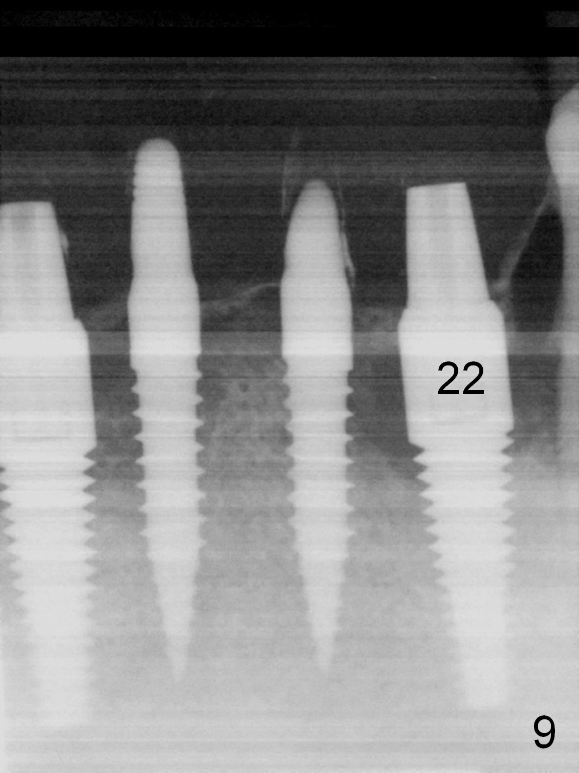
The patient returns for restoration 4.5 months postop (Fig.7-9). There appears bone growth around the implants. Impression is taken together with the implants at #4 and 6.
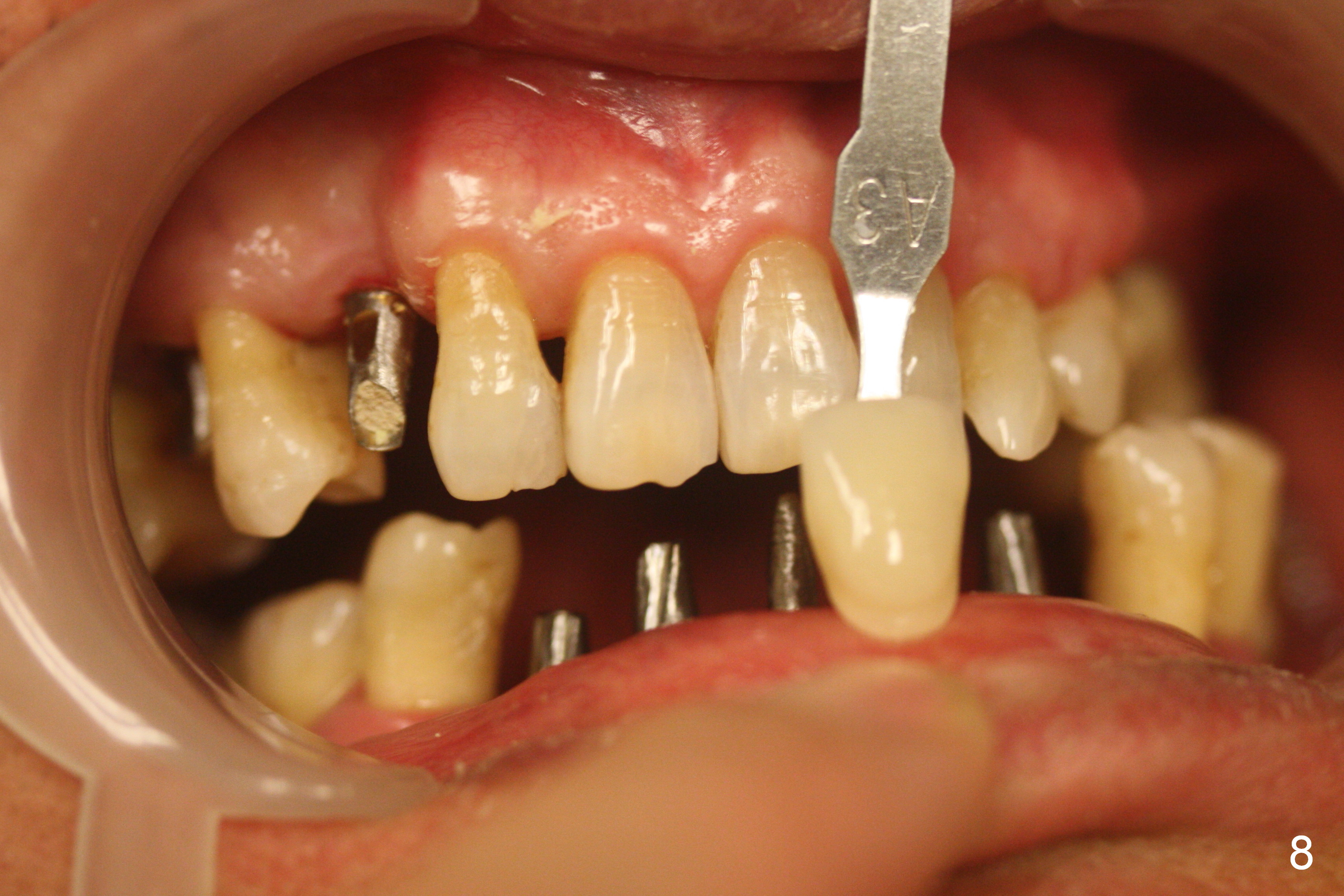
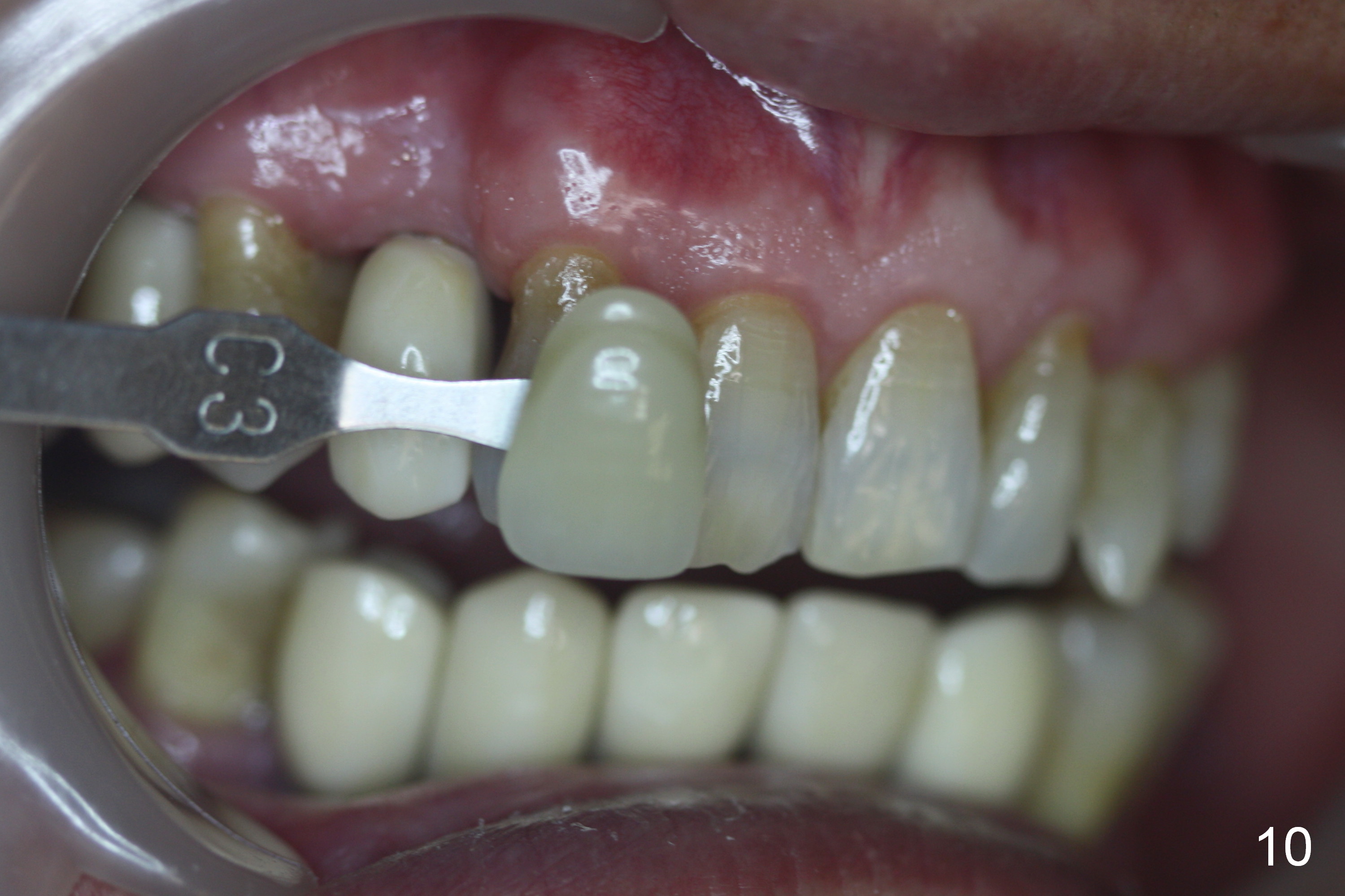
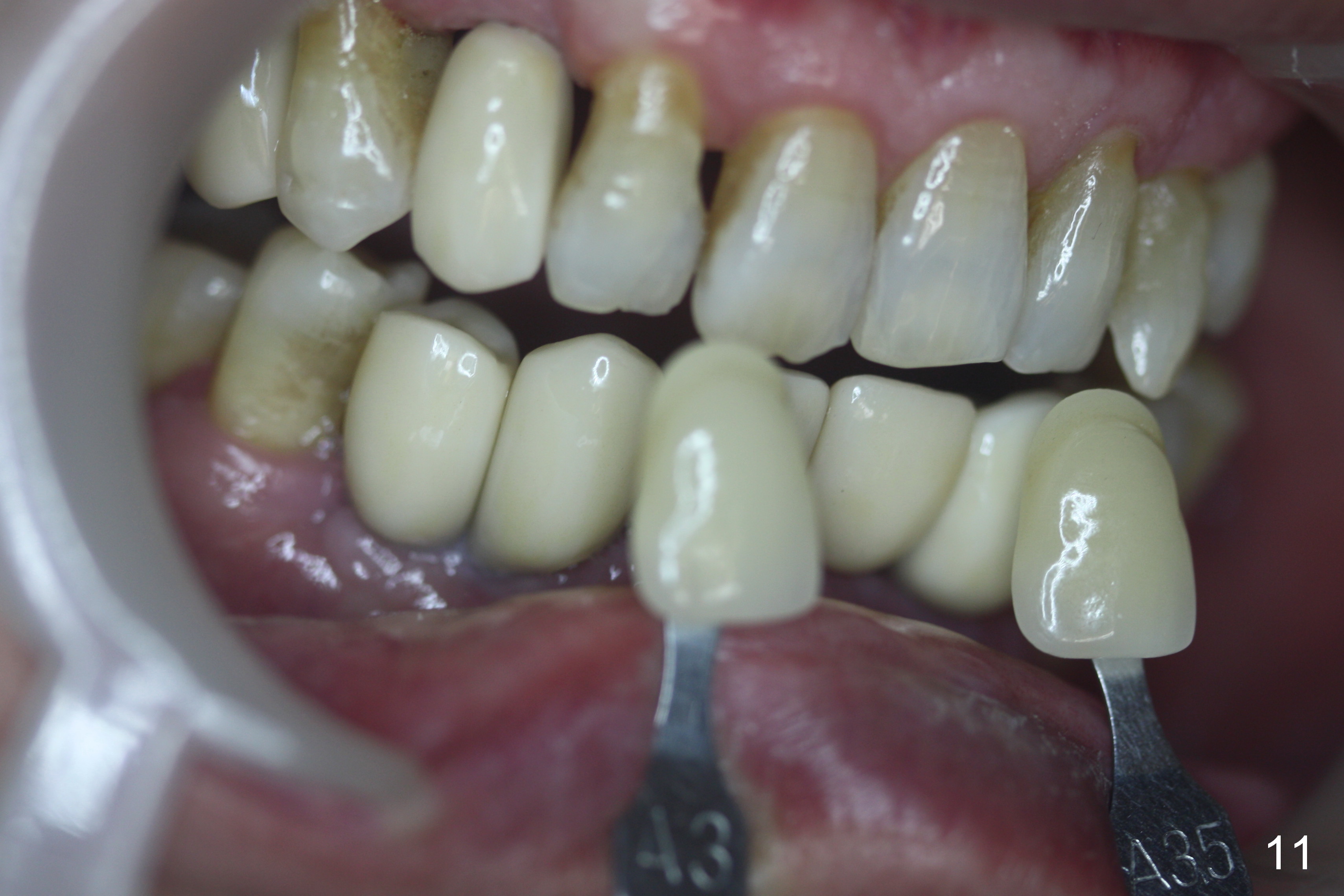
When the final crowns are seated, the shade is off. It appears that C3 is appropriate (Fig.10,11). Please make the crowns more transparent.
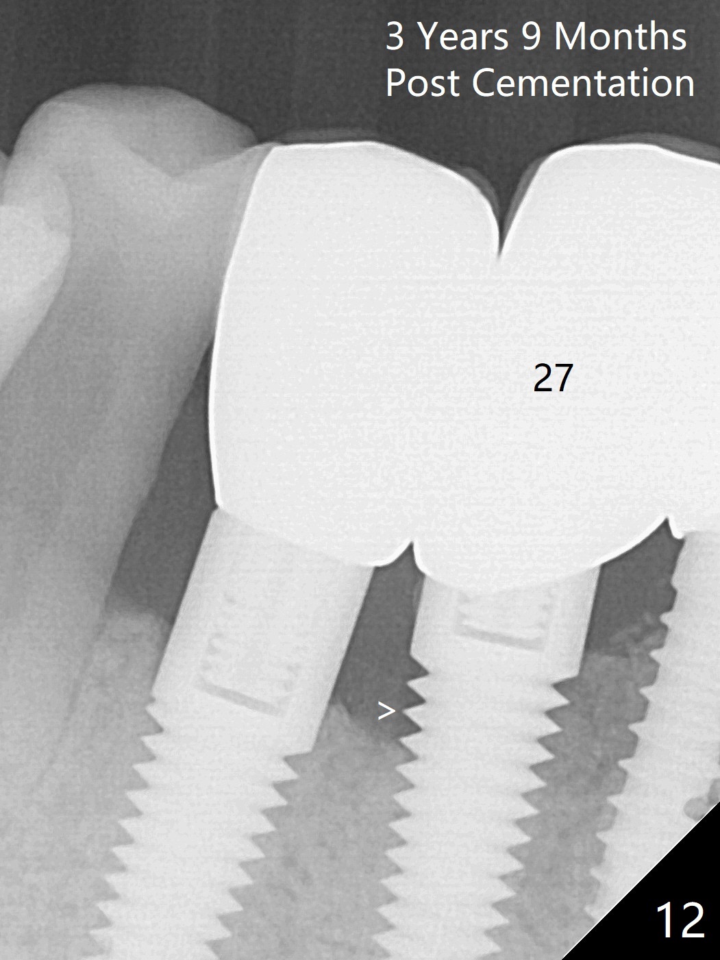
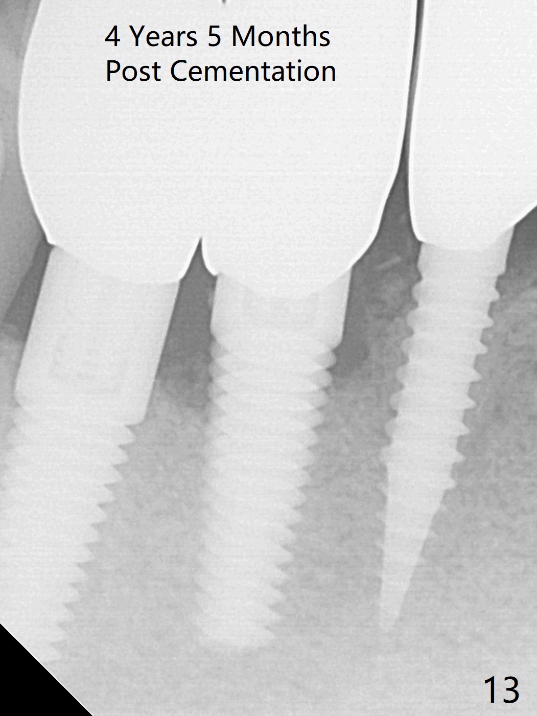
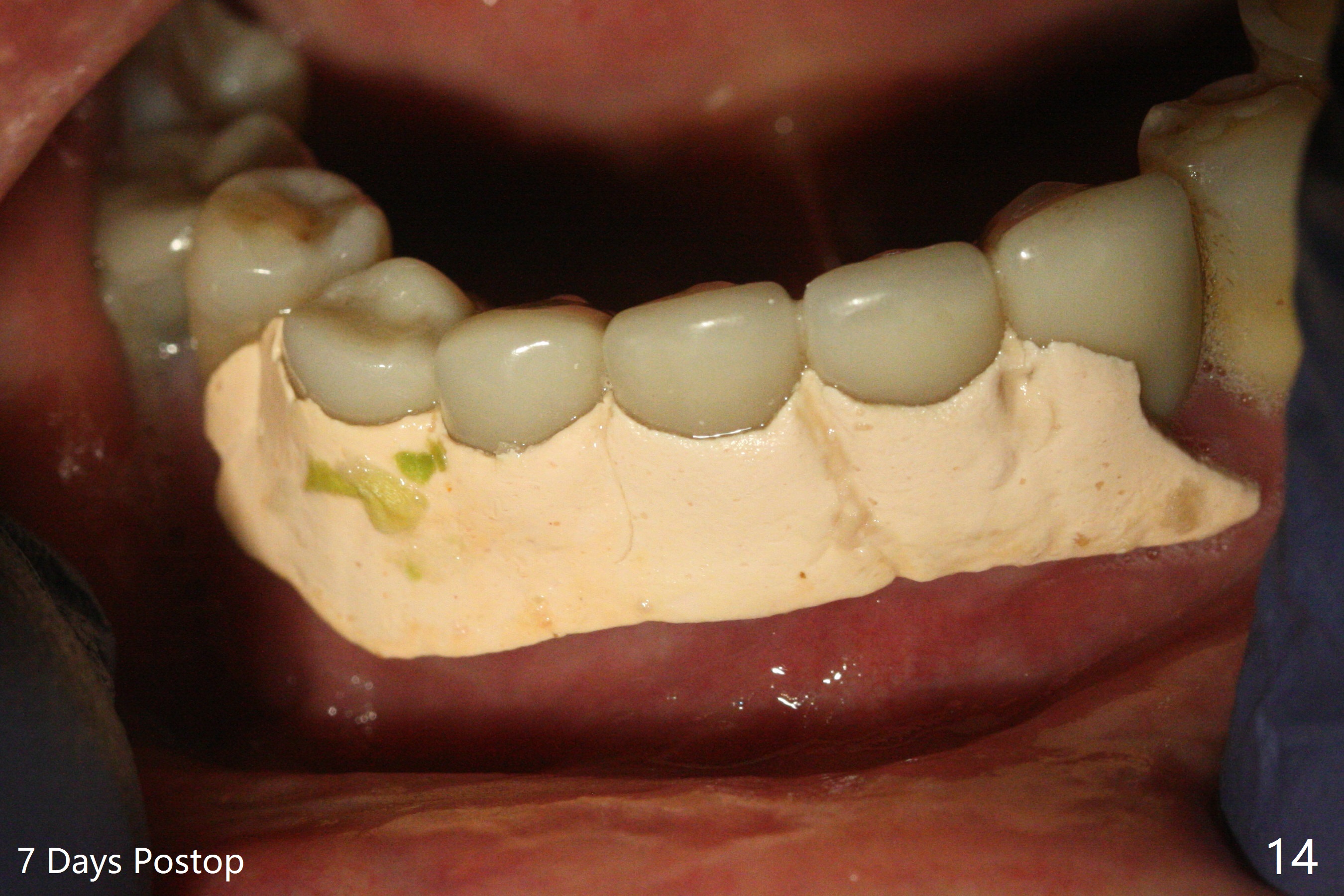
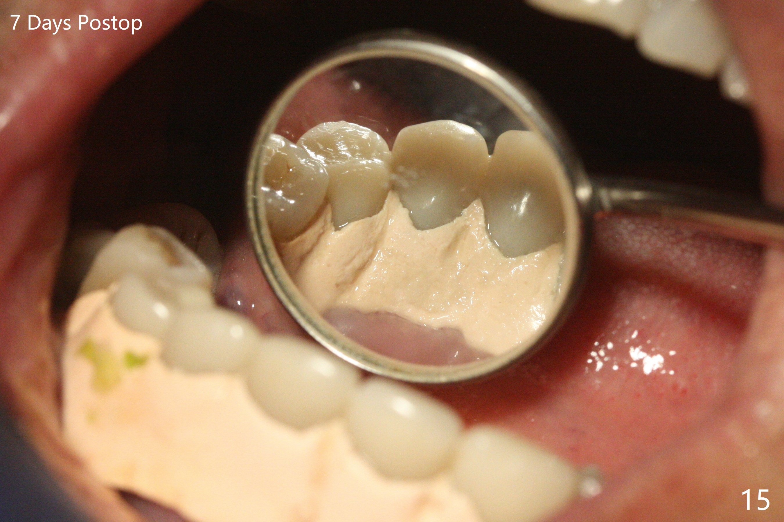
The implants remain asymptomatic years months post cementation (Fig.12), while the one at #27 is associated with buccal and lingual swelling and hemorrhage and bone loss (Fig.13). There is bone loss distal to #27 without symptom 3 years 9 months post cementation (Fig.12). Periimplantitis develops buccal and lingual 4 years 5 months post cementation (coronavirus, Fig.13). Sticky bone and PRF are used after debridement. There is no discomfort 7 days postop (Fig.14,15).
Return to Lower Arch Immediate Implant,
#4,6, 19 Immediate Implants,
Technicians
No Deviation
Xin Wei, DDS, PhD, MS 1st edition 09/27/2015, last revision 08/21/2020