
|
|
|
|
|
|
|
|
|
|
|
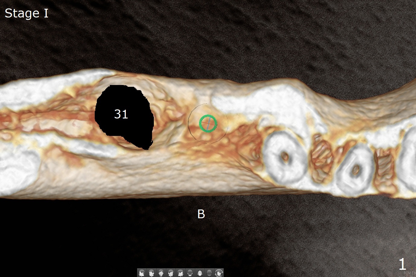
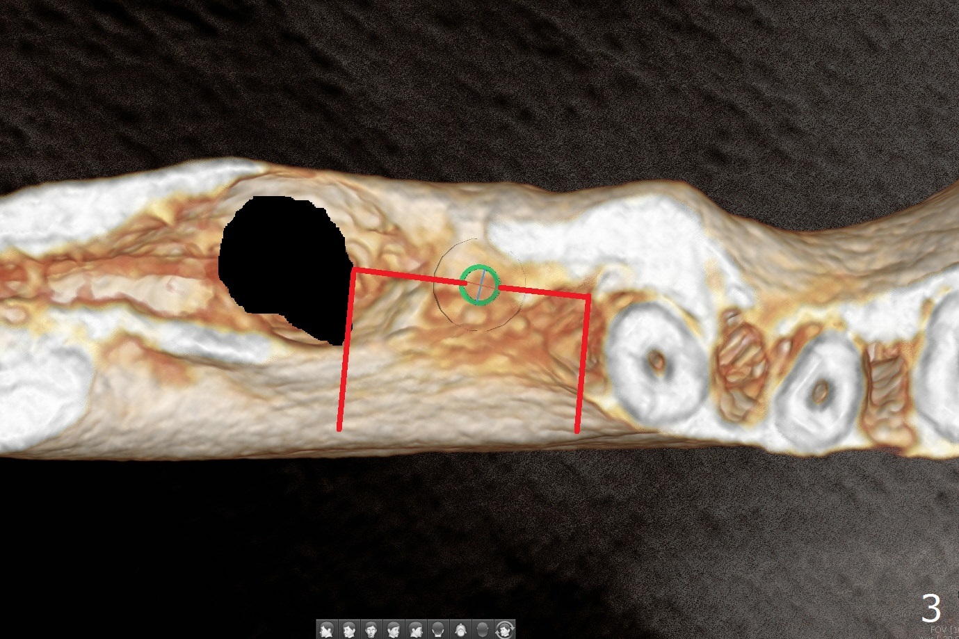
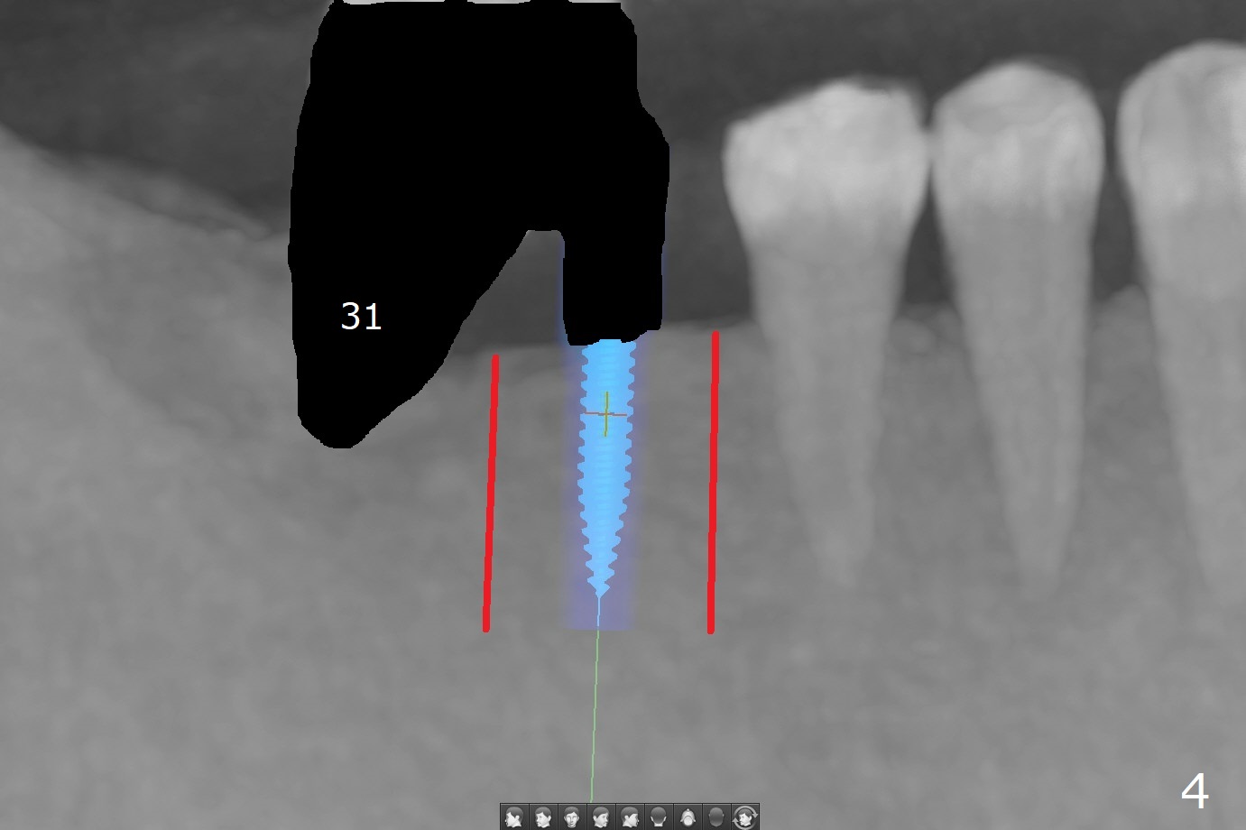
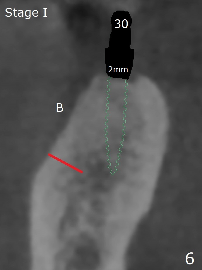
2-Stage Ridge Split with Guide M
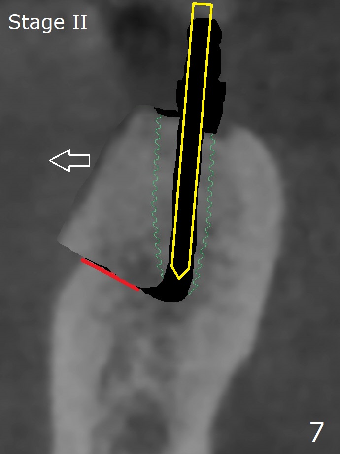
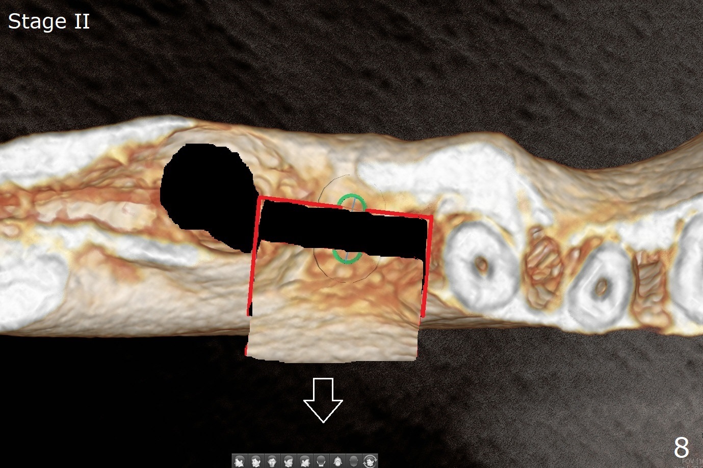
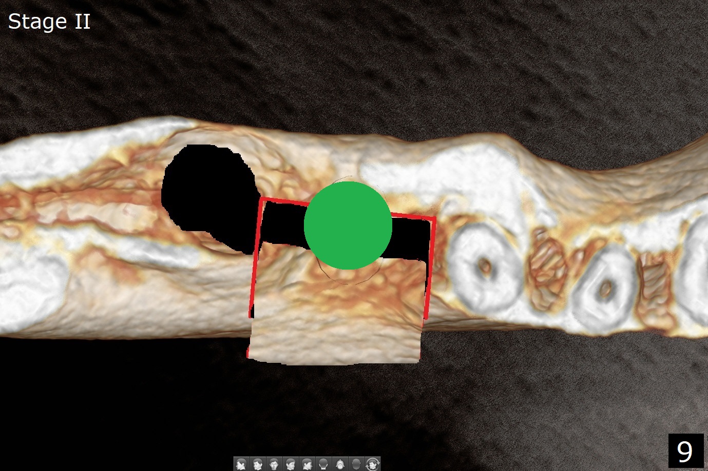
Several months post #31 socket preservation, the narrow ridge of #30 (Fig.1 (CT 3-D occlusal view), 6 (coronal section)) will be exposed with a crestal and 2 oblique incisions, followed by osteotomy with a 2 (2.2) mm drill (green) and a surgical guide. A crestal transverse cut will be conducted using a surgical fissure bur (Fig.2 red), followed by 2 vertical cuts (Fig.3,4 (3-D X-ray mode, buccal view) and an apical transverse one (Fig.5,6). The incision will be closed. Three weeks later, a crestal incision will be made. The buccal plate will be fractured and pushed buccal (Fig.7,8 arrow) using chisel (yellow). The guide will be reseated for osteotomy, implant placement (Fig.9 green) and bone graft. In fact there is not much problem when the osteotomy moves lingual.
Return to
Trajectory II
No Deviation
Cement
GEM21S
Xin Wei, DDS, PhD, MS 1st edition
04/14/2020, last revision
08/25/2020