,%20Vanilla.jpg)
,%20more%20Vanilla.jpg)


 |
,%20Vanilla.jpg) |
,%20more%20Vanilla.jpg) |
 |
 |
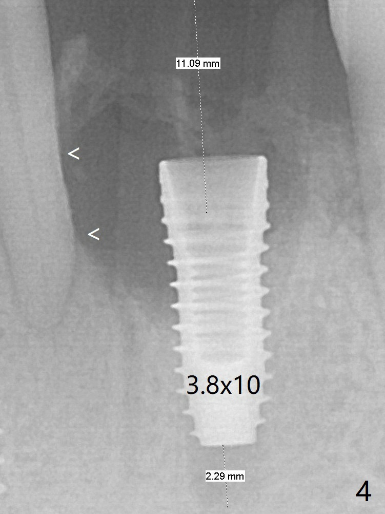
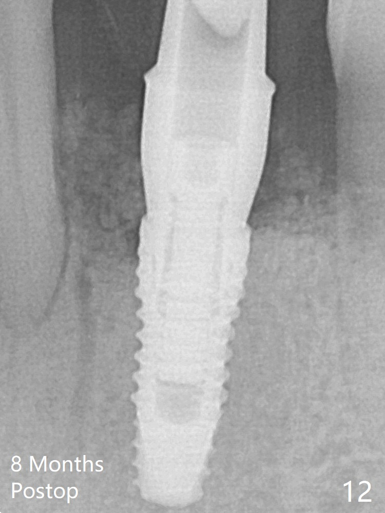
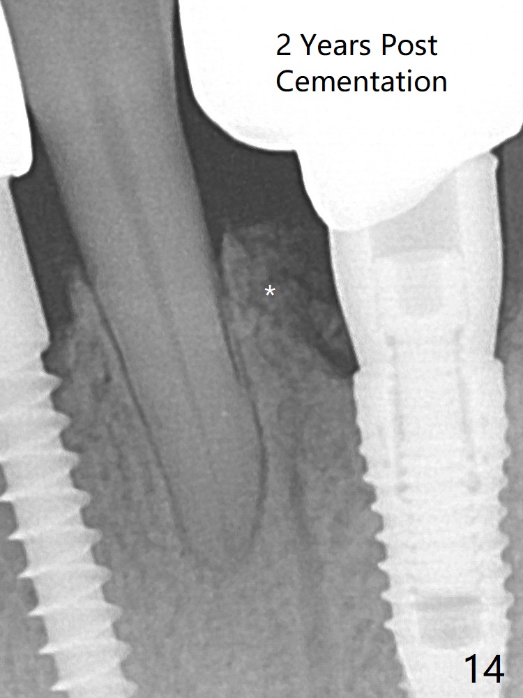
A 3.8x10 mm dummy implant is placed tentatively with an apical space (Fig.4 (the distal root surface of the lateral incisor is denuded (<)). When a same dimension definitive implant is placed with 40 Ncm, it is 2 mm below the lingual gingival margin, whereas 6-7 mm below the buccal one (Fig.5). Vanilla graft is placed before placement of a 5.5x4(5) mm abutment (Fig.6). The root surface of the lateral incisor is covered by the bone graft. Later the abutment is changed to a longer and smaller one (Fig.8) with more of the allograft (*). The short implant is chosen because it has to be placed deep to prevent periimplantitis, especially lingually, in spite of the fact of the unfavorable crown/implant ratio (Fig.4). The diameter of the implant is small so that there is ample space to pack bone graft both buccally and lingually. The majority of the bone graft seems to be in place 8 months postop (Fig.12). The ridge completely regenerates 2 years post cementation (Fig.14).
2-Piece Implant Last Fig.5 Fig.7 Fig.9, Fig.11, Fig.13 Next Xin Wei, DDS, PhD, MS 1st edition 12/12/2018, last revision 12/13/2020