,%20Vanilla.jpg)

,%20more%20Vanilla.jpg)
|
|
|
|
|
|
|
|
|
|
|
|
|
|
|
|
|
|
|
|
A Small and Short 2-Piece Implant for 2 Fused Teeth
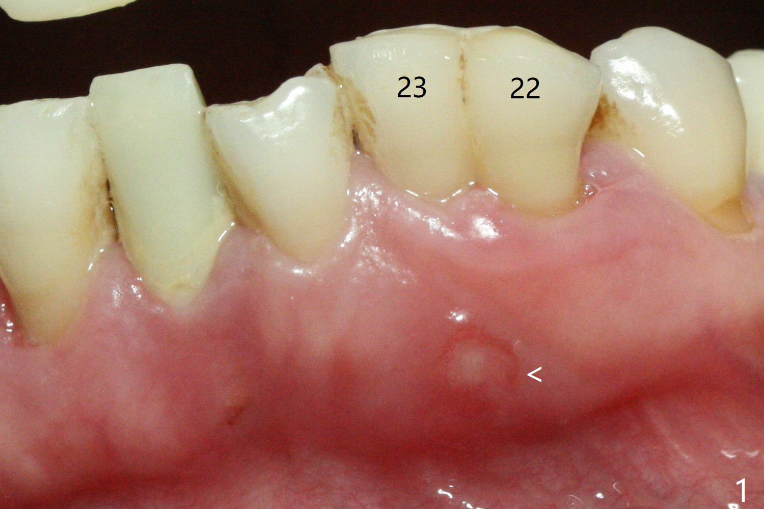
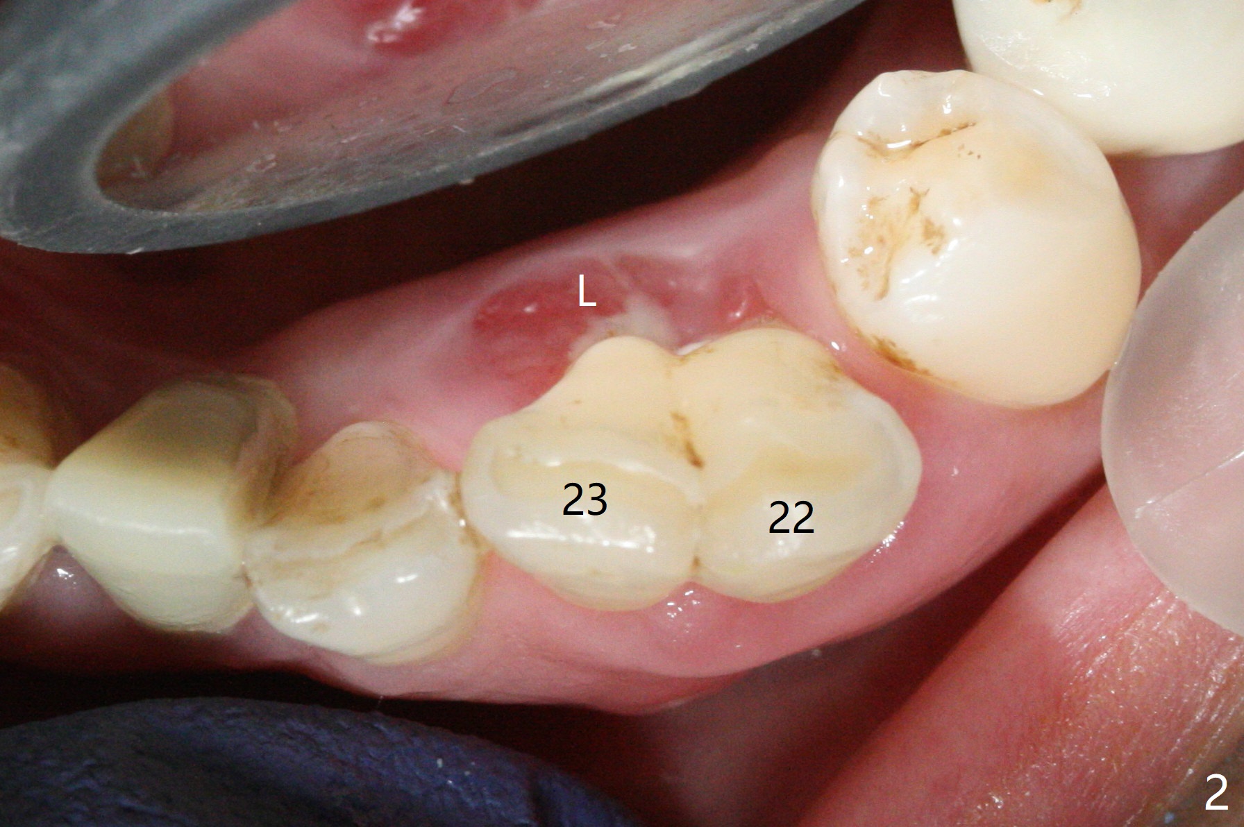
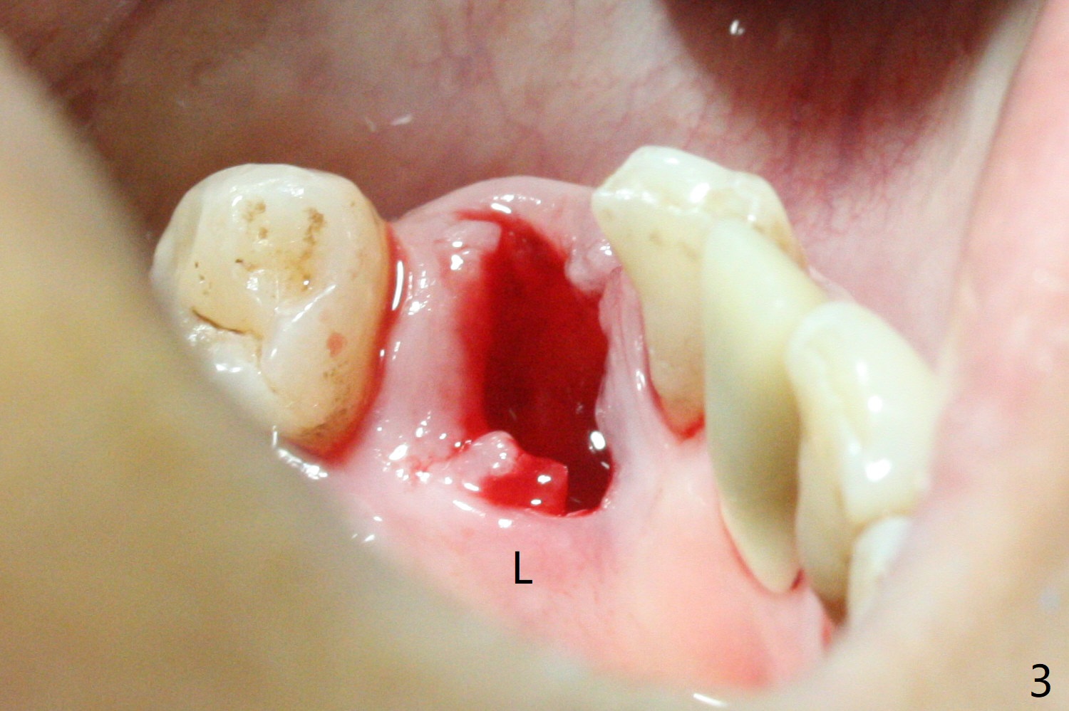
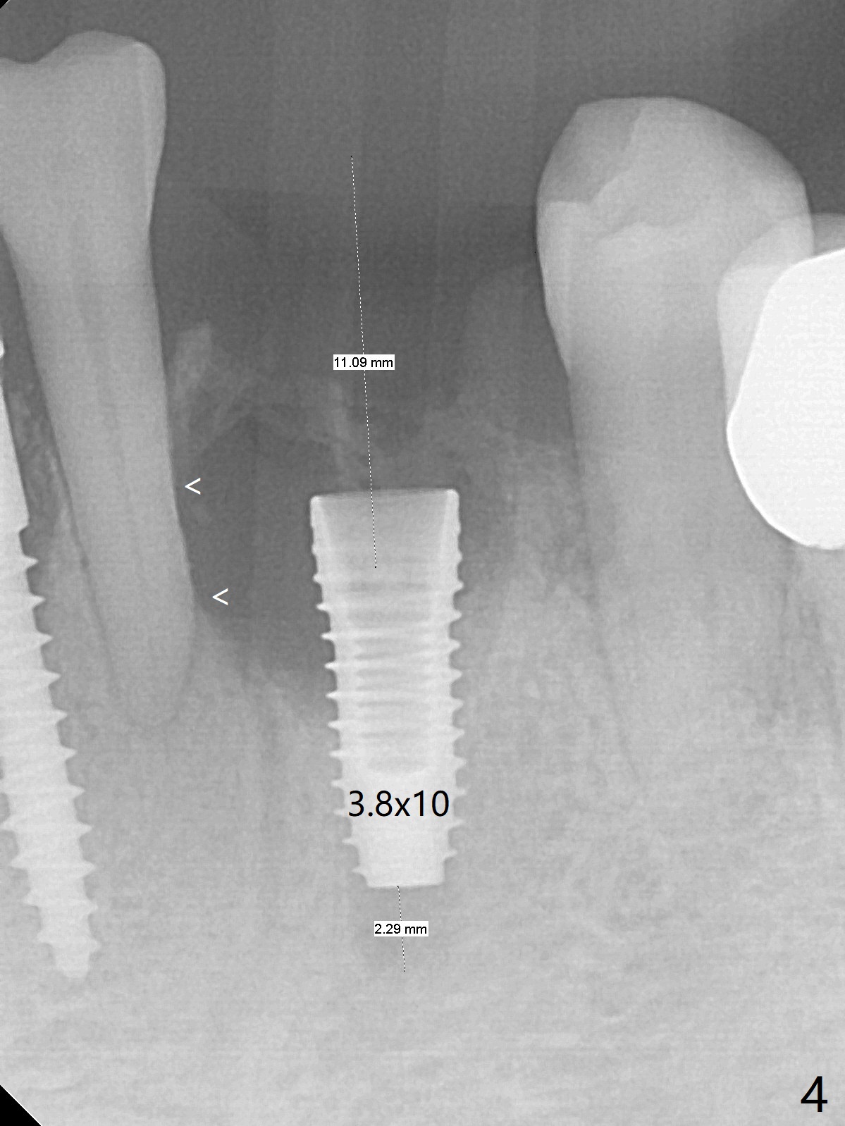
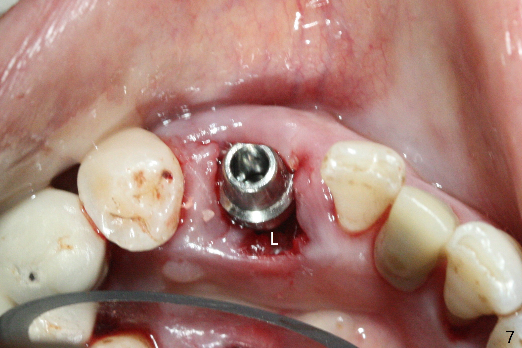
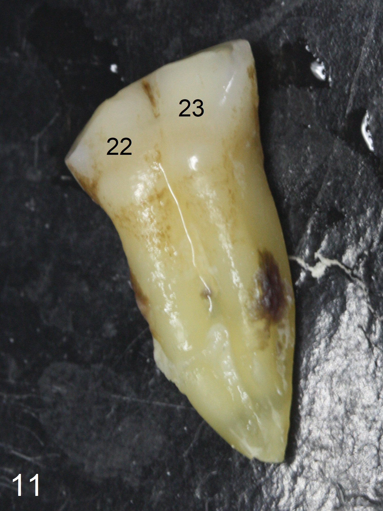
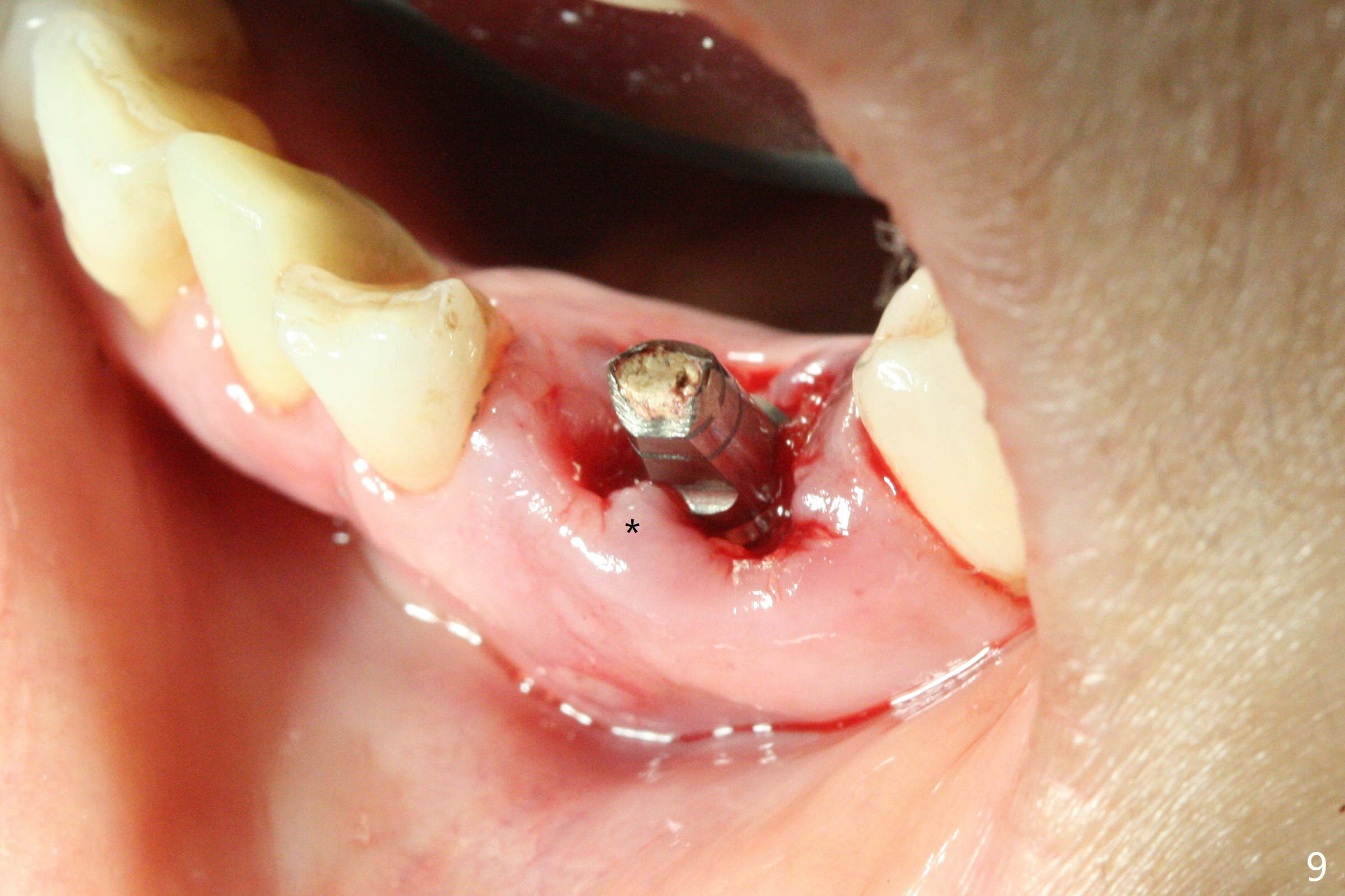
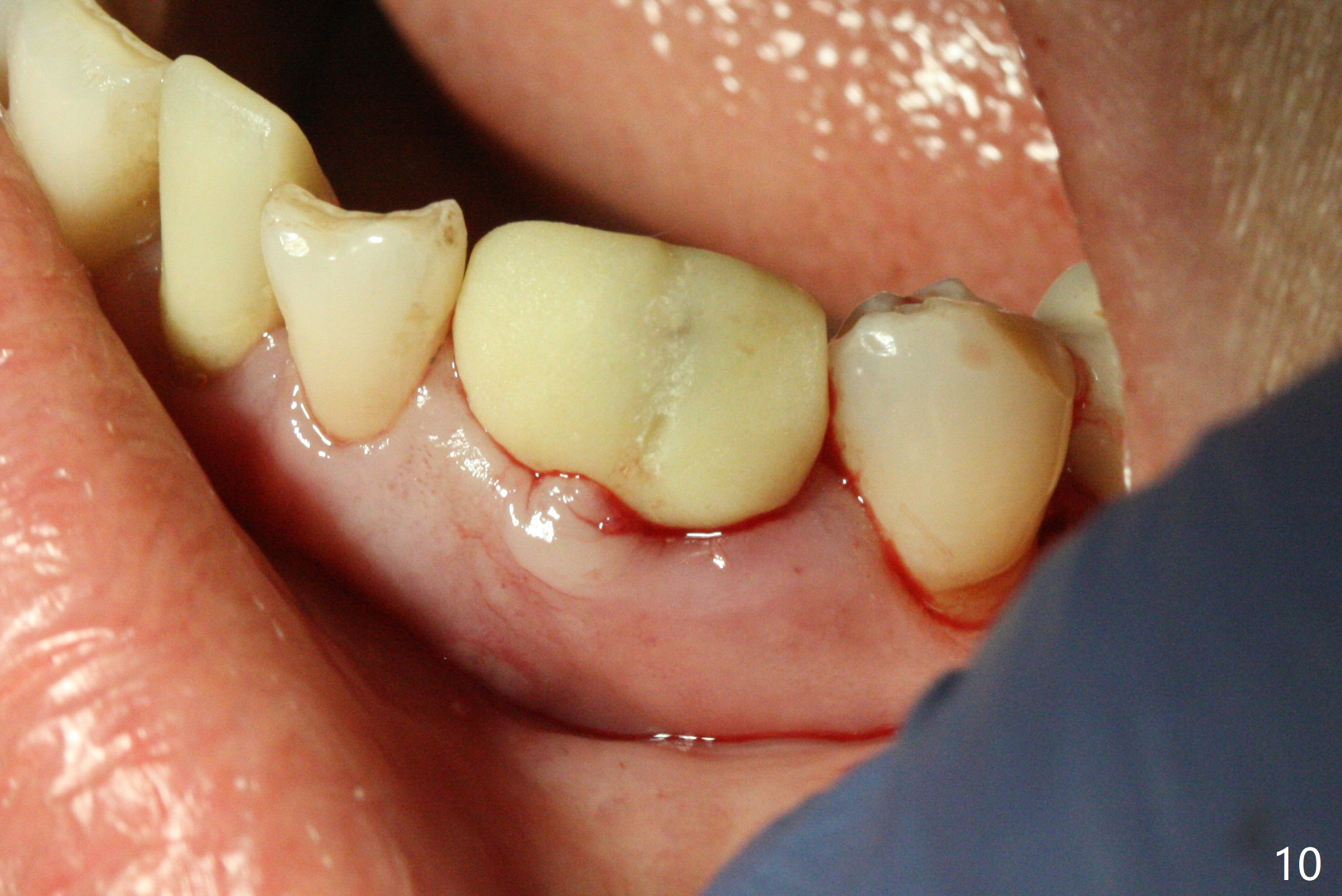
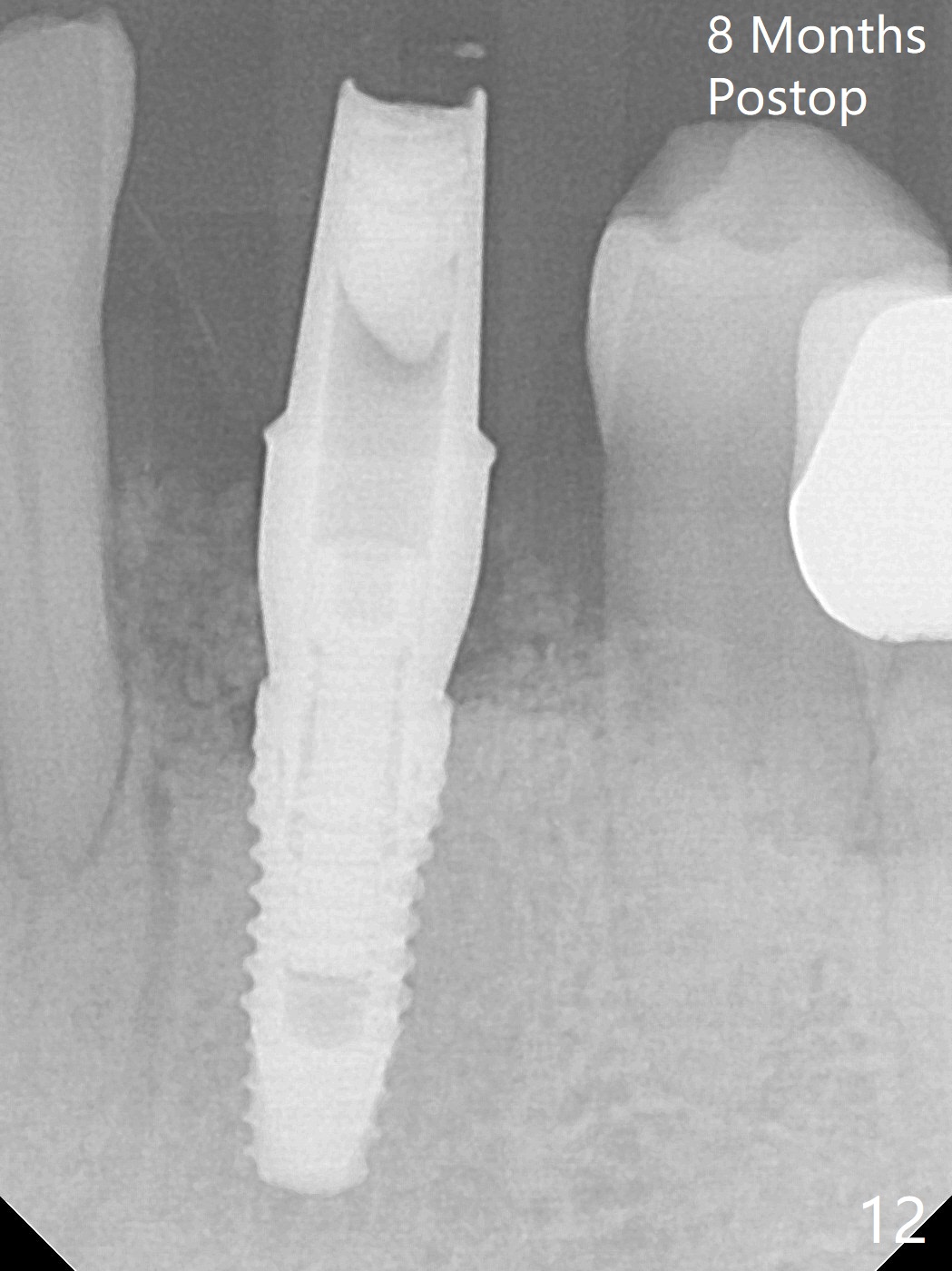
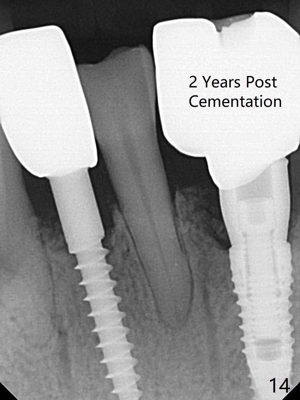
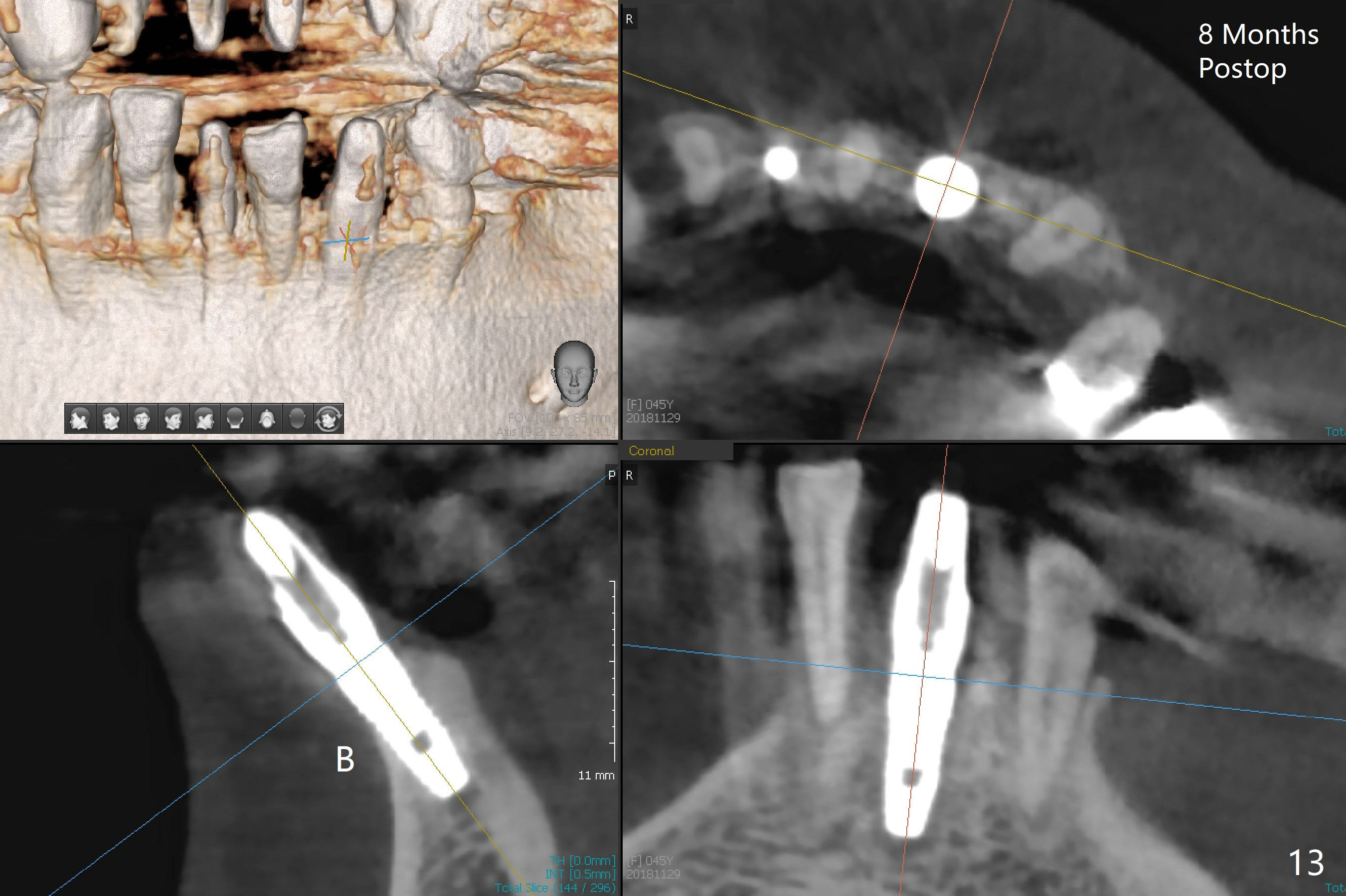
There is a fistula buccal to the apex of the tooth #23 preoperatively (Fig.1), which is related to loss of the buccal plate of the socket of #23. Therefore an implant is placed mainly in the socket of #22 (Fig.3). The lingual (Fig.2 L) gingiva appears to have more extensive inflammation. After extraction, the lingual (Fig.3 L) gingival margin is significantly lower than the buccal one. The lingual crest is ~ 4 mm lower than the buccal one. A 3.8x10 mm dummy implant is placed tentatively with an apical space (Fig.4). When a same dimension definitive implant is placed with 40 Ncm, it is 2 mm below the lingual gingival margin, whereas 6-7 mm below the buccal one (Fig.5). Vanilla graft is placed before placement of a 5.5x4(5) mm abutment (Fig.6,7). There is a 2-3 mm lingual (L) gap to be filled with the allograft secondarily to prevent periimplantitis (Fig.7). Later the abutment is changed to a longer and smaller one (Fig.8) with more of the allograft (*). After trimming of the abutment (Fig.9 (*: papilla between the fused teeth)), an immediate provisional is fabricated to close the socket (Fig.10, similar to Fig.1). The majority of the bone graft seems to be in place 8 months postop (Fig.12). The implant appears to have been placed buccal, consistent with the thin and slightly erythematous buccal gingiva (Fig.13). The ridge completely regenerates 2 years post cementation (Fig.14).
Lower Incisor Canine Immediate Implant, Armaments, 10 Redo 25
Xin Wei, DDS, PhD, MS 1st Version 03/20/2018, Last Update 12/13/2020