,%206.5x4(2).jpg)
,%206.5x4(2)%20Vanilla%20with%20PRF,%20sticky%20bone.jpg)
|
|
|
|
||
|
|
|
|
||
|
|
|
|
|
|
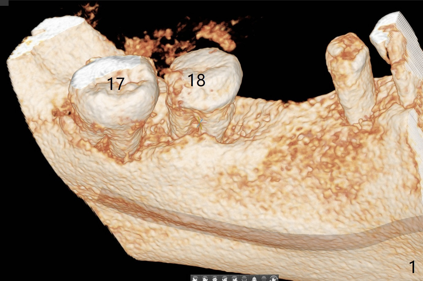
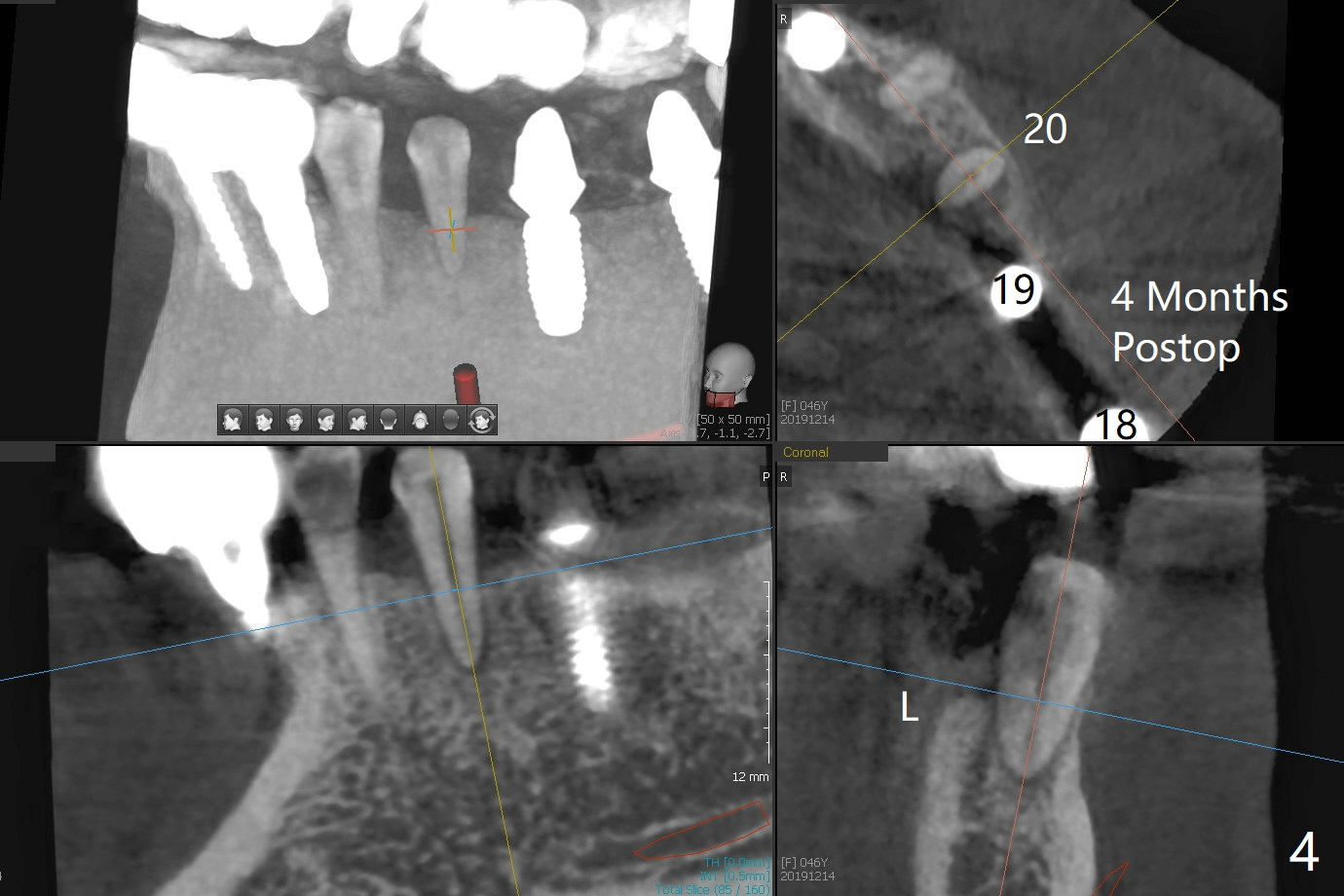
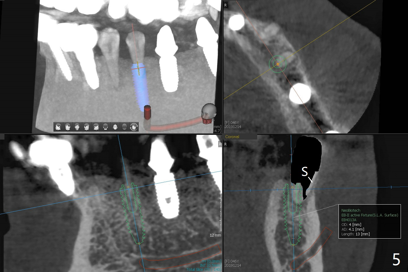
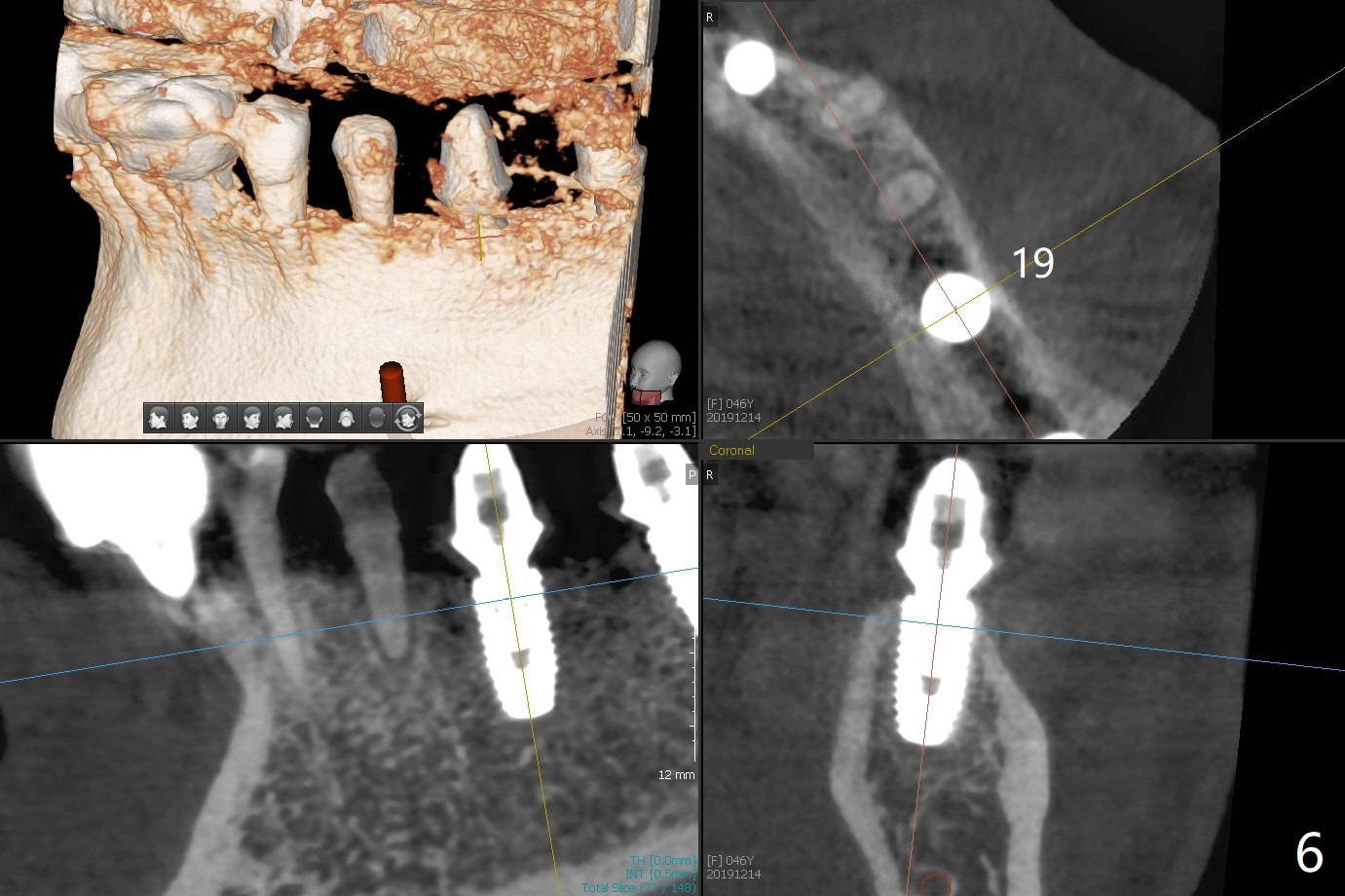
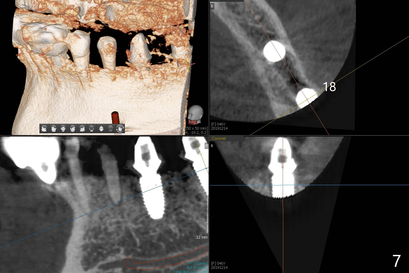
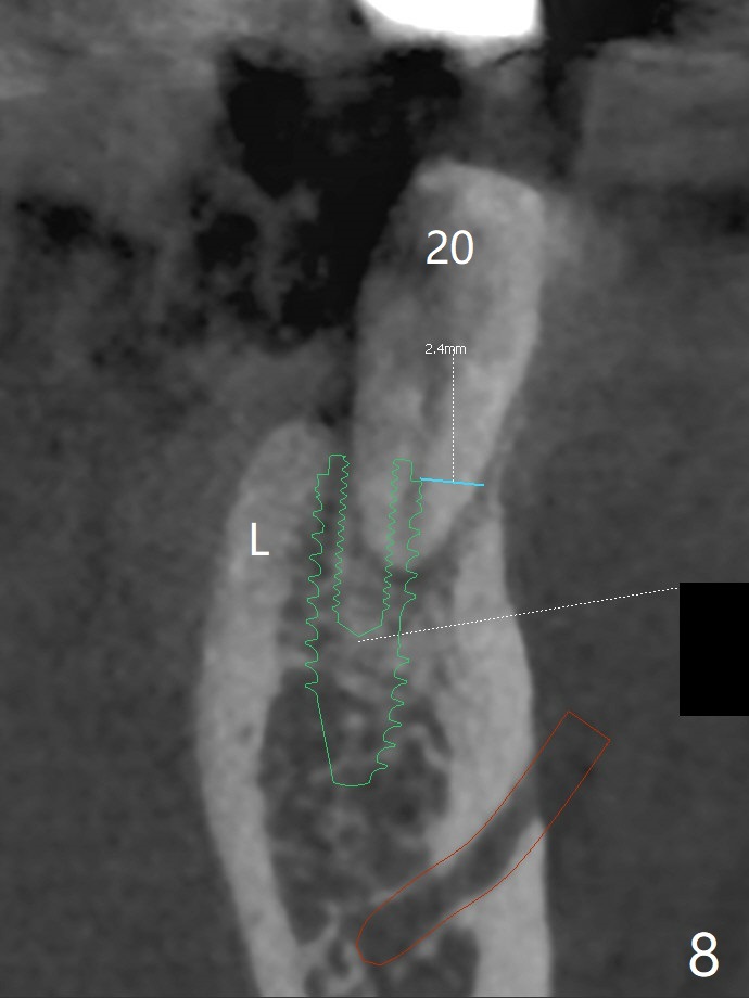
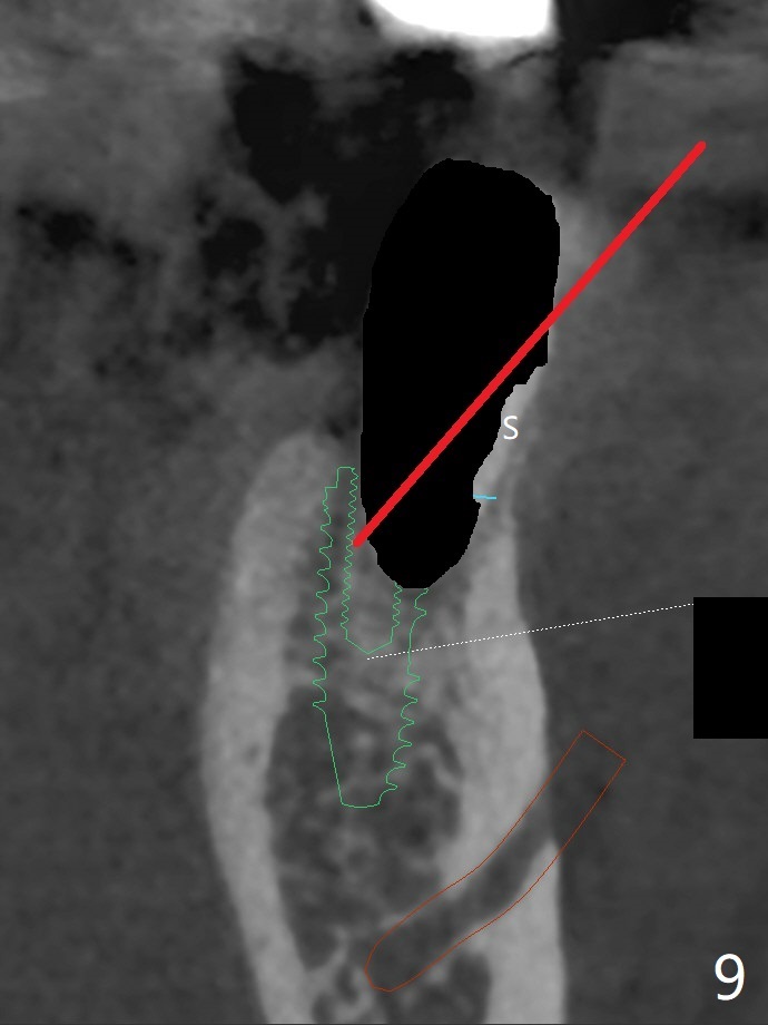
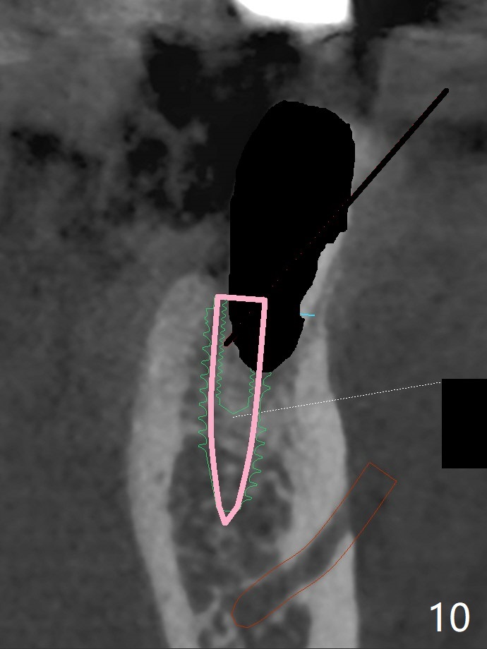
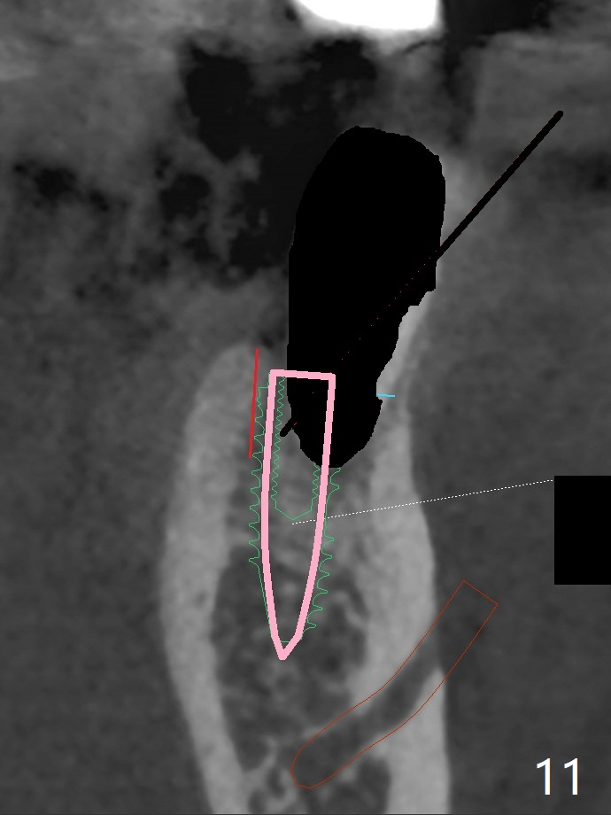
Re-analysis of preop CT reveals extensive bone loss around #18 (Fig.1 (lingual view)). Blood is withdrawn for sticky bone. After implant placement (Fig.2), sticky bone is placed at #18 (Fig.3 red dashed line (yellow: superior border of the Inferior Alveolar Canal)). PRF membrane and an immediate provisional FPD (#18-20) further keep the bone graft in place for healing. Four months postop, the patient reports difficulty in mastication on the left and requests extraction of the tooth #20 for implant (Fig.4). The implant will be placed lingually, while socket shield will be performed buccally (Fig.5 S) to keep bone graft in place. The implant at #19 is equicrestal (Fig.6); the one at #18 is apparently supracrestal buccally (Fig.7). Since there is a lot of scattering from nearby crowns, the implant at #20 will be placed free hand. To overcome the thick dense lingual plate (Fig.8 L), osteotomy is initiated (Fig.9 red line) in the middle of the lingual wall of the extraction socket (black area) on the top of the socket shield (S). After the last drill (3.5x11.5 mm, Fig.10 pink) and before 4x11.5 mm implant, use Lindamann bur to remove the coronal portion of the lingual plate (Fig.11 red line) to prevent implant buccal deviation.
Return to
Lower
Molar Immediate Implant,
Prevent Molar Periimplantitis (Protocols,
Table),
Trajectory II No Deviation
Metronidazole
Next Case
Xin Wei, DDS, PhD, MS 1st edition
08/05/2019, last revision
12/22/2019