.jpg)
|
|
|
|
|
||
|
|
|
|
|
|
|
|
|
|
||||
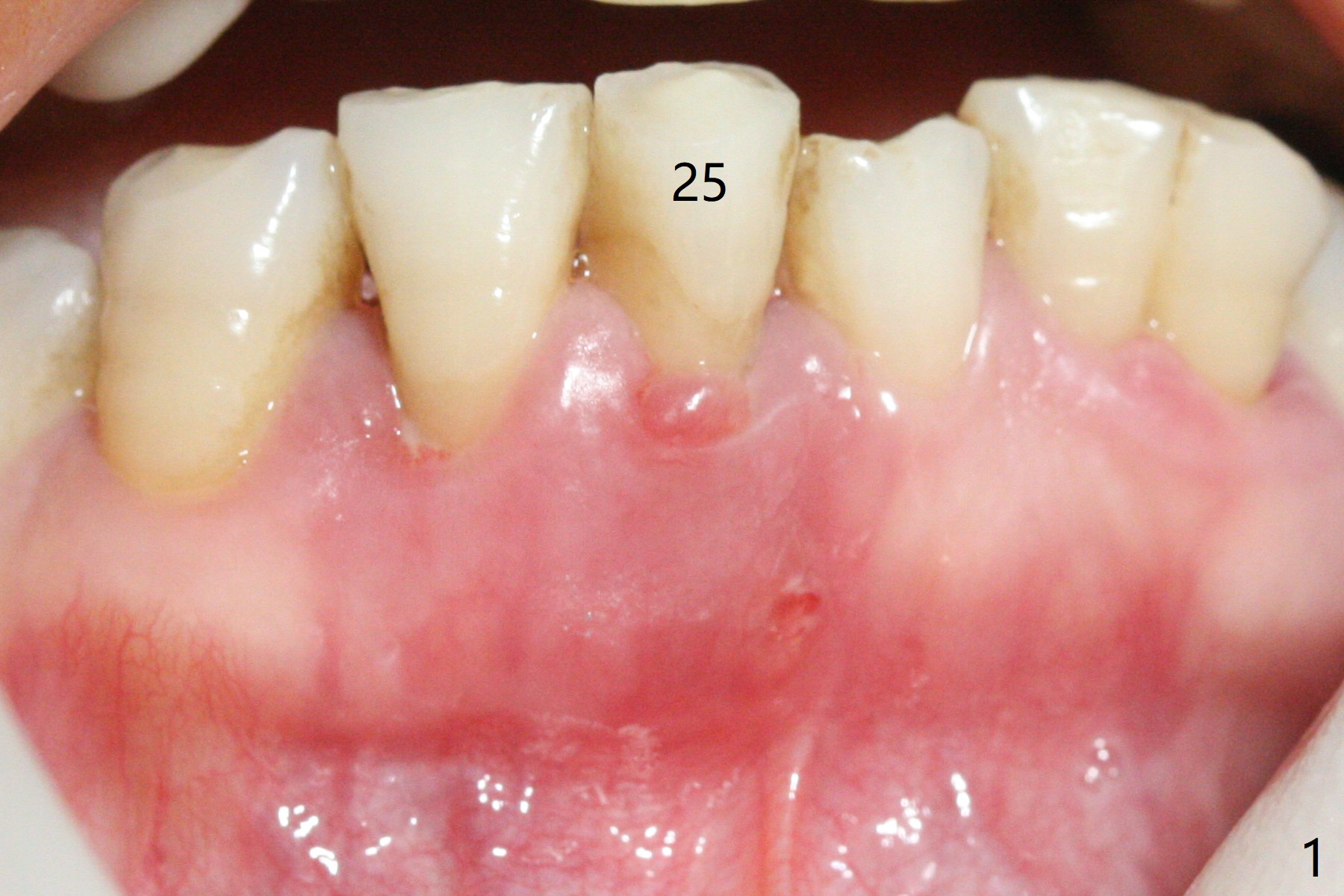
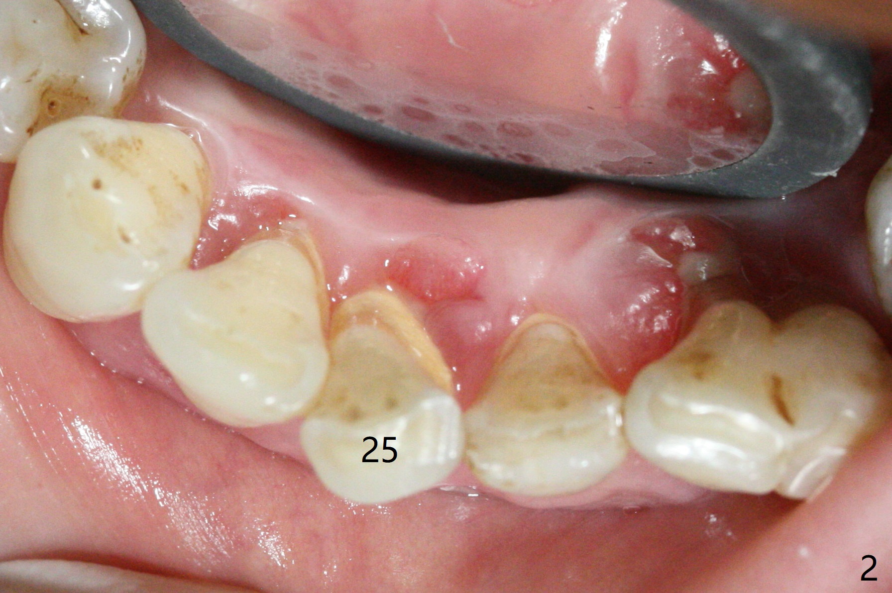
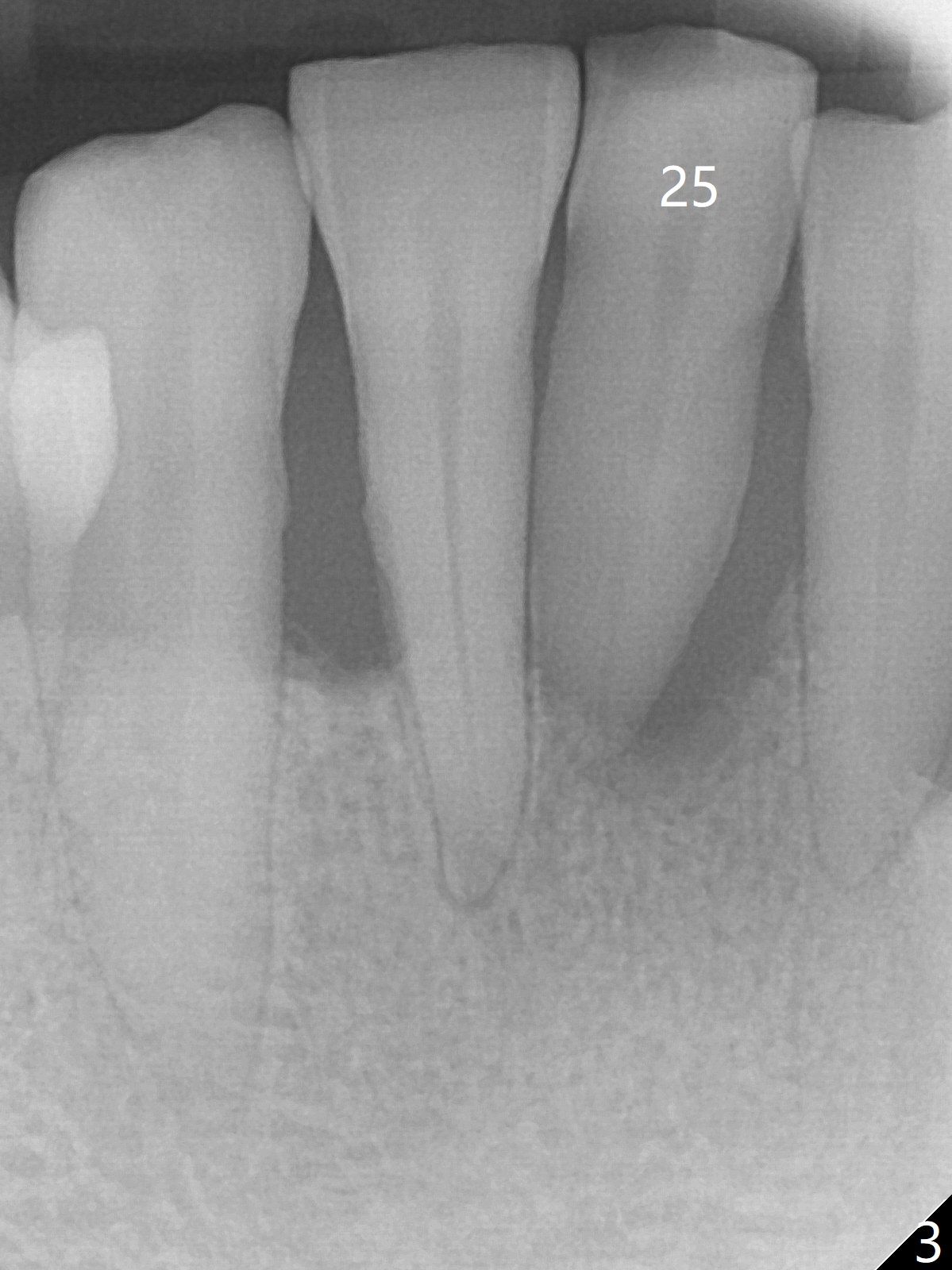
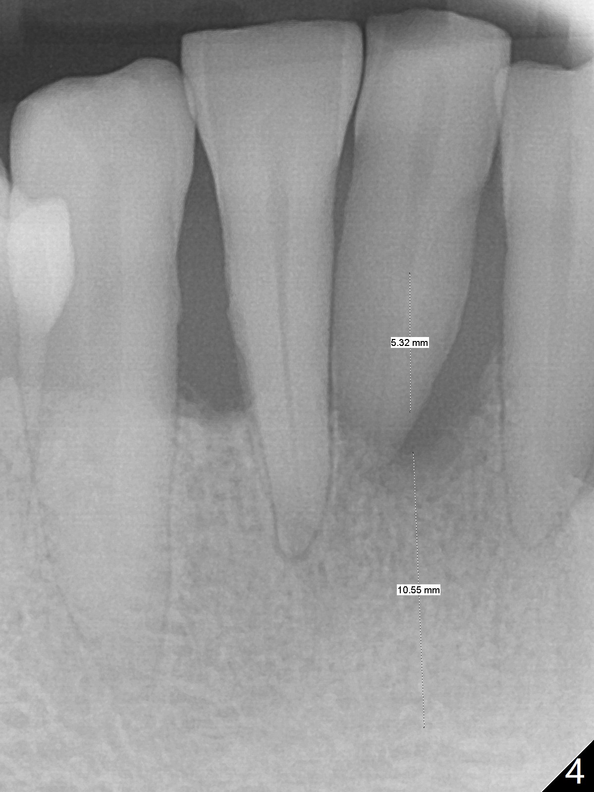
New Osteotomy
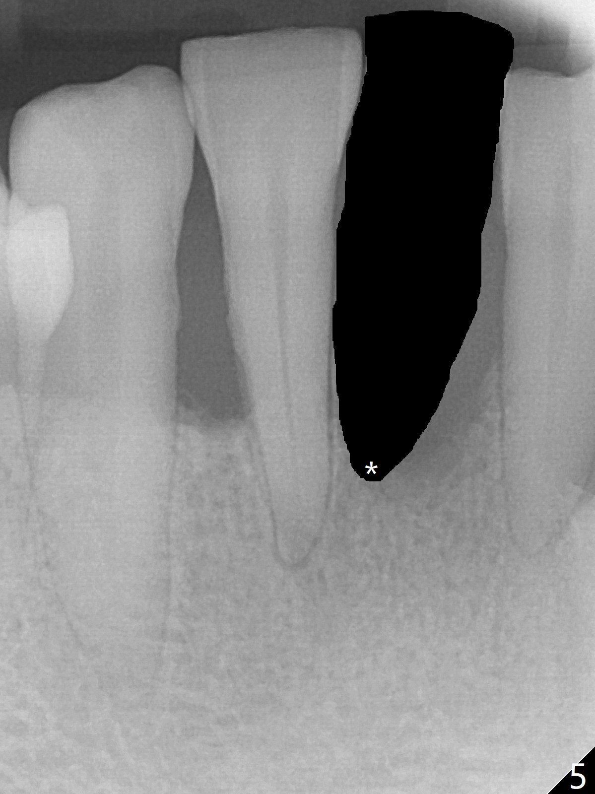
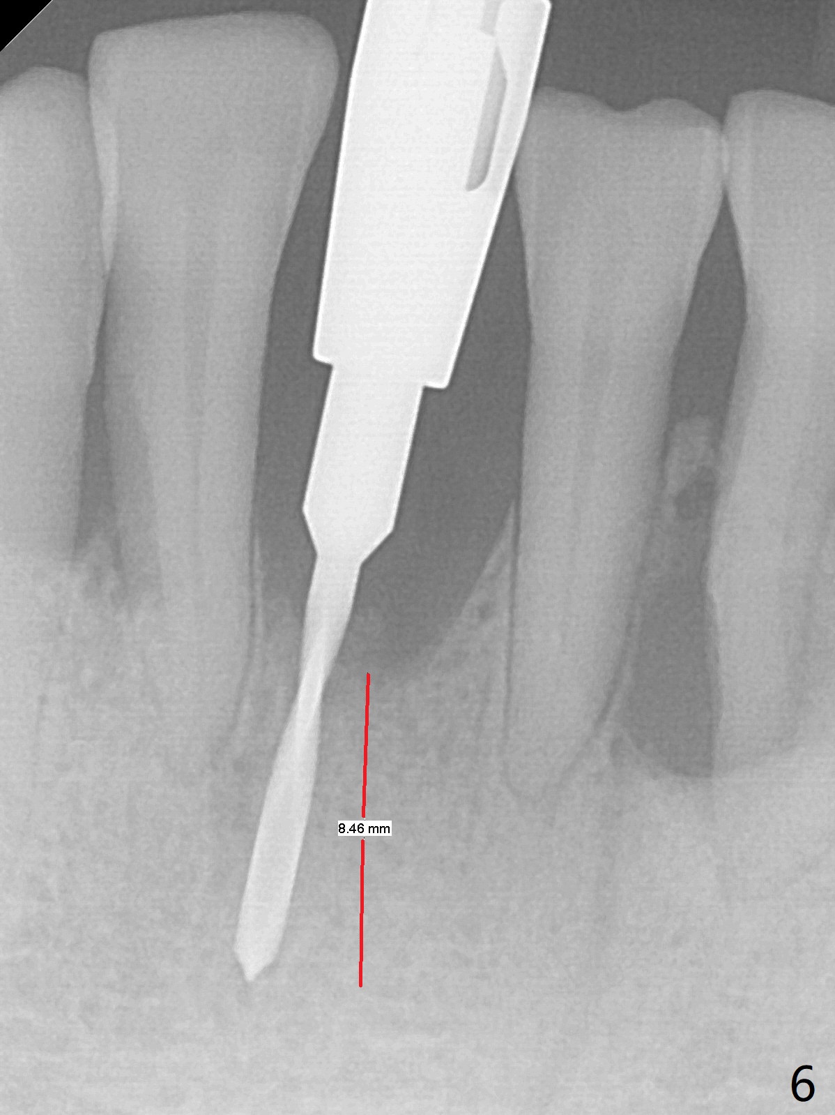
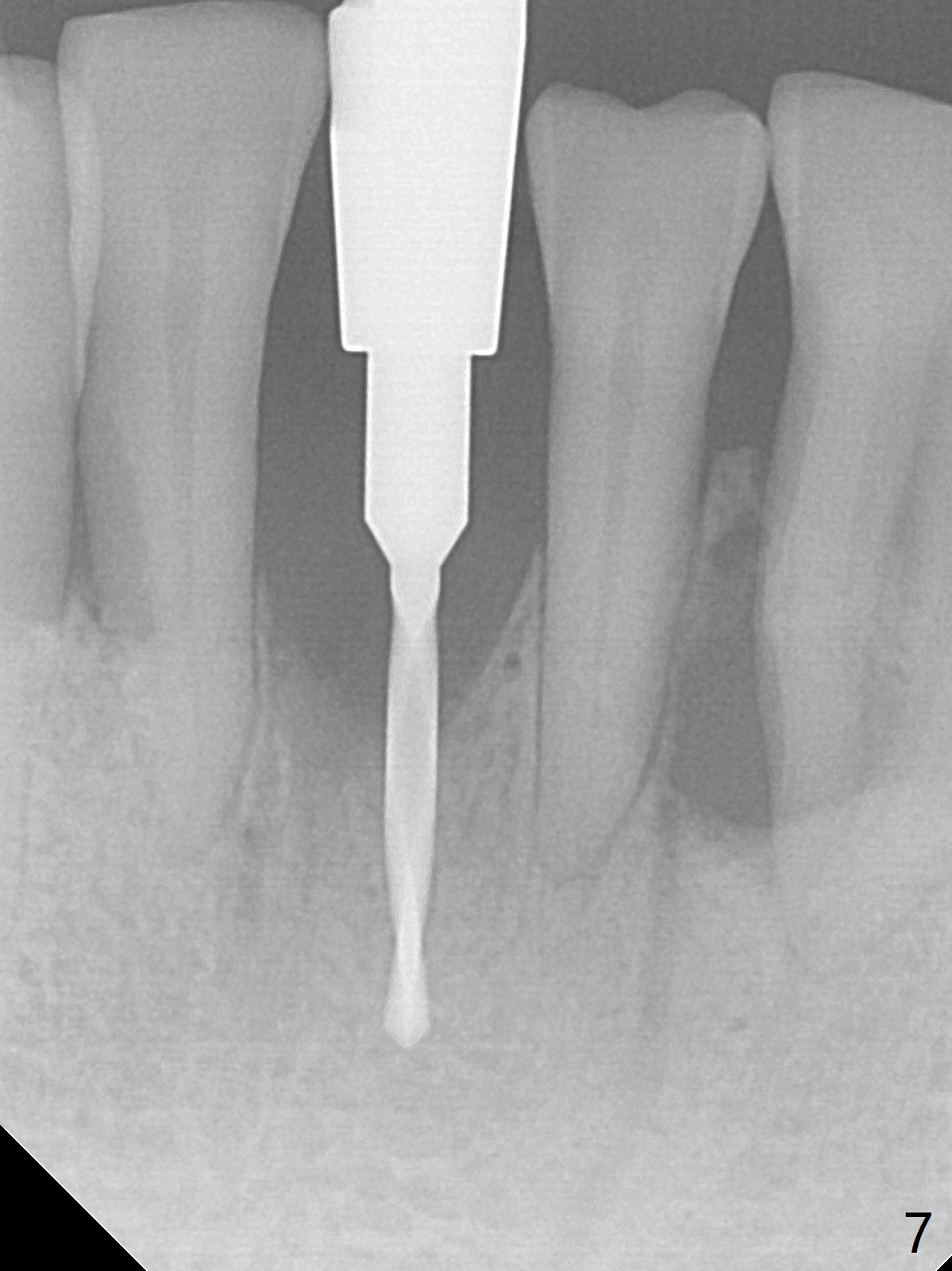
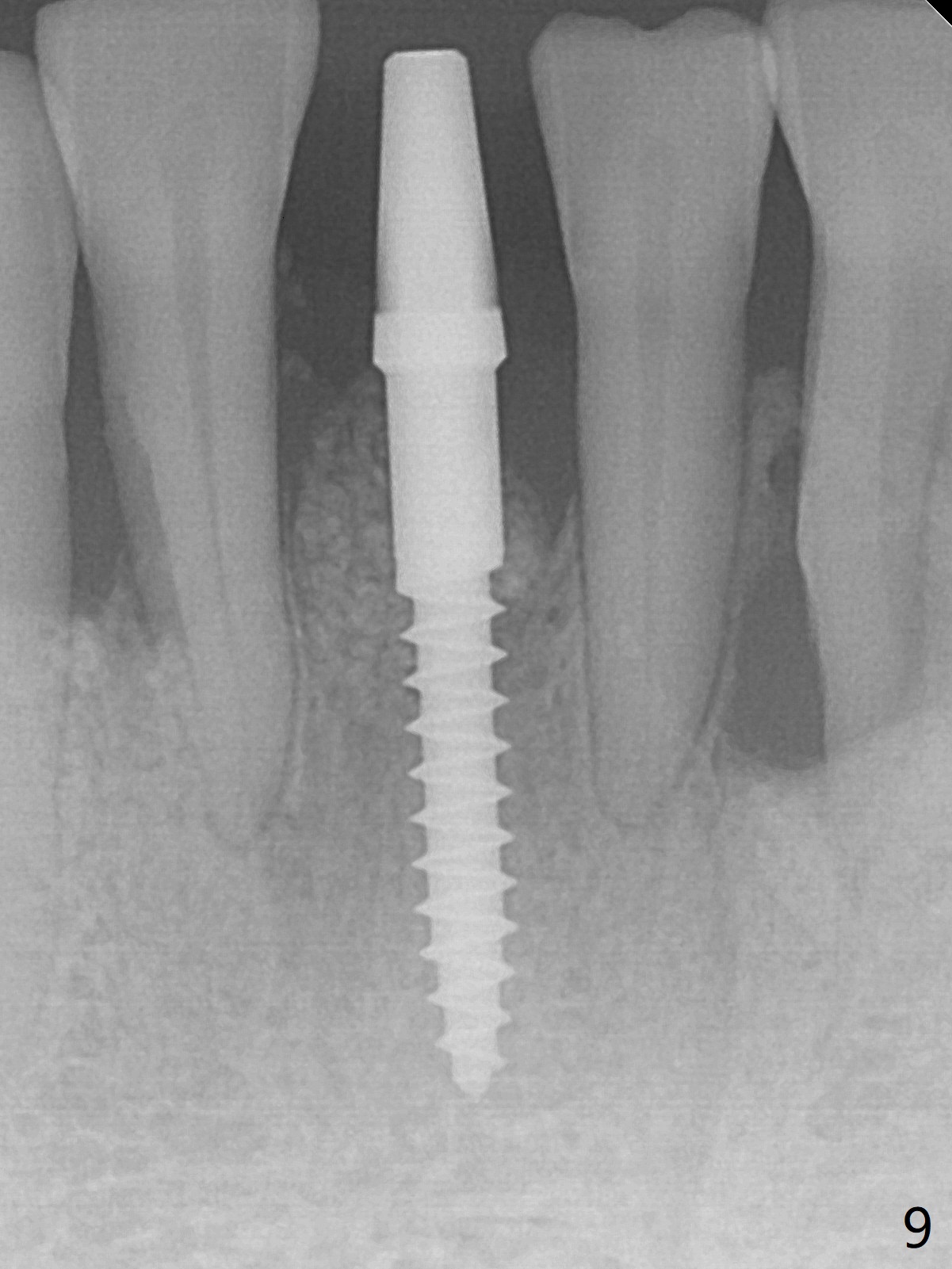
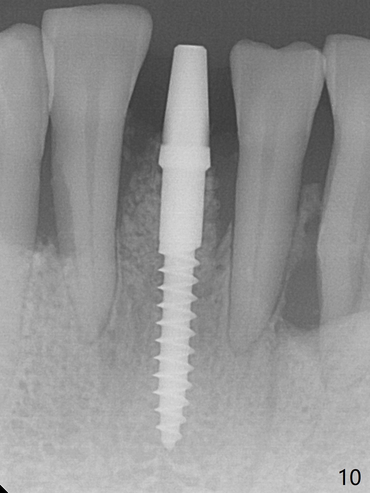
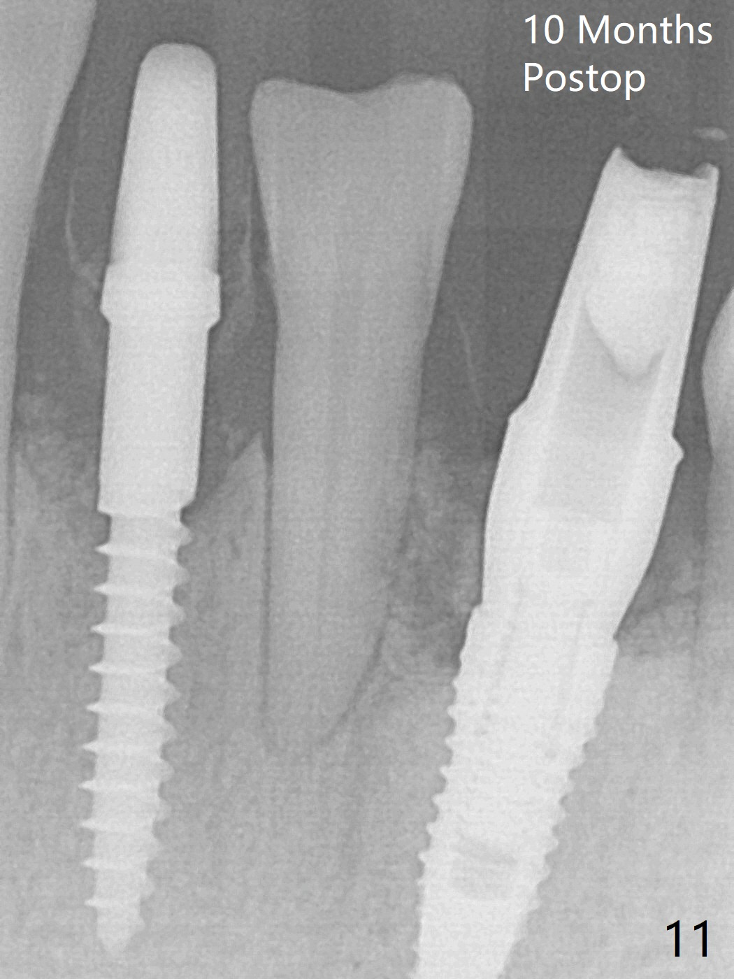
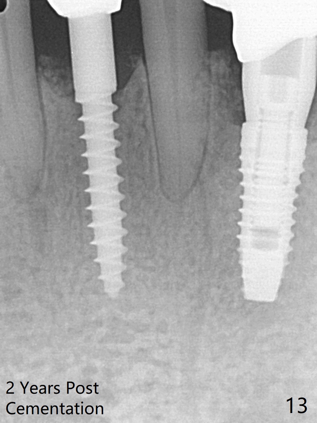
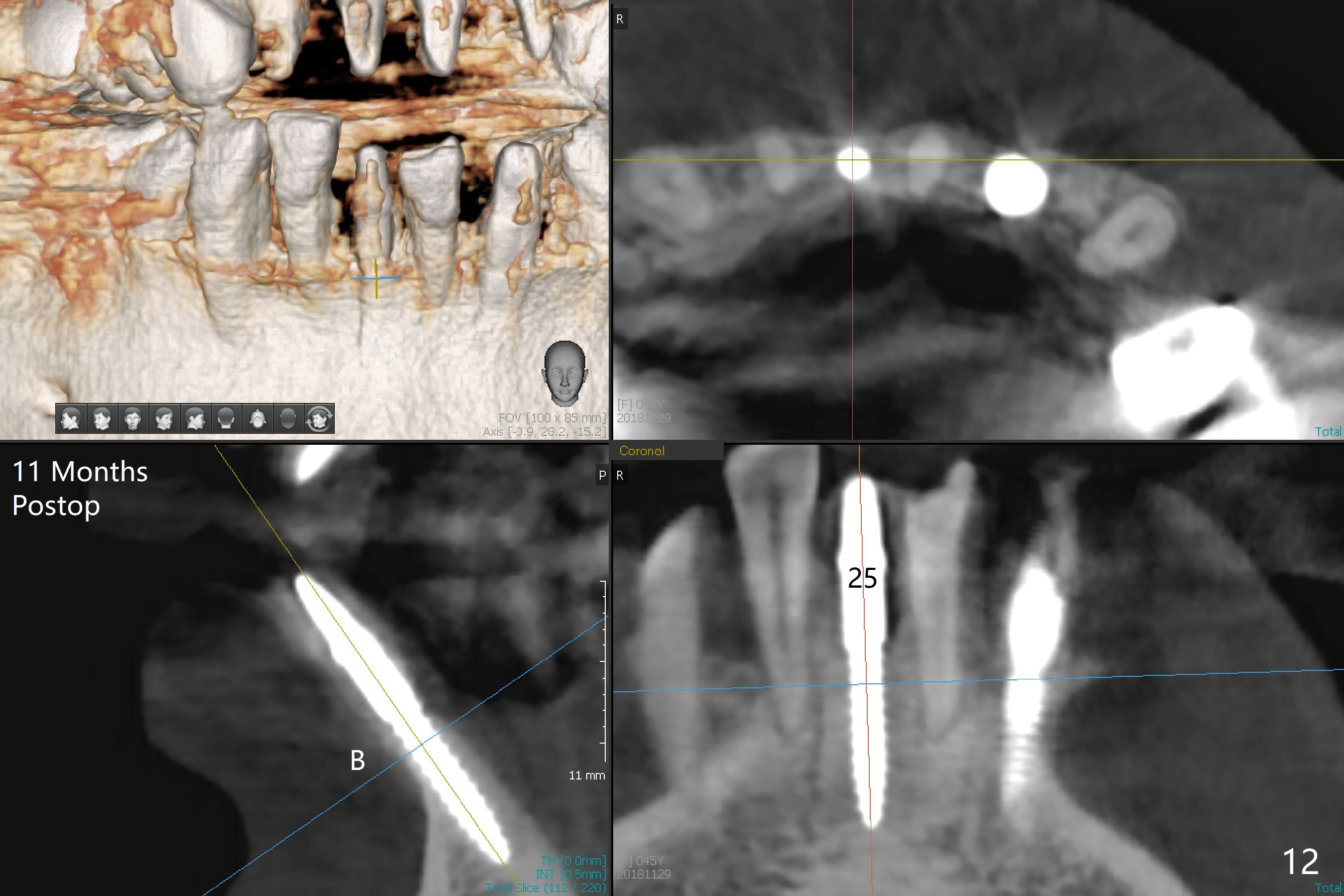
There is gingival inflammation at #25 buccally (Fig.1) and lingually (Fig.2). The bone loss is severe (Fig.3). Soft and hard tissue heights are 5 mm (cuff will be 4 mm) and 10 mm (implant will be 12 mm with 2 mm outside the native bone, Fig.4). The apex of the affected tooth appears deviated distal (Fig.5 *). The initial osteotomy happens to follow the long axis of the socket (Fig.6); to establish a correct trajectory, a new osteotomy should be made at the site labeled as a red line. In fact it is executed as planned (Fig.7). Because of the narrow flat ridge buccolingually, a 2.5x12(4) mm 1-piece implant is placed with >40 Ncm (Fig.8). With deeper placement of the implant, Vanilla graft is placed in 2 steps (Fig.9,10). The patient will return 2.5 months for extraction and implant of the fused teeth #22 and 23. No implant threads are exposed 10 months postop (Fig.11). CT taken 11 months postop shows that the 2.5 mm implant is in the middle of the bone (Fig.12) or 2 years post cementation (Fig.13).
Return to
Lower
Incisor Immediate Implant,
Armaments
22/23手术
Xin Wei, DDS, PhD, MS 1st edition 01/04/2018, last revision 12/13/2020