
|
|
|
|
|
||
|
|
|
|
|
|
|
PRF and
Implant-Assisted Bone Graft M
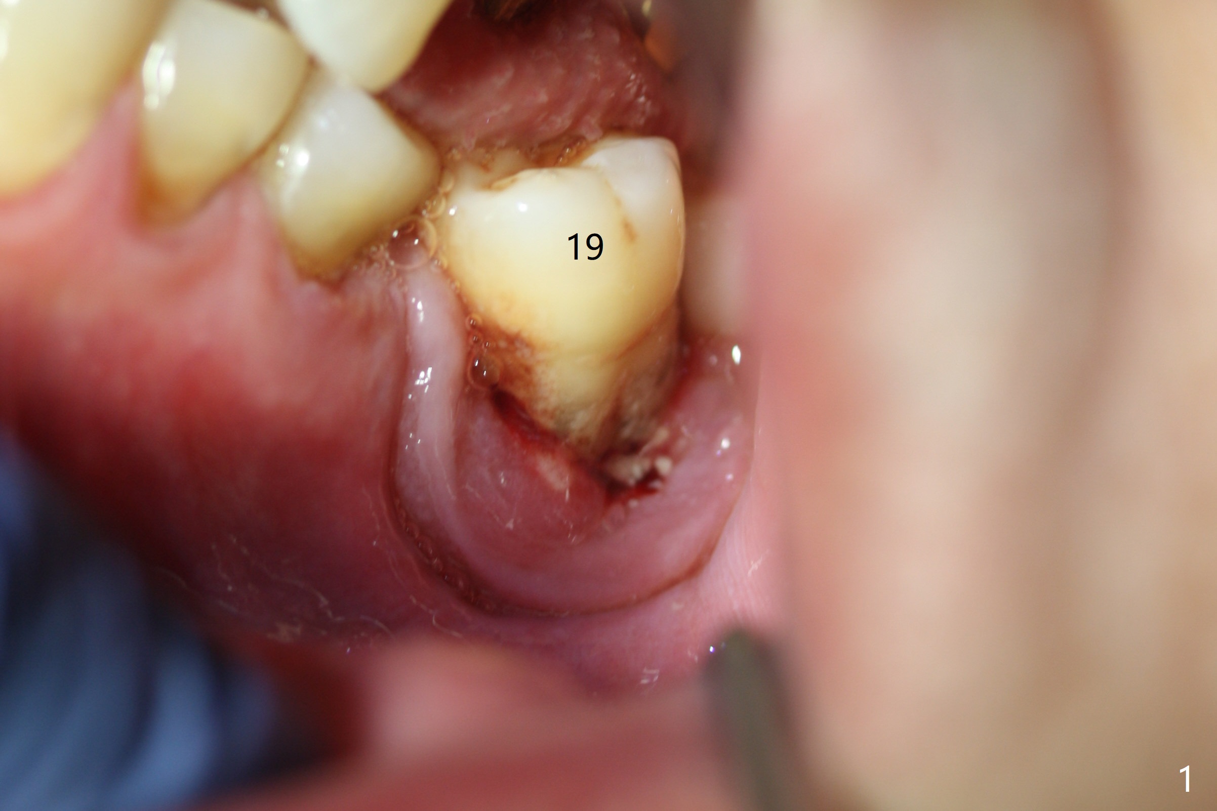
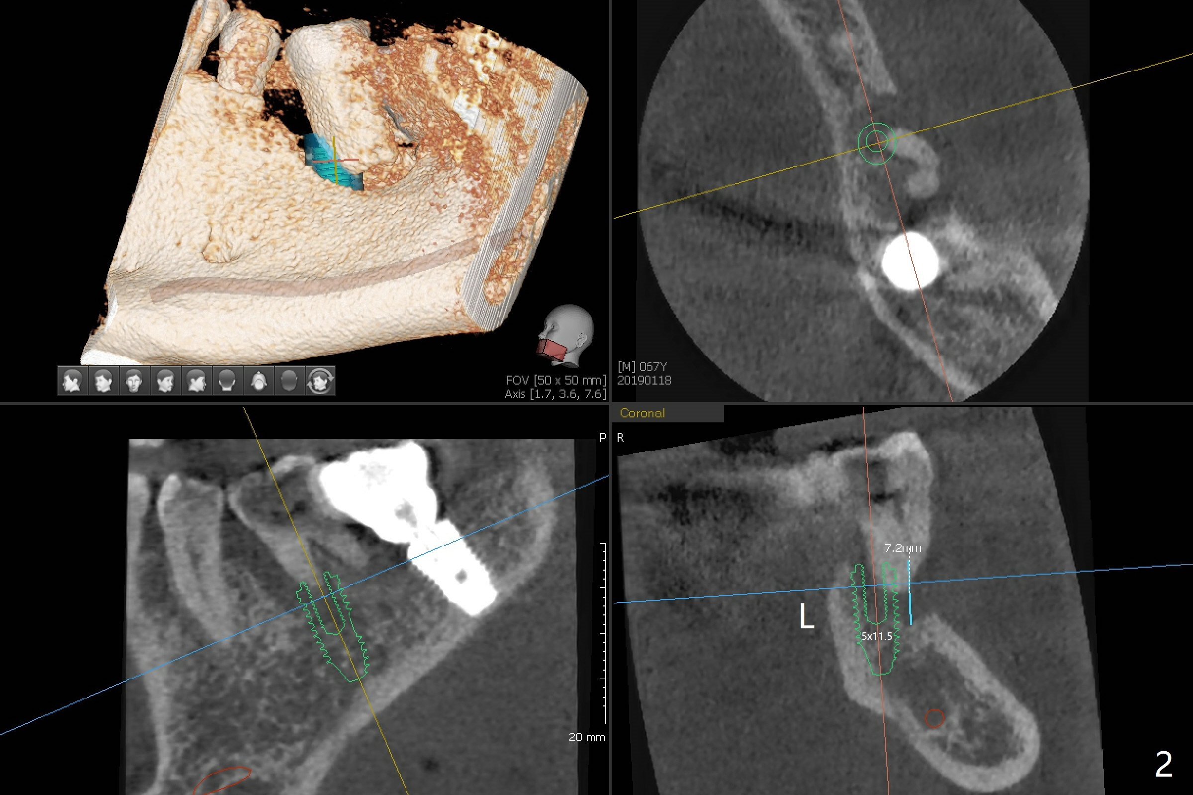
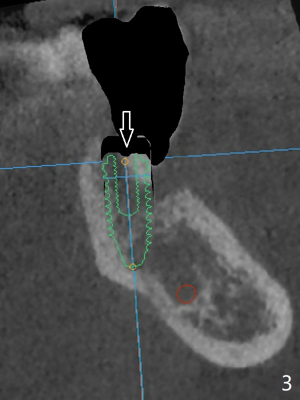
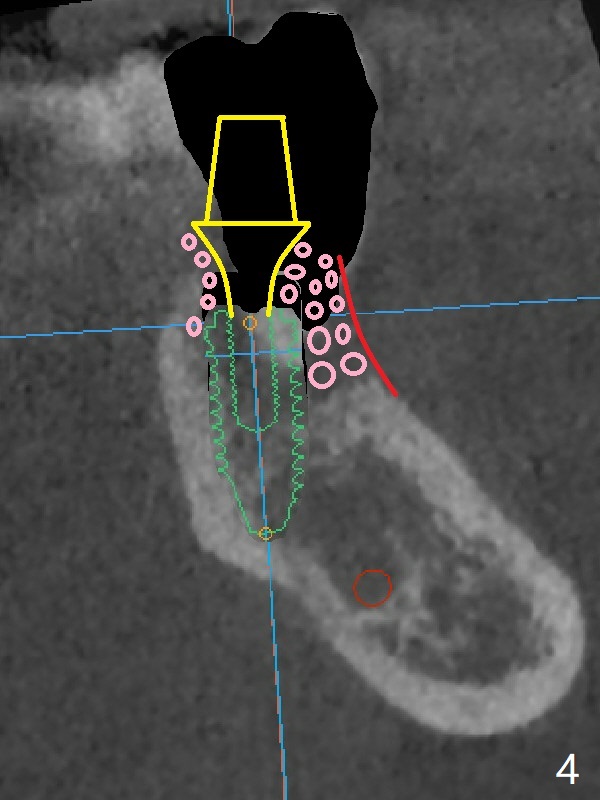
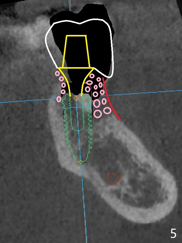
A 67-year-old man requests treatment for the tooth #19 with severe buccal gingival recession (Fig.1). After extraction, a 5x11.5 mm implant will be placed as lingual as possible (Fig.2 L) with ~7 mm buccal implant thread exposure. To reduce the exposure, the implant will be placed deeper, as shown by arrow in Fig.3. PRF membrane and collagen membrane (Fig.4 red line) will be placed against the buccal gingiva, while PRF associated sticky bone (allograft, pink circle) will be packed around the exposed implant threads and a long-cuff abutment (yellow). Finally an immediate provisional (Fig.5 white) will be fabricated to cover the bone graft.
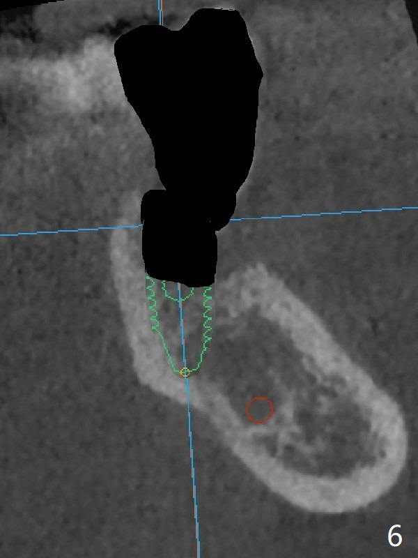
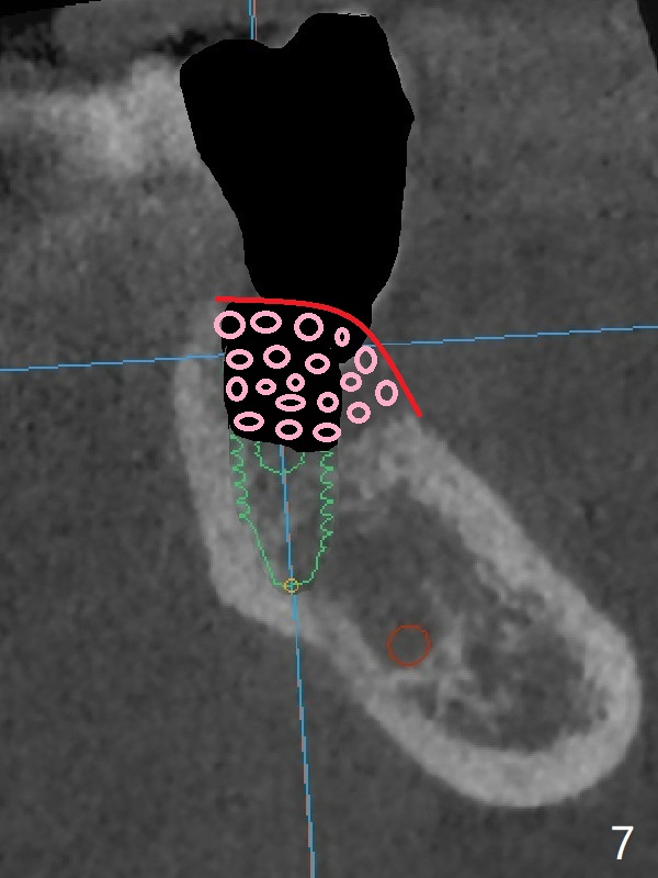
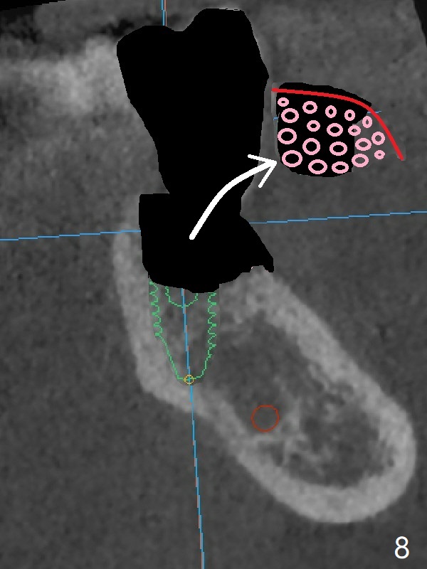
In contrast without implantation after extraction (Fig.6 (ignore green)), the socket may be grafted (Fig.7 pink circles) and covered by PRF membrane (red line). Due to the large socket with severe buccal defect, the graft is more likely to be lost (Fig.8). Several months later, an implant to be placed (Fig.9 green) will be short with unfavorable crown/implant ratio.
Return to Lower Molar Immediate Implant, Prevent Molar Periimplantitis (Protocols, Table), Trajectory, Clindamycin Metronidazole No Antibiotic, Weichat 第一磨牙即种 Xin Wei, DDS, PhD, MS 1st edition 01/20/2019, last revision 04/28/2021