.jpg)
|
|
|
|
|
|
|
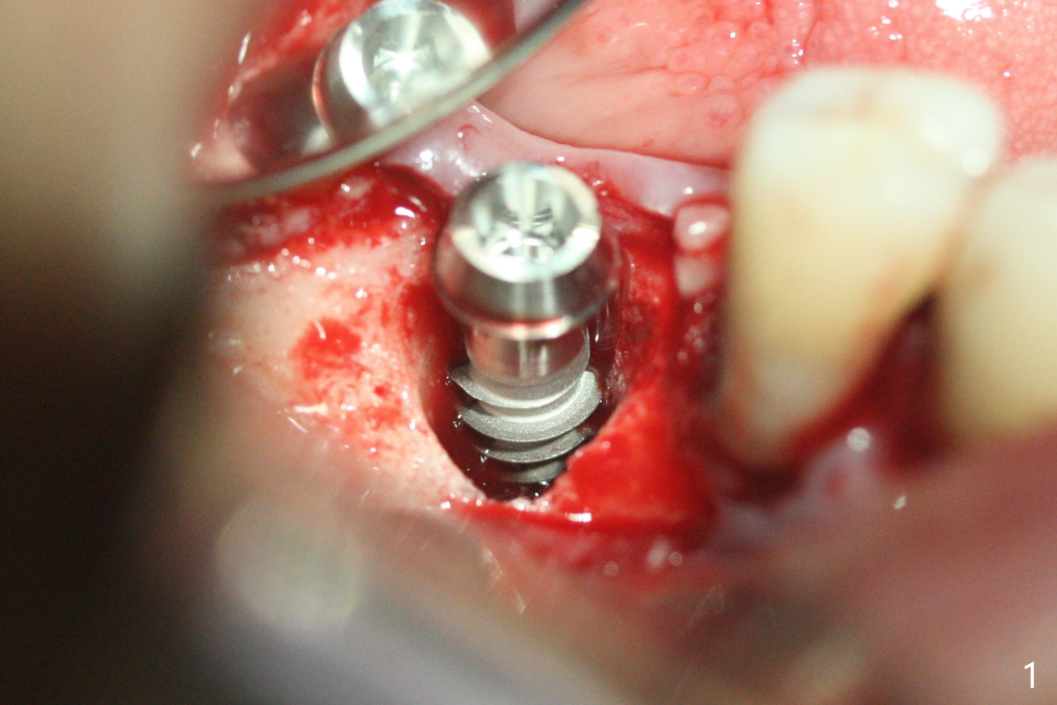
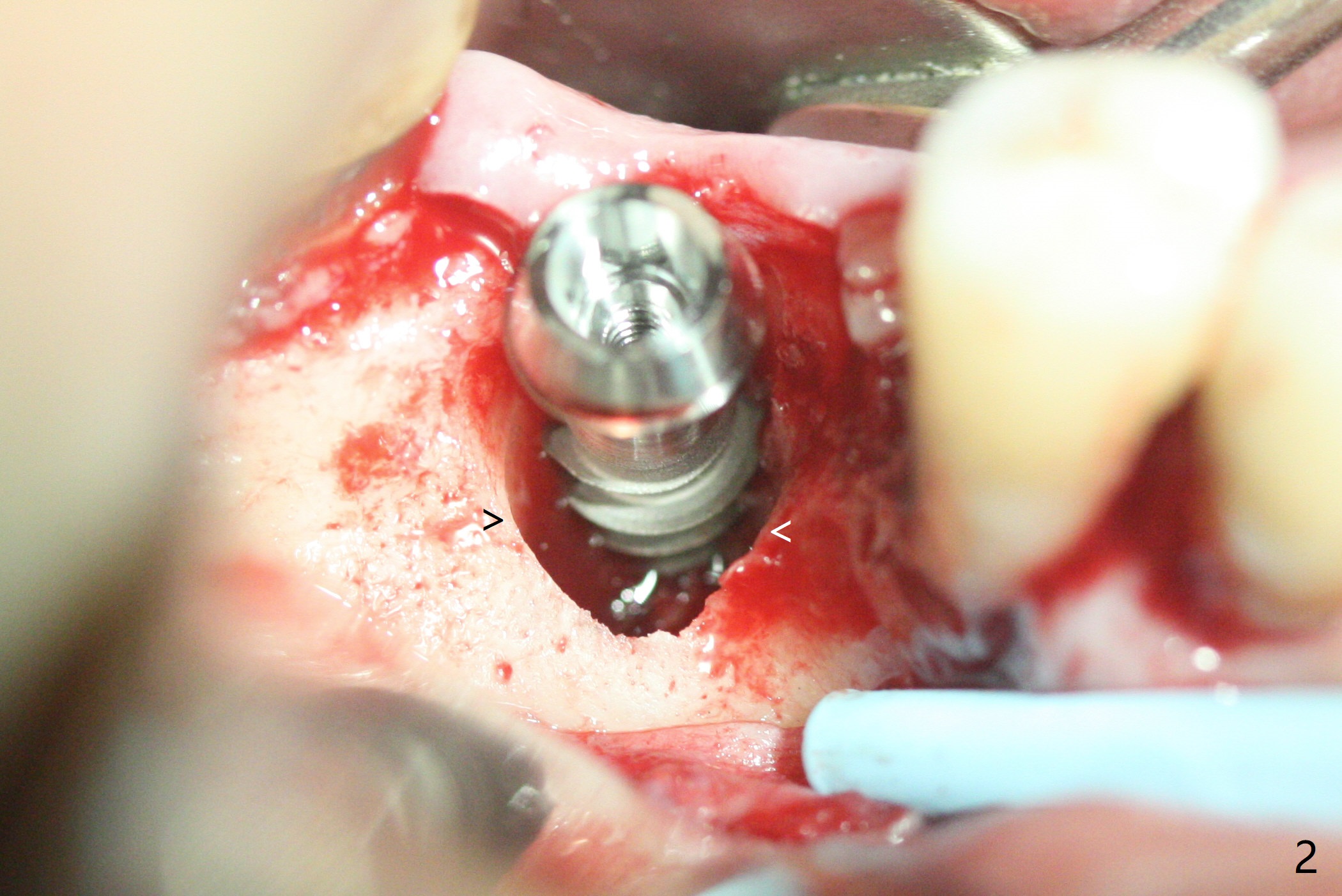
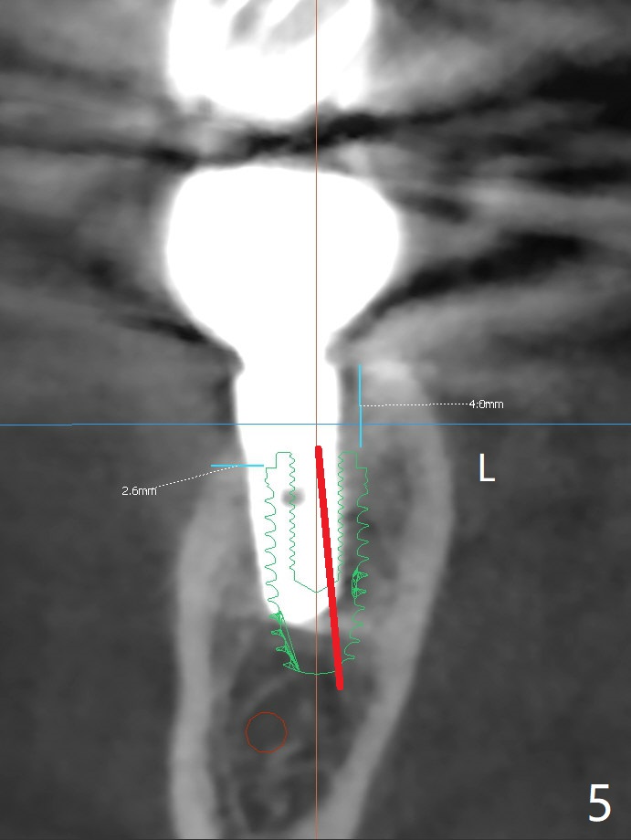
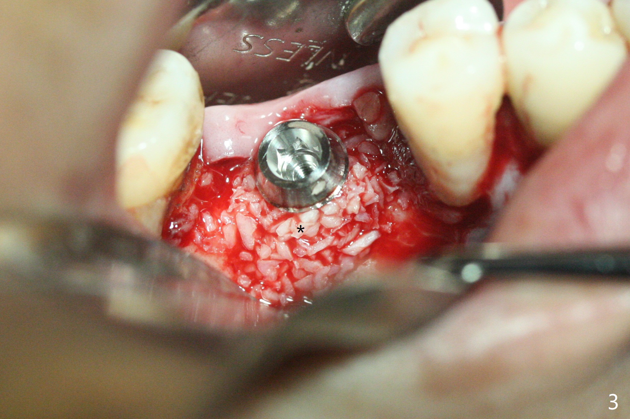
Free Hand Immediate Redo
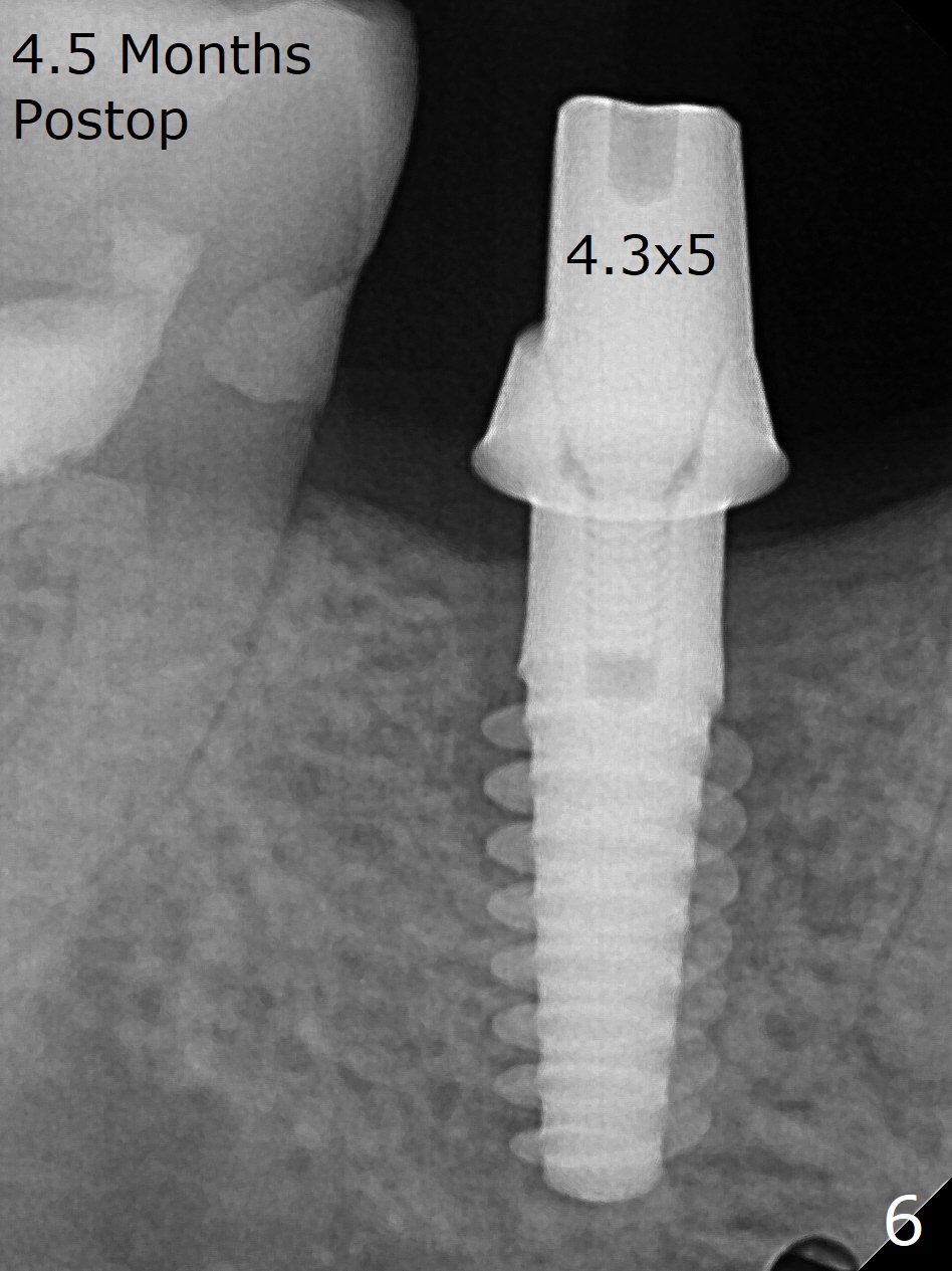
A 5x12 mm SM implant with periimplantitis at #30 is removed with a 5/6 mm trephine bur, small elevator and implant driver. A 3.8 mm Magic drill is used to start a new osteotomy in the lingual wall of the old one free hand. With the final drill (4.8x13 mm), a 5x11(3) mm Magicore is placed (Fig.1-4). The large buccal gap (Fig.2 arrowheads) is filled with allograft (Fig.3 *). The latter is covered by PRF, followed by suture. Periodontal dressing is applied around the 4.2x3 mm solid abutment and the coronal portion of the Magicore (Fig.4 <). The green outline in Fig.5 (CT coronal section) represents a new 5x10 mm implant (design), while the thick red line is approximately the long axis of the new implant. The implant heals in spite of buccal recession. Impression is taken with placement of a 4.3x5 mm solid abutment 4.5 months postop (Fig.6). Take follow-up CT to determine buccal bone formation.
Return to Lower Molar Immediate Implant, Trajectory II Magicore Cases Prevent Screw Loosening 植体取出后处理之二 Xin Wei, DDS, PhD, MS 1st edition 08/26/2019, last revision 03/06/2021