|
|
|
|
|||
|
|
|
|
|||
|
|
|
|
|
|
|
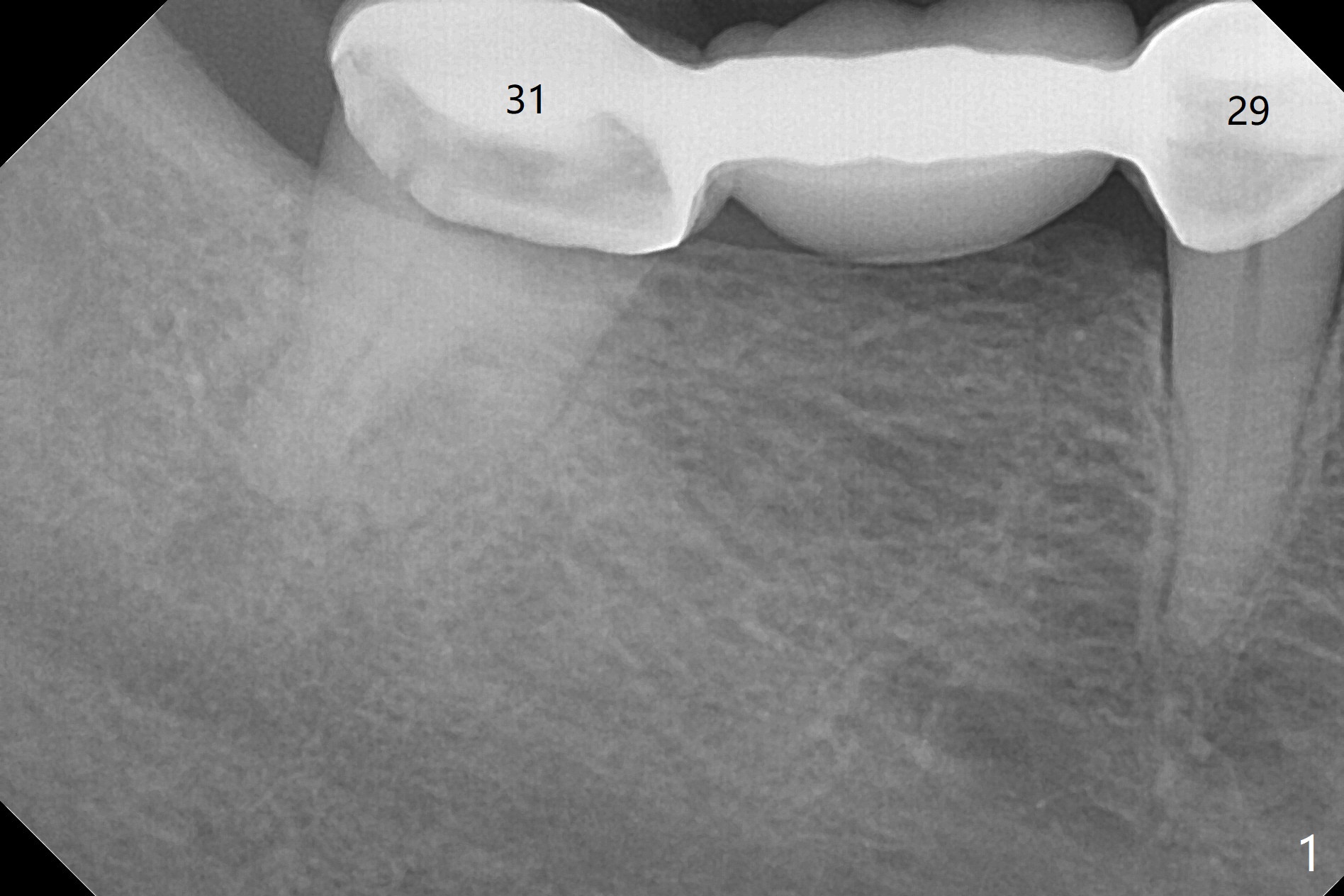
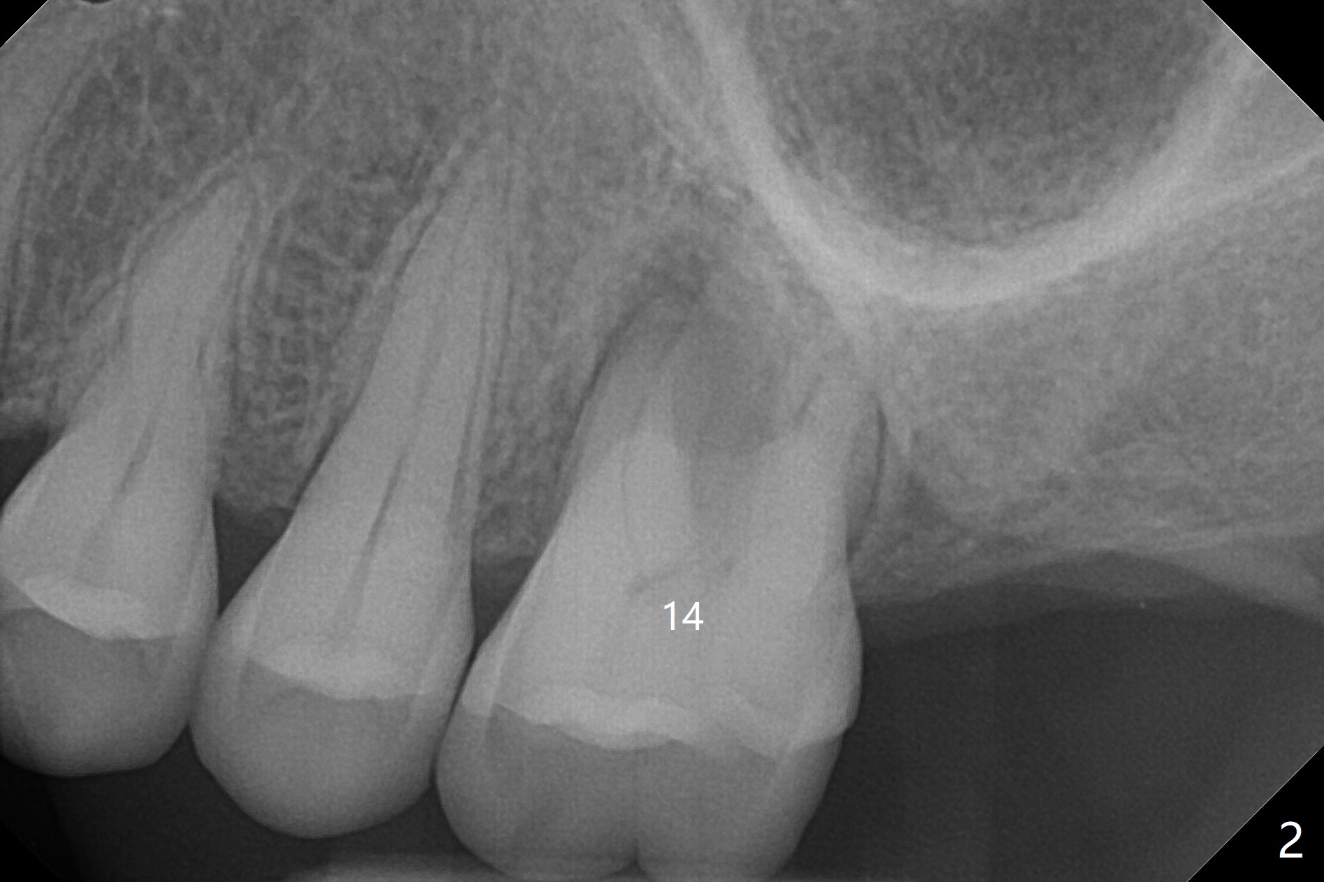
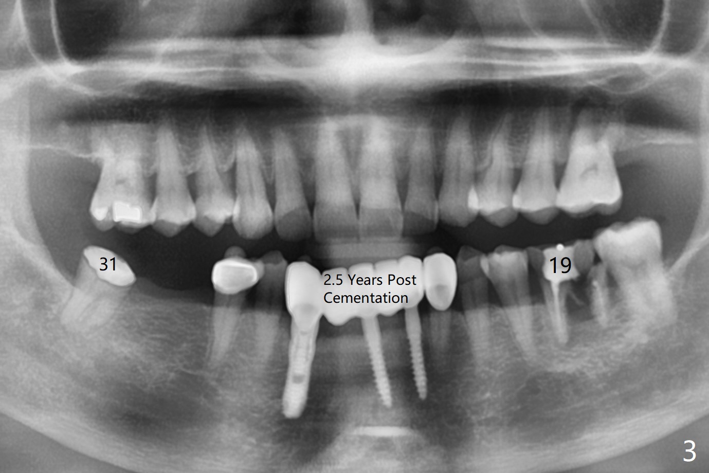
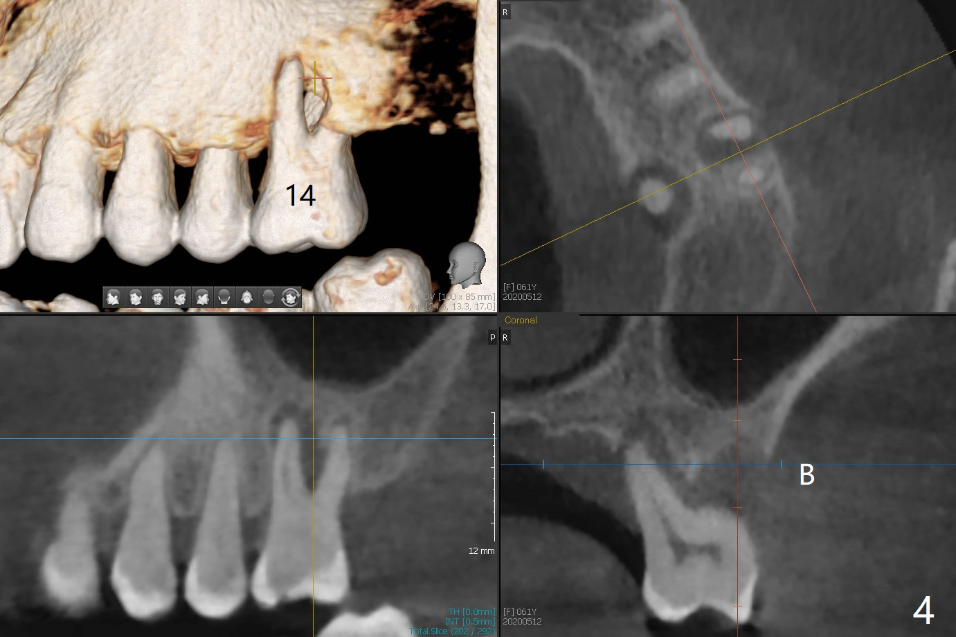
FPD Failure
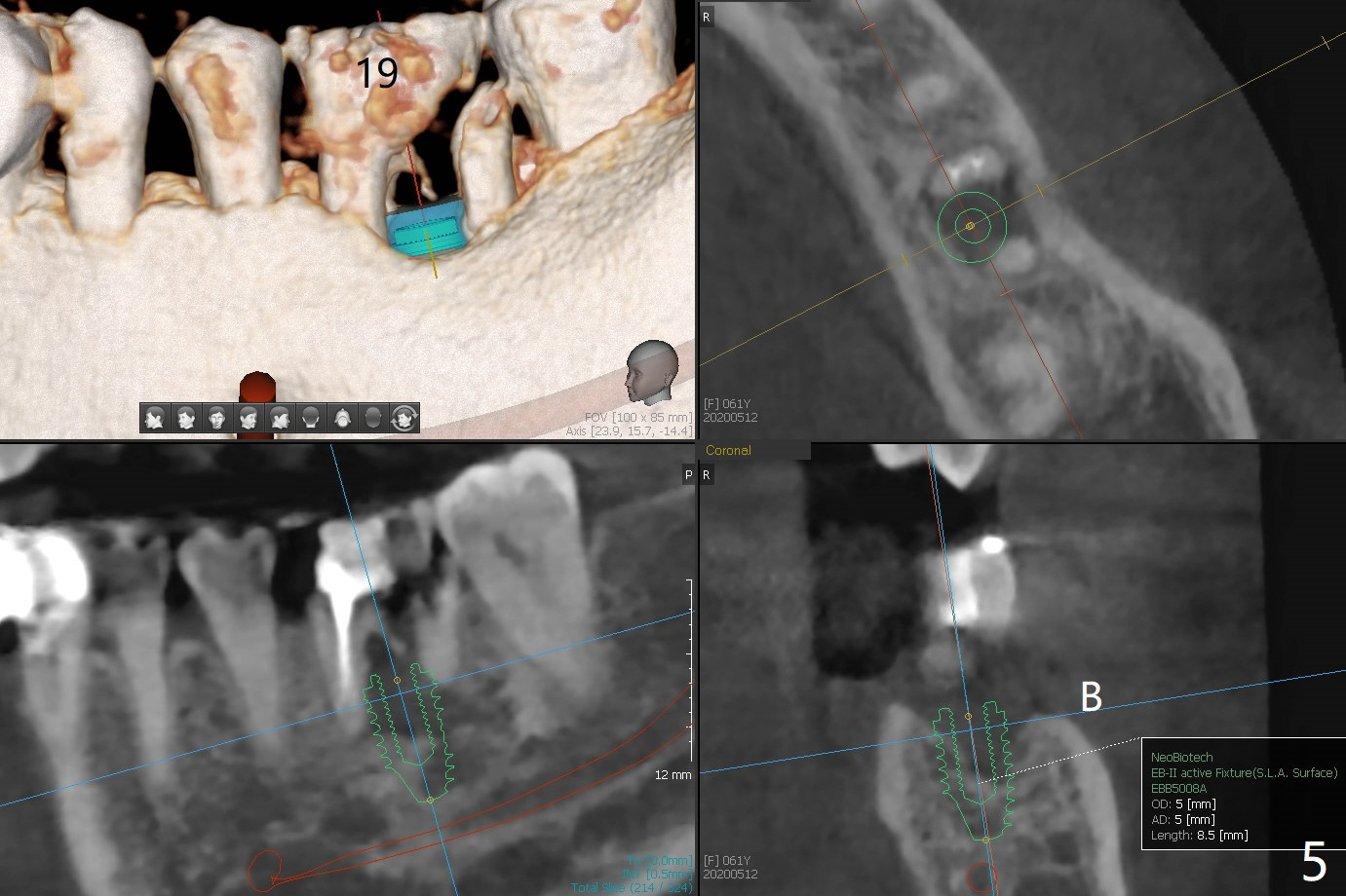
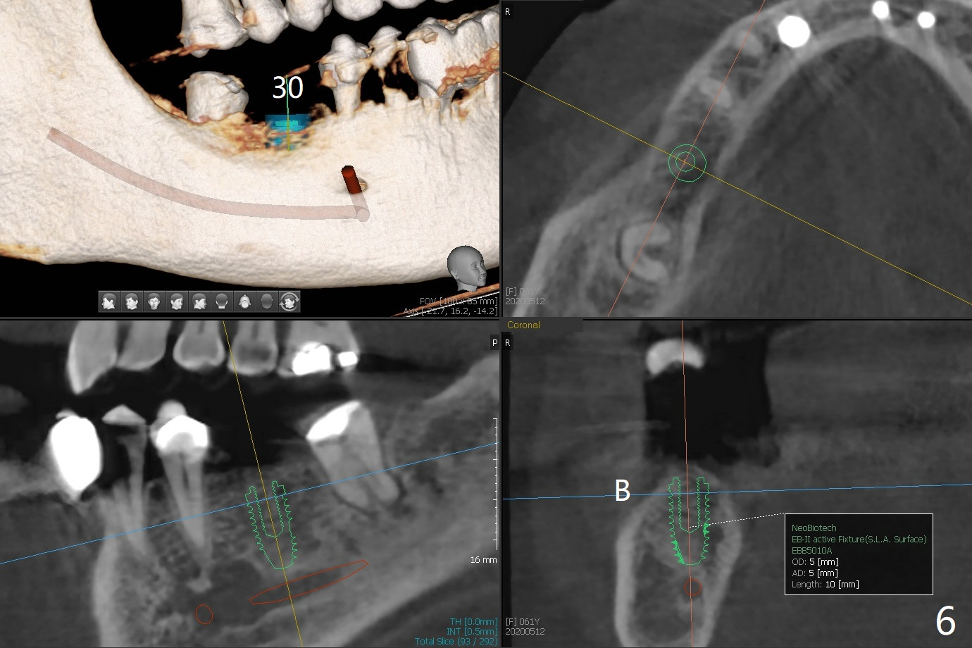
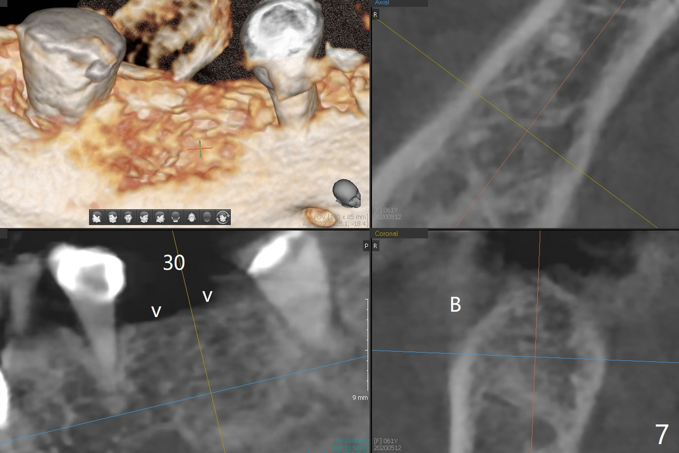
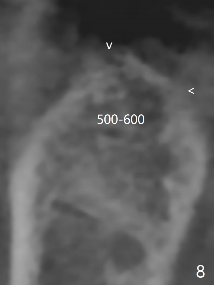
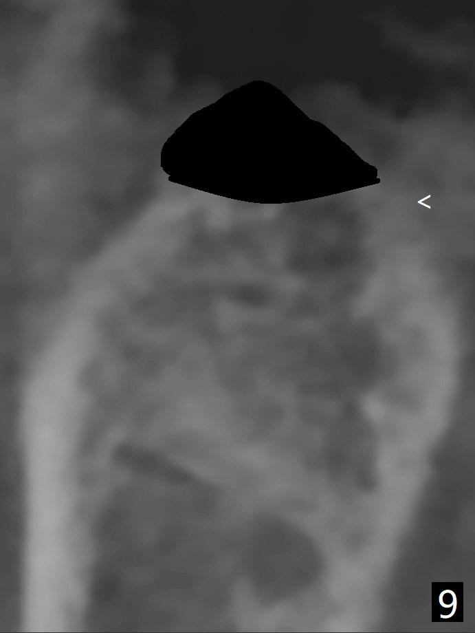
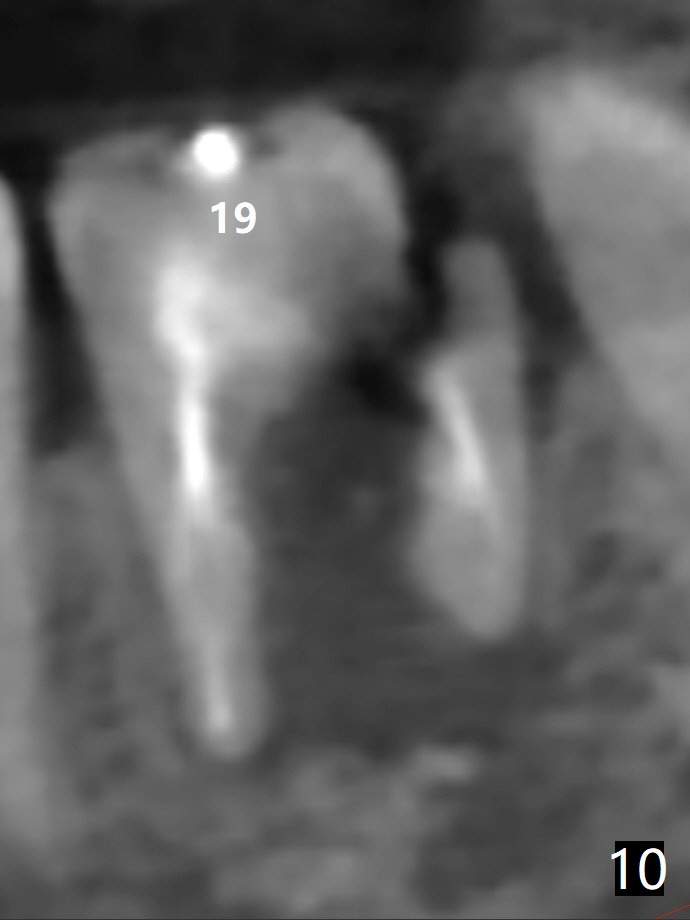
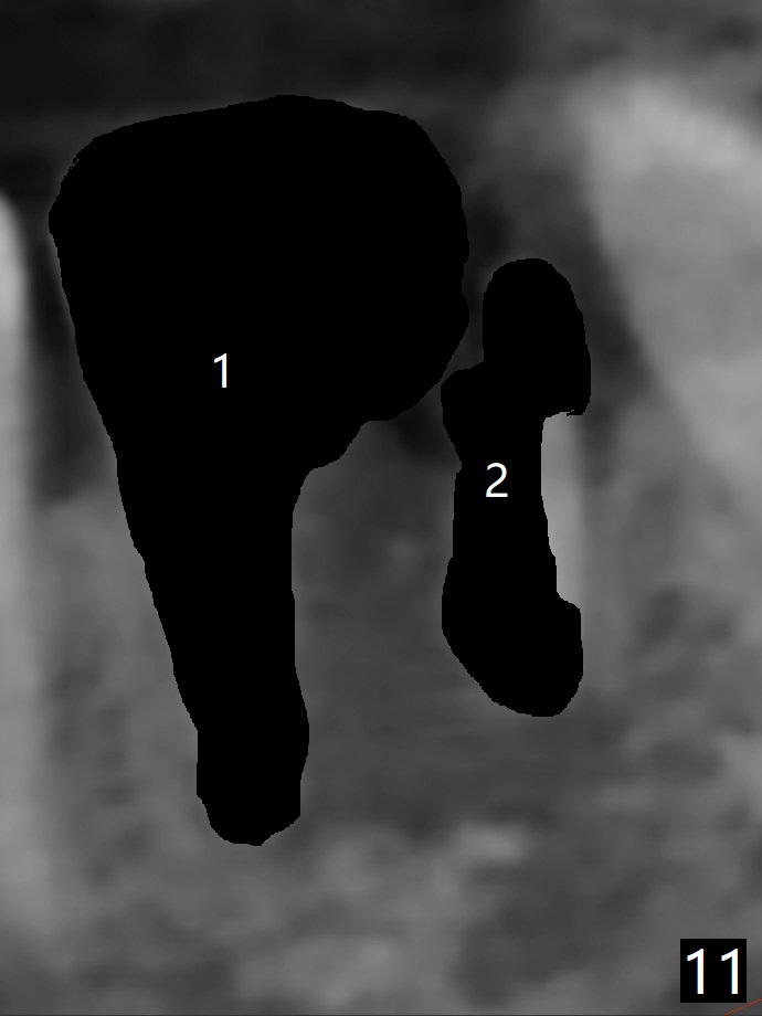
A 61-year-old woman with history of bruxism returns to clinic with chief complaint "I cannot chew bottom right. Top left has had pain and swelling before". Exam shows loose FPD at #31 (Fig.1) and necrosis of #14 (Fig.2). When the FPD is sectioned, the tooth #31 is found to have subgingival caries, filled with IRM (Fig.3). CT shows large PARL around MB and P roots of the tooth #14 (Fig.4), the fractured distal root of the tooth #19 with the low buccal plate (Fig.5) and a 5x10 mm implant being able to be placed at #30 (Fig.6). The crestal cortex (Fig.7 arrowheads) is thin in the edentulous area for several decades. The bone density is low (Fig.8, underprep). To place an implant over the pointed ridge, it should be trimmed prior to osteotomy (Fig.9). The bone loss associated with the fractured distal root is severe at #19 (Fig.10). After removal of the mesial root (Fig.11: 1), perform distal socket shield (2).
Return to
No Deviation
Clindamycin Metronidazole
No Antibiotic
23/25/27
Flap Surgery
Graft or
Not
Xin Wei, DDS, PhD, MS 1st edition
05/12/2020, last revision
06/04/2020