|
|
|
|
|
|
|
|
|
|
|
|
|
|
|
|
Bone Graft Before Implant I
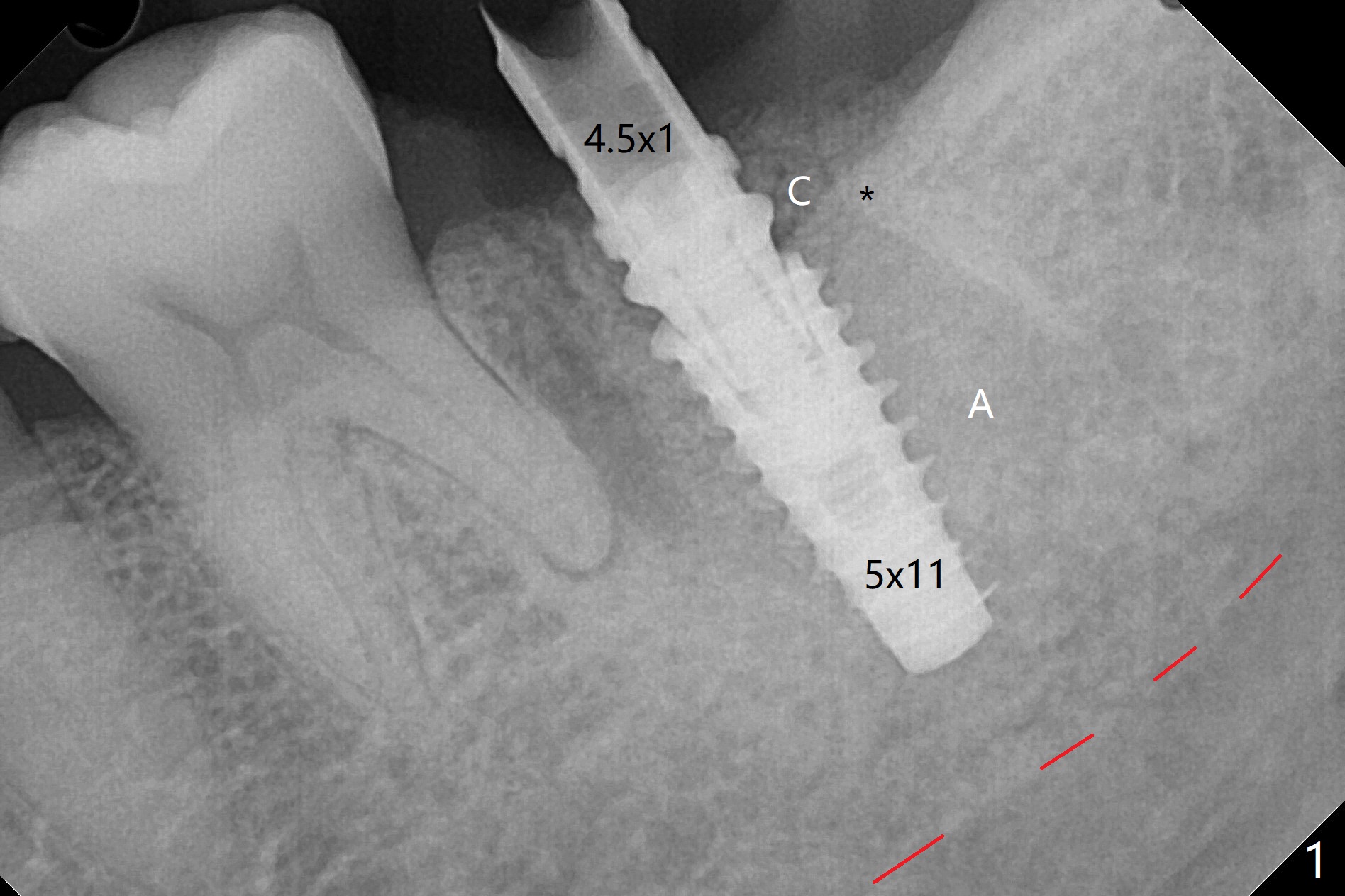
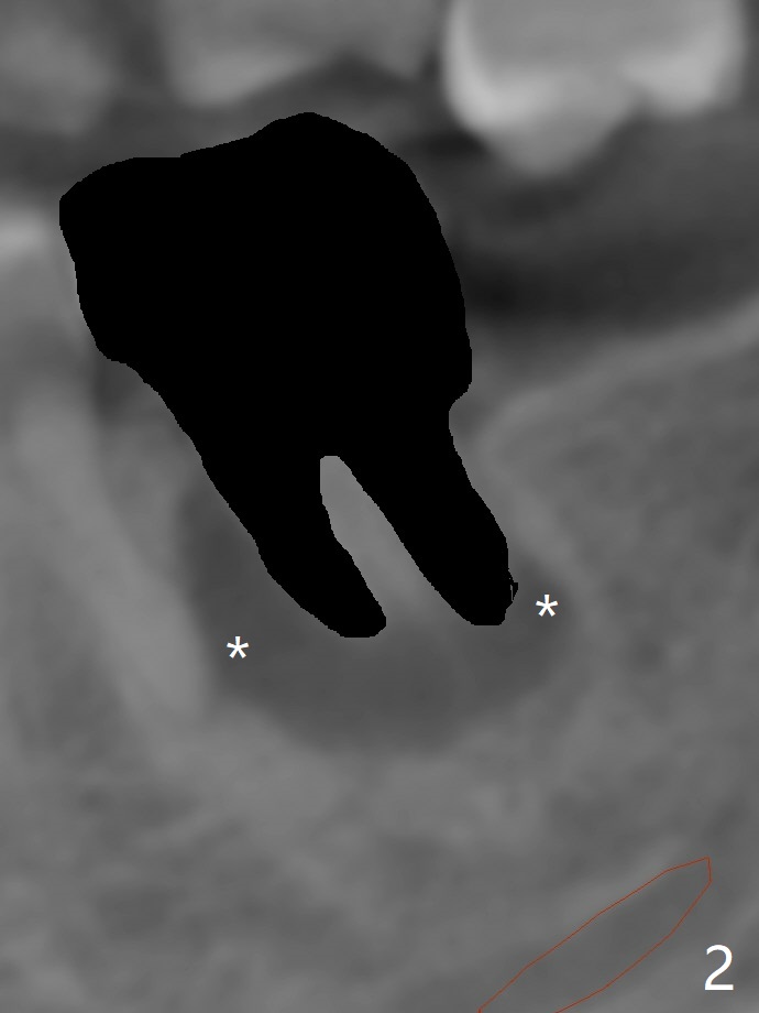
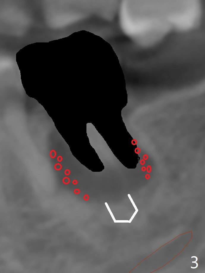
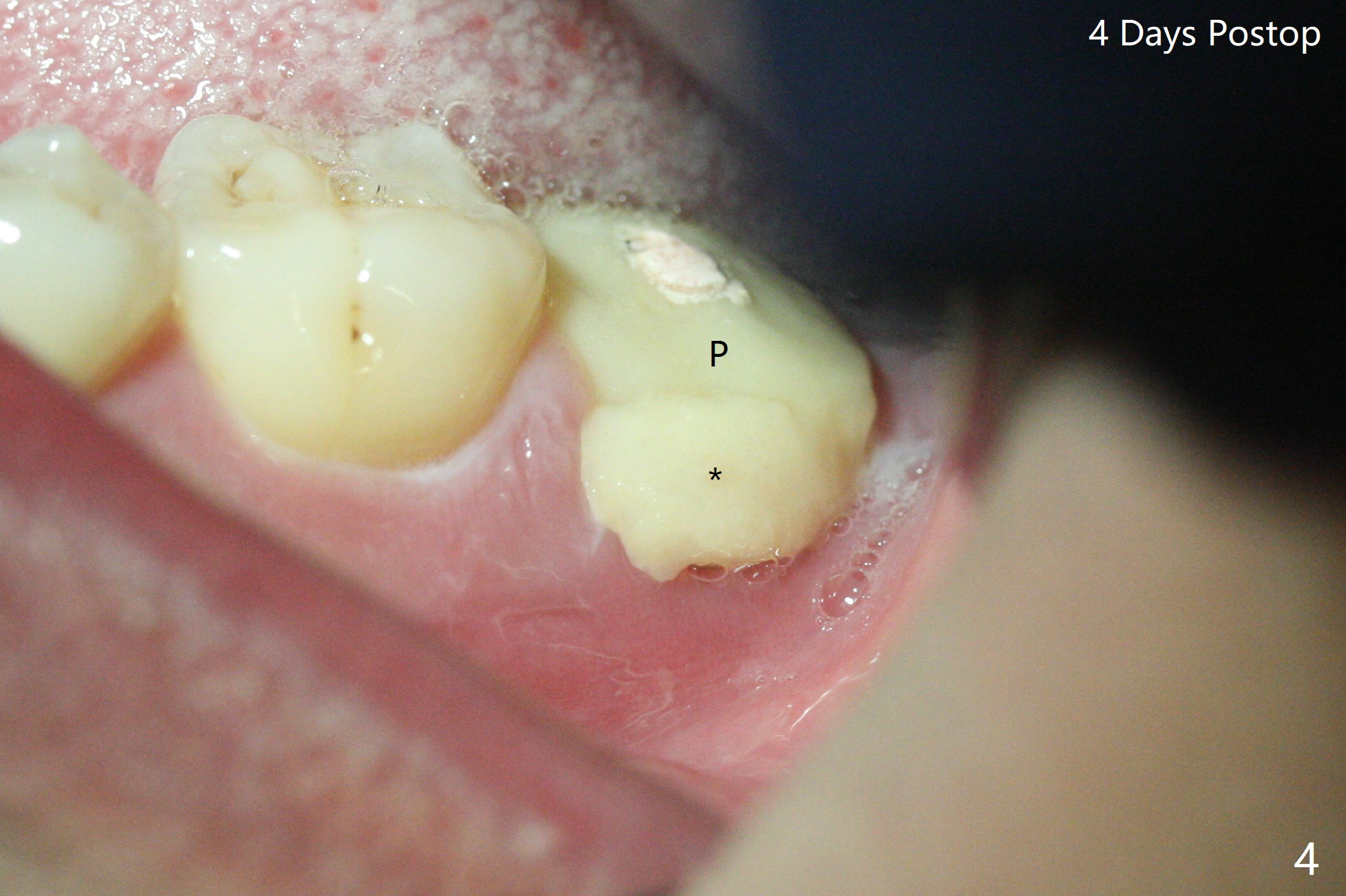
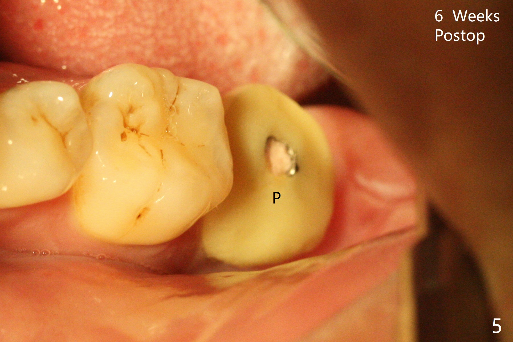
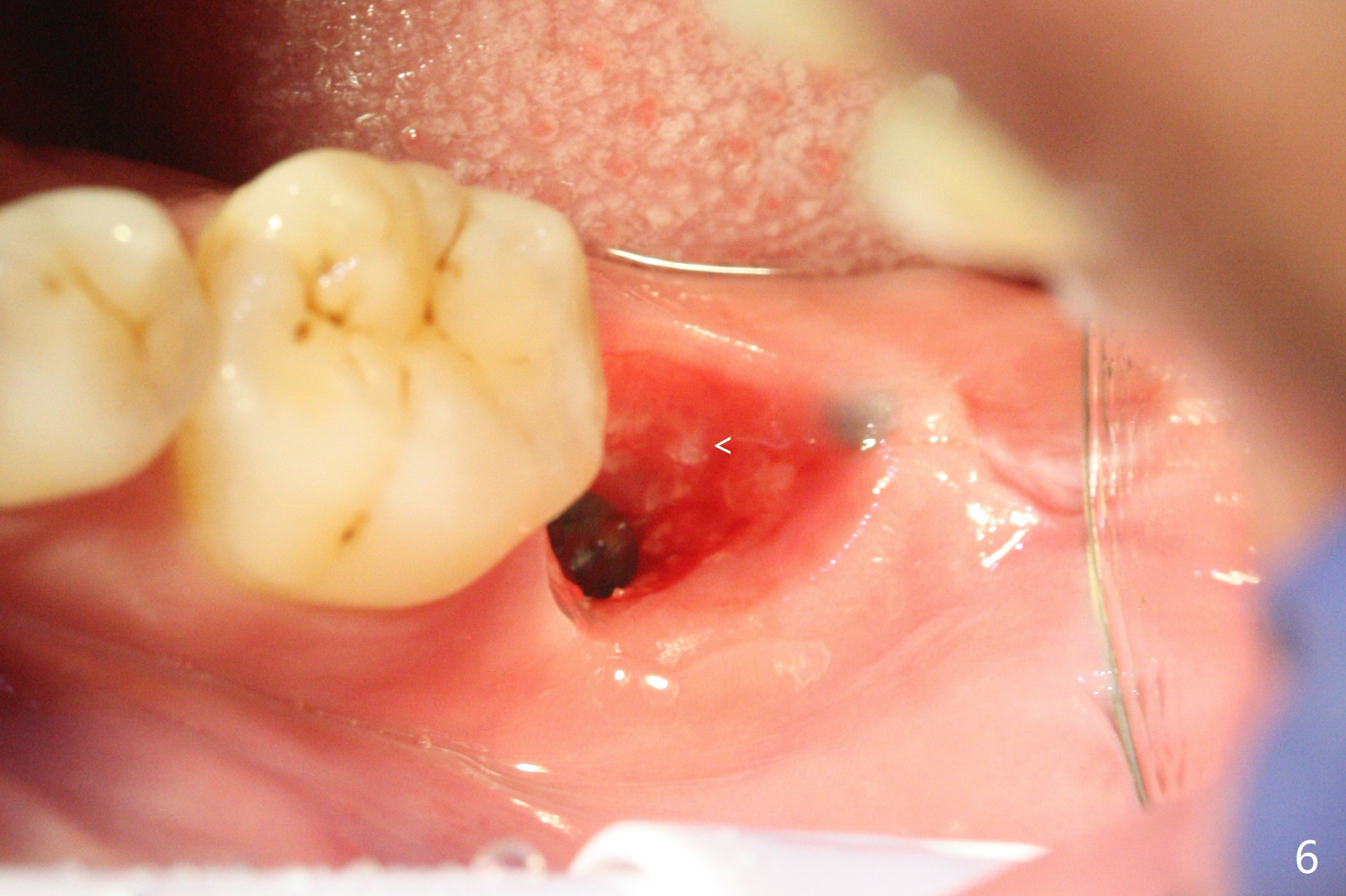
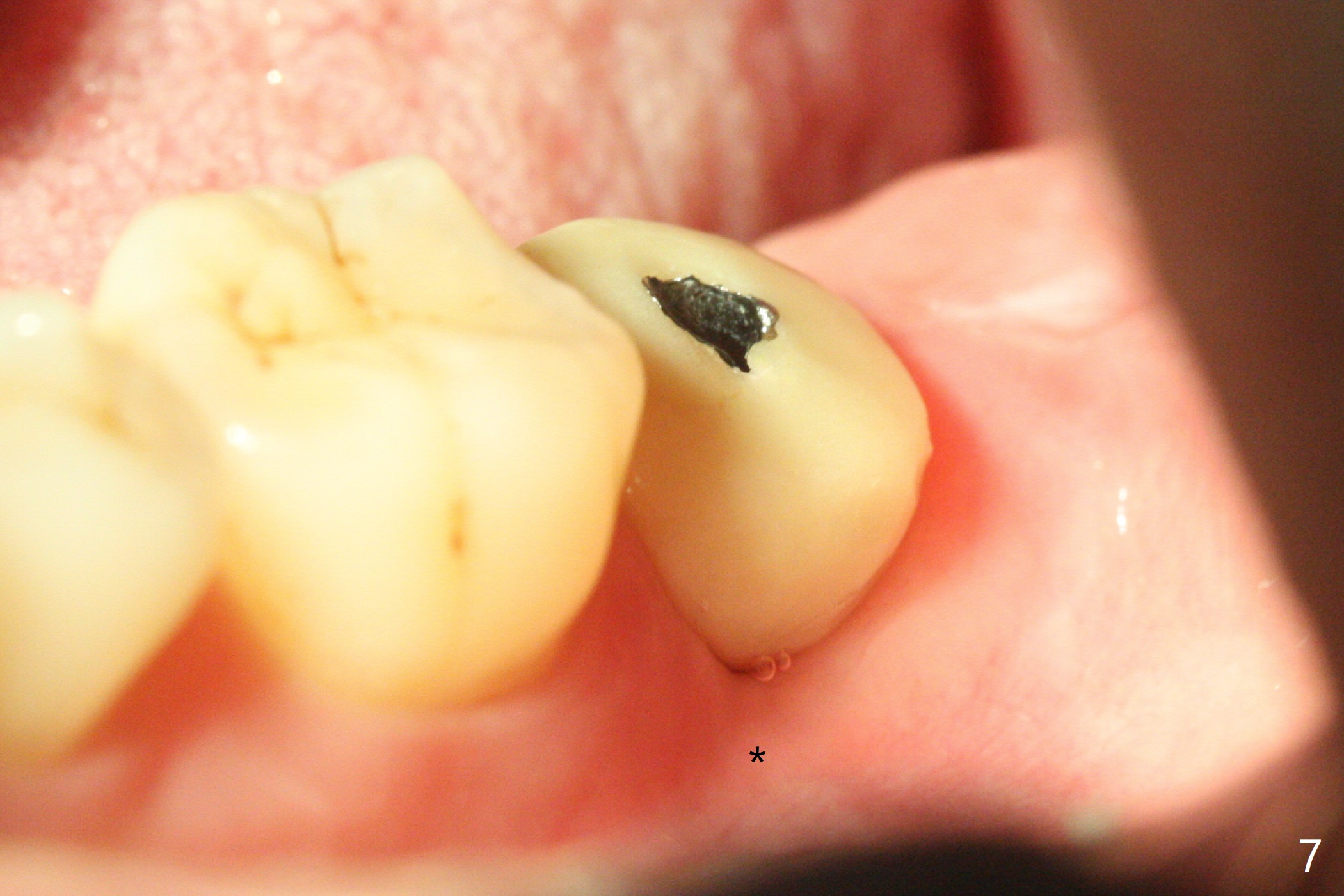
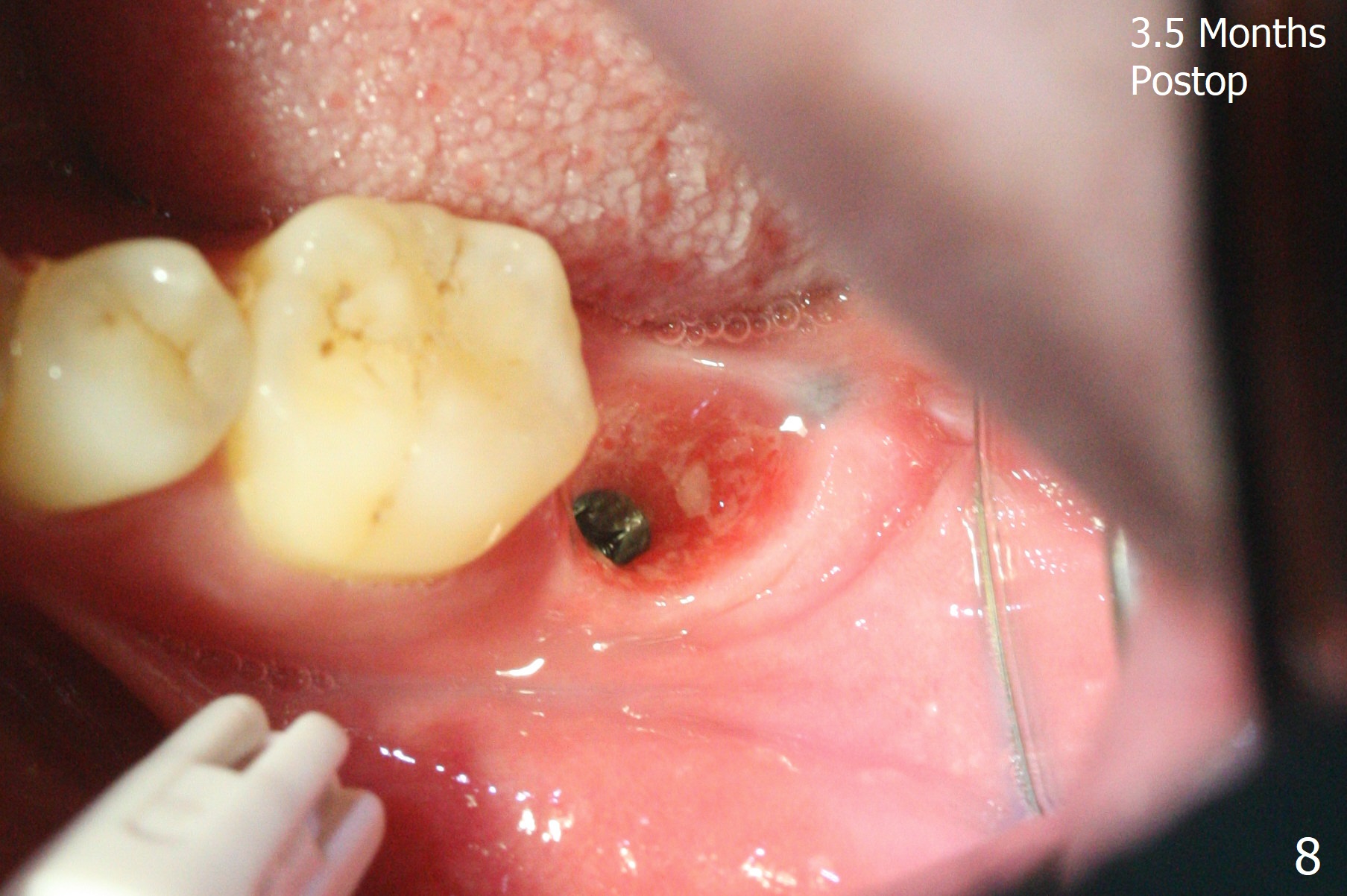
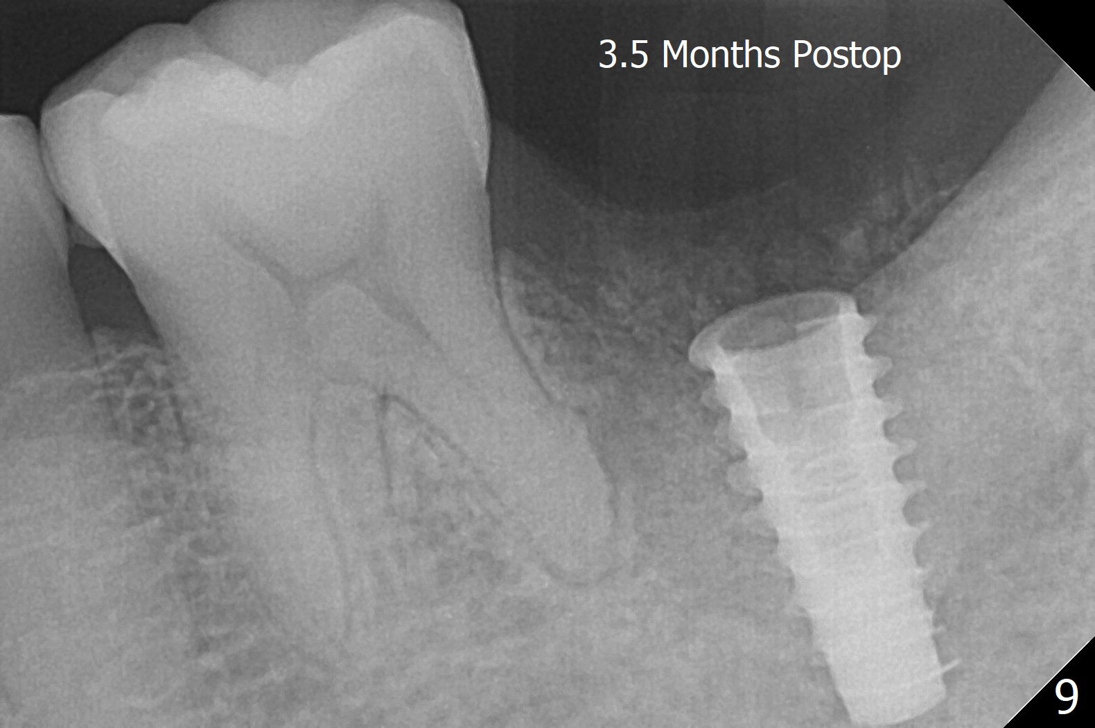
After placement of a 5x11 mm implant and cover screw at #18, allograft mixed with PRF (sticky bone) is placed in the peri-implant space, but it appears that bone density in the apical region (Fig.1 A) is lower than that in the coronal one (C) because of a constriction between the implant and the distal crest (*). Ideally the undercuts (Fig.2 *) of the extraction socket (black area) should be identified. Place bone graft in the undercuts (Fig.3 red circles) after osteotomy (white outlines) and before implant placement! A 4.5x1 mm temporary abutment is placed for an immediate provisional. The 2nd shortcoming of this case is that the implant is placed a little buccal. The papilla mesial to the immediate provisional (Fig.4 P) looks normal 4 days postop (no food impaction is expected when a final restoration is cemented). Additional acrylic (*) was placed to close a buccal gap when the provisional was seated with the temporary abutment. The patient reports loss of a piece of material in 4 days. It must be the additional acrylic, since it is absent 6 weeks postop (Fig.5). The provisional (Fig.5 P) looks wide, probably related to post-extraction gingiva and bone atrophy (Fig.6, 7 *). Bone graft (Fig.6 <) becomes a component of the gingival cuff. The provisional is re-trimmed for better oral hygiene. The gingival cuff is basically healthy 3.5 months postop, although the temporary abutment is loose and the implant is tender to rewinding and winding (Fig.8). The implant seems to remain non-osteointegrated 3.5 months postop (Fig.9).
Return to
Lower
Molar Immediate Implant,
Prevent Molar Periimplantitis (Protocols,
Table)
Trajectory II
Xin Wei, DDS, PhD, MS 1st edition
11/21/2019, last revision
03/13/2020