|
|
|
|
|
|
|
|
|
|
|
Onlay Graft Immediate Post Implant Removal
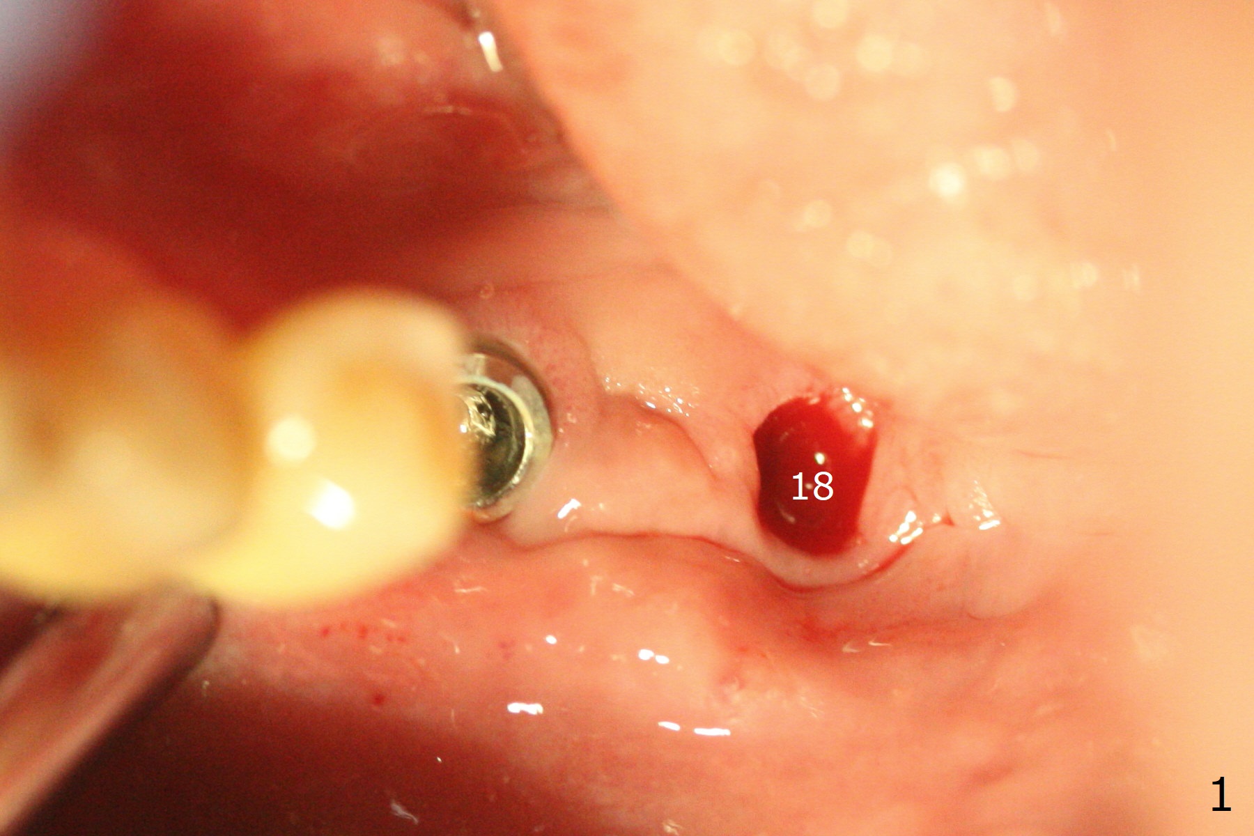
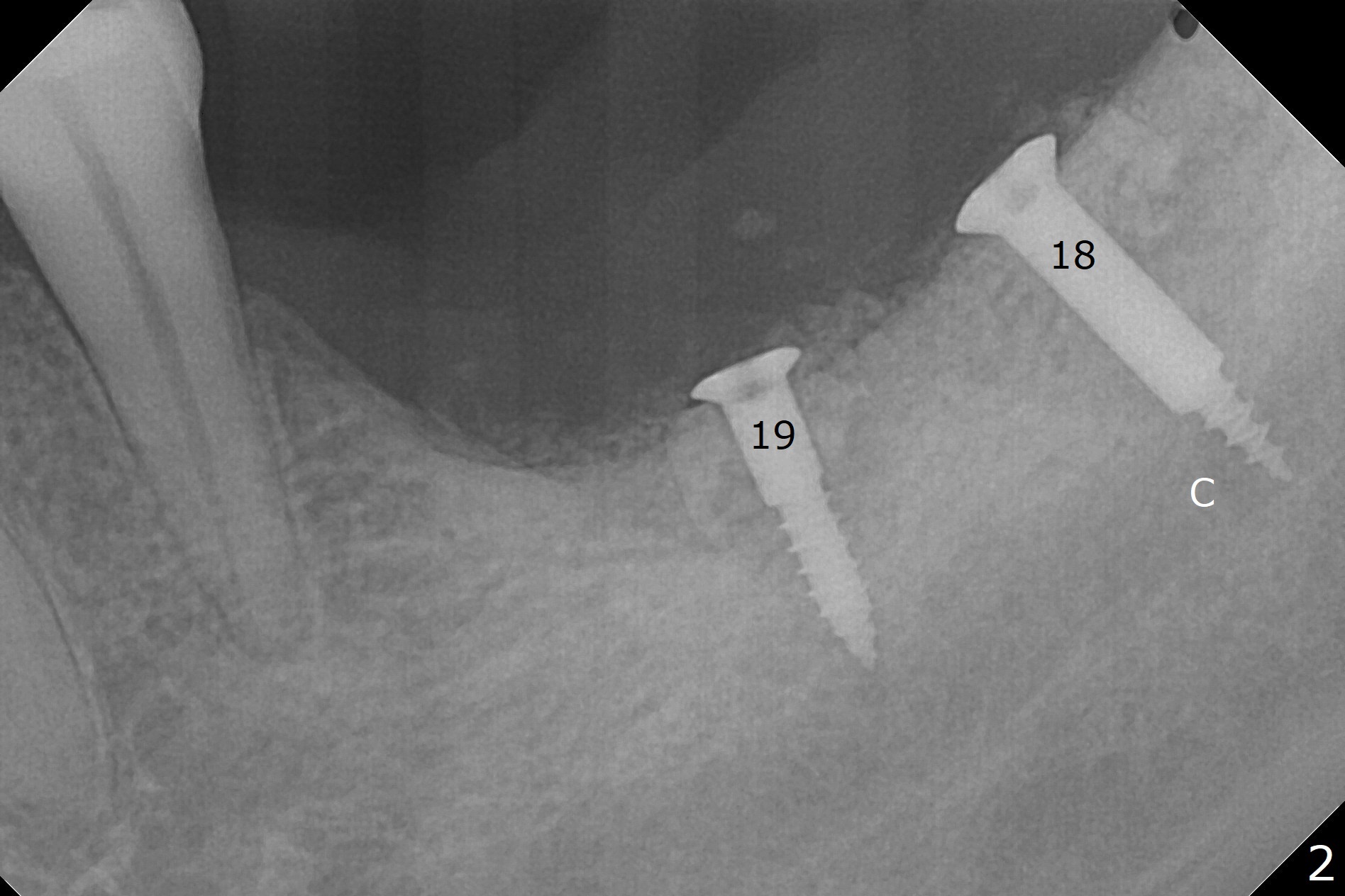
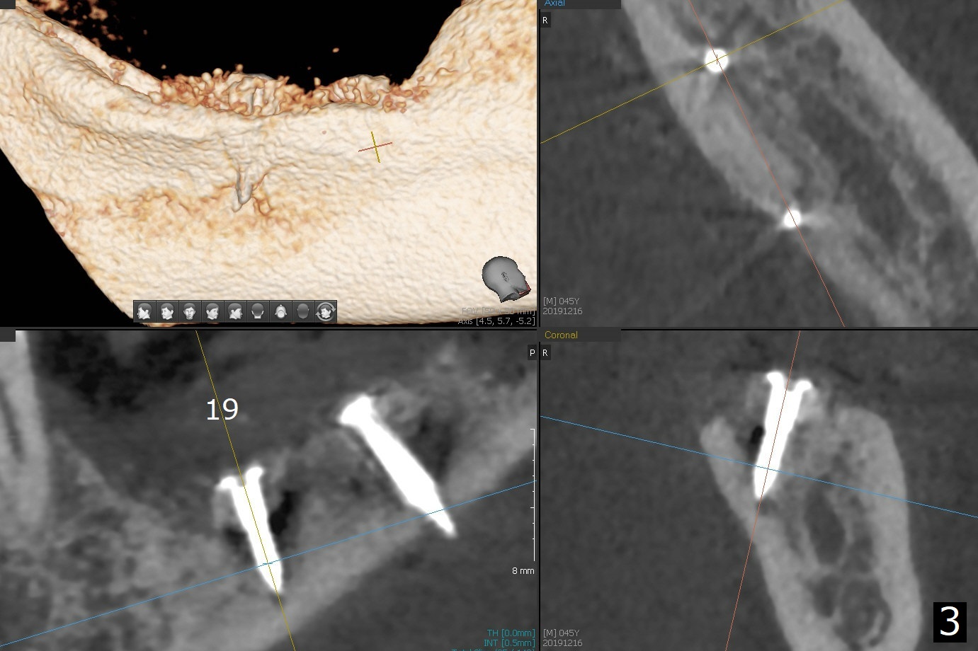
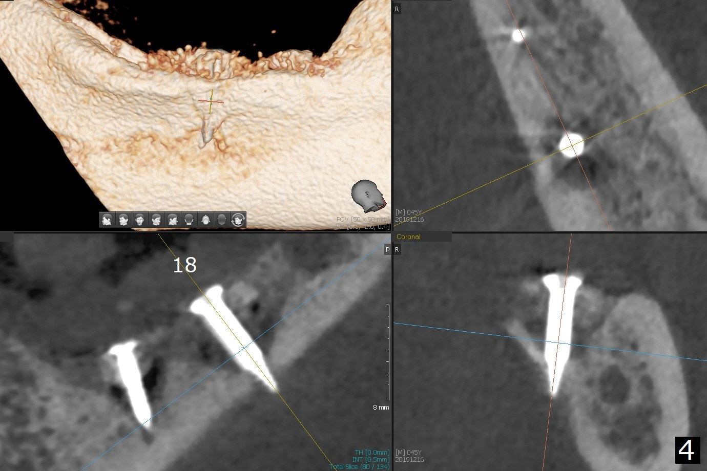
While the implant at #18 is easily removed, the one at #19 is superficial with buccal nonkeratinized gingiva (Fig.1). The latter is removed. Two small pieces of onlay graft is harvested from the left ramus and fixed in the defects with pins (Fig.2), surrounded by allograft with PRF. Since the pin at #18 looks violating the Inferior Alveolar Canal (Fig.2 C), CT is taken. In fact the pin perforates the lingual plate at #18 (Fig.4, as compared to Fig.3 (#19)). It leaves in place. Postop the wound dehisces with loss of allograft and exposure of the screws. The wound heals gradually mesiodistal with exposure of #18 screw 2.5 months postop. Follow up is disrupted by coronavirus pandemic.
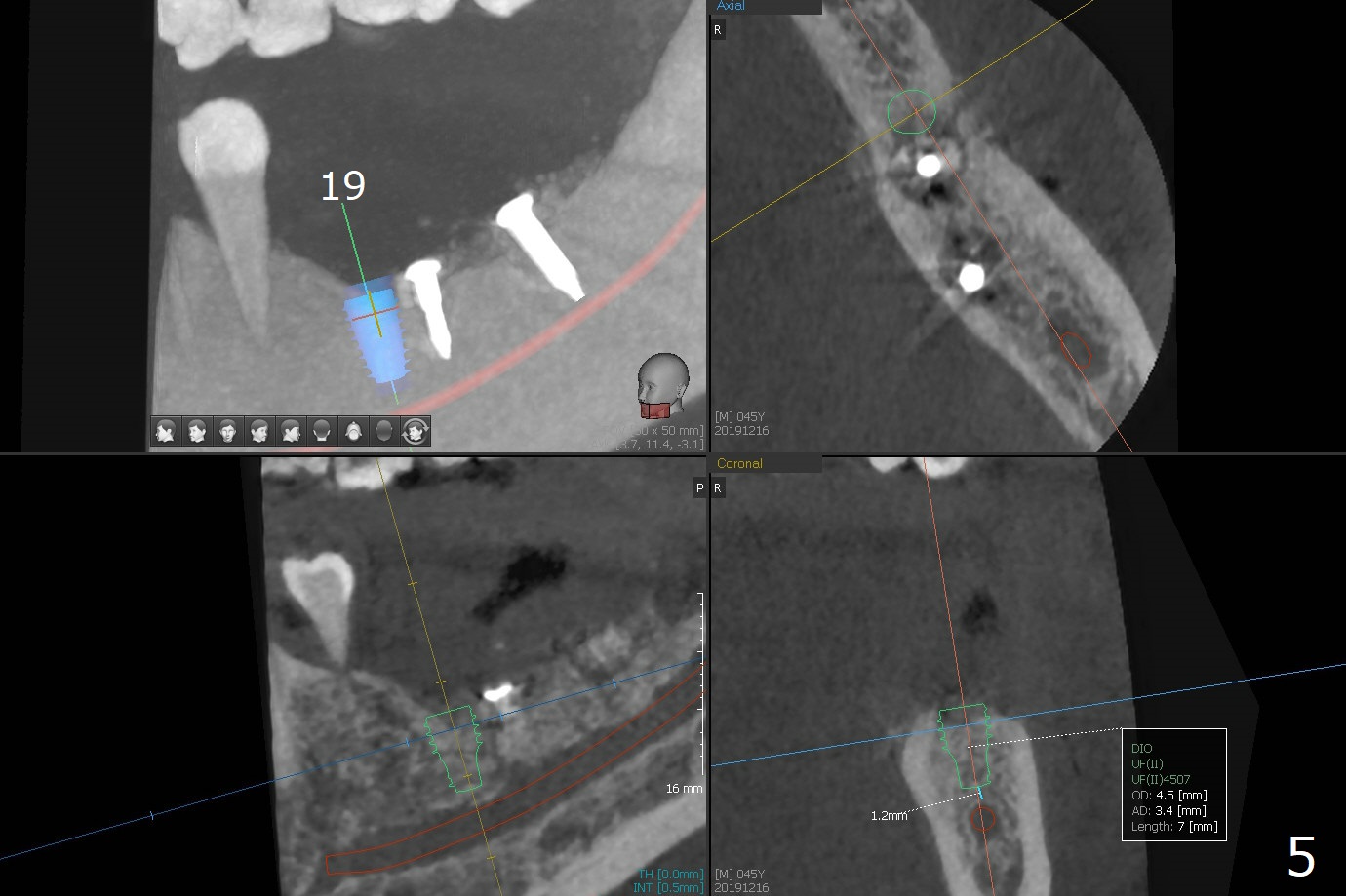
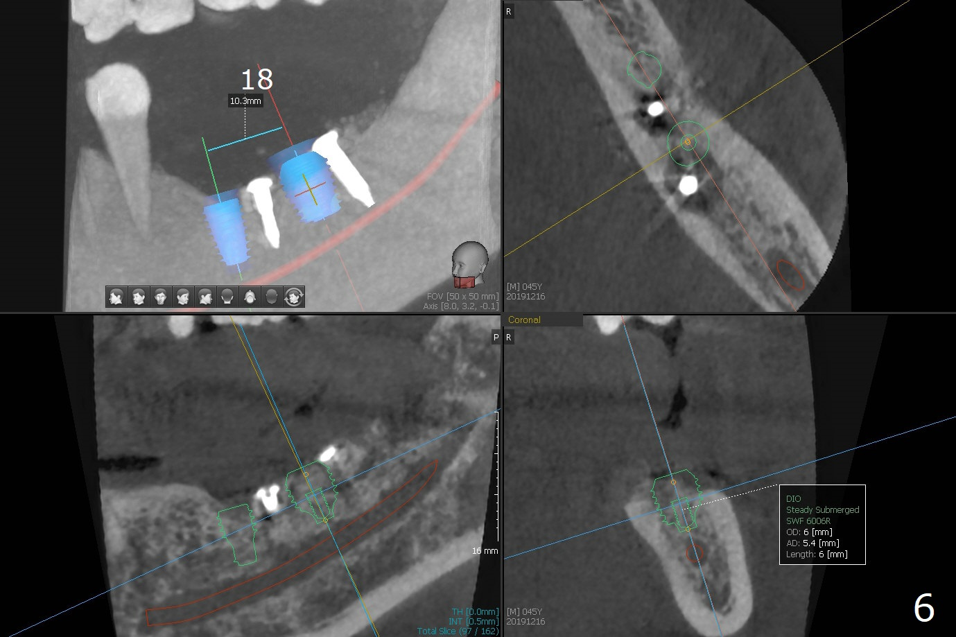
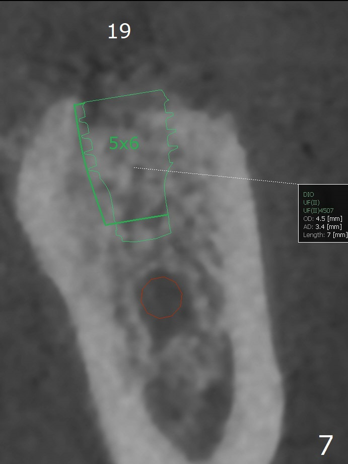
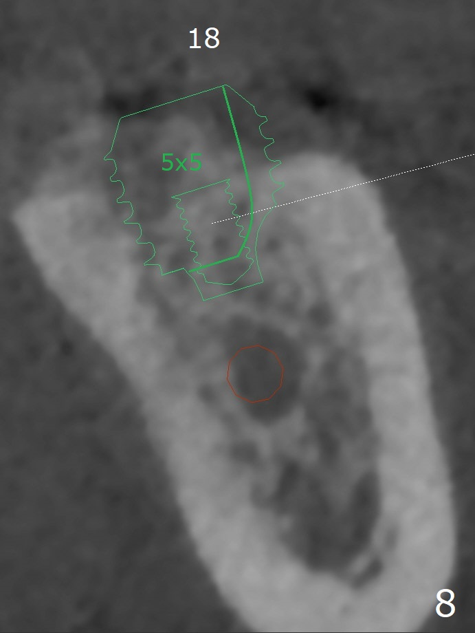
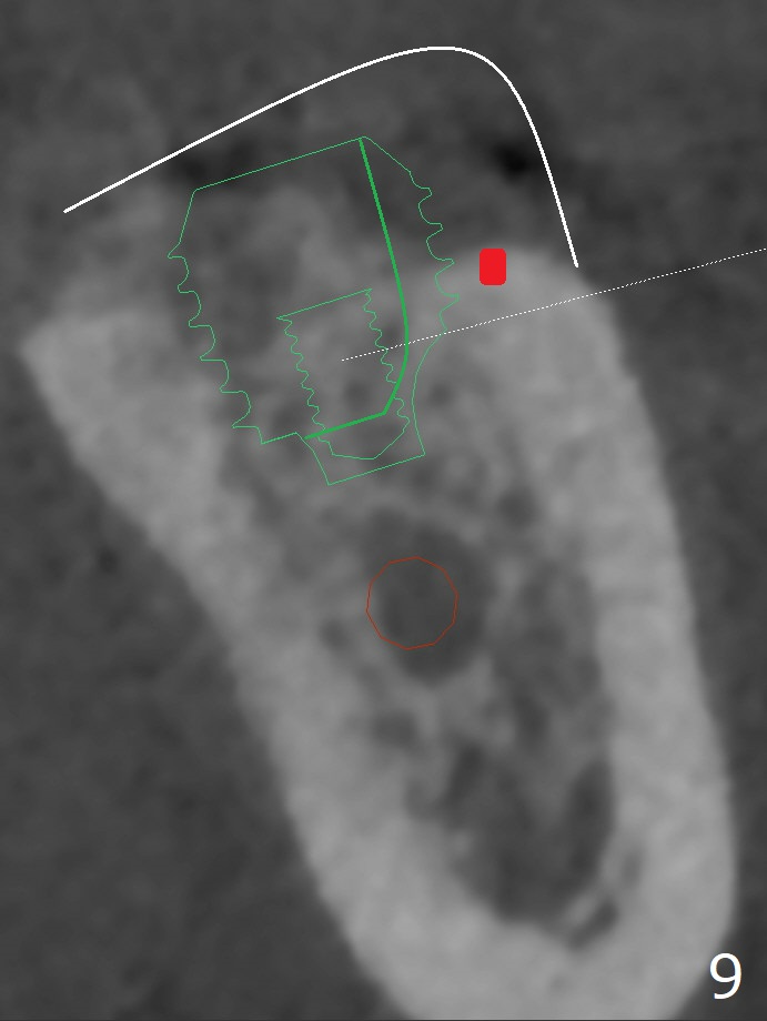
It appears that short implants could be placed mesial to the original sites (Fig.5,6), 5x6 and 5x5 mm at #19 and 18, respectively, with guide as lingual as possible. The exposed buccal threads will be covered by 3-D Bond (Fig.9 white line, post decortication (red)), collagen plug and PGA suture. Healing screws are most likely used, although healing abutments should be prepared if they help wound closure.
Return to
Trajectory II
Surgery
Xin Wei, DDS, PhD, MS 1st edition
12/19/2019, last revision
07/22/2021