 |
 |
 |
 |
 |
,%20Vera.jpg)  |
||
Narrow Mesio-distal Space
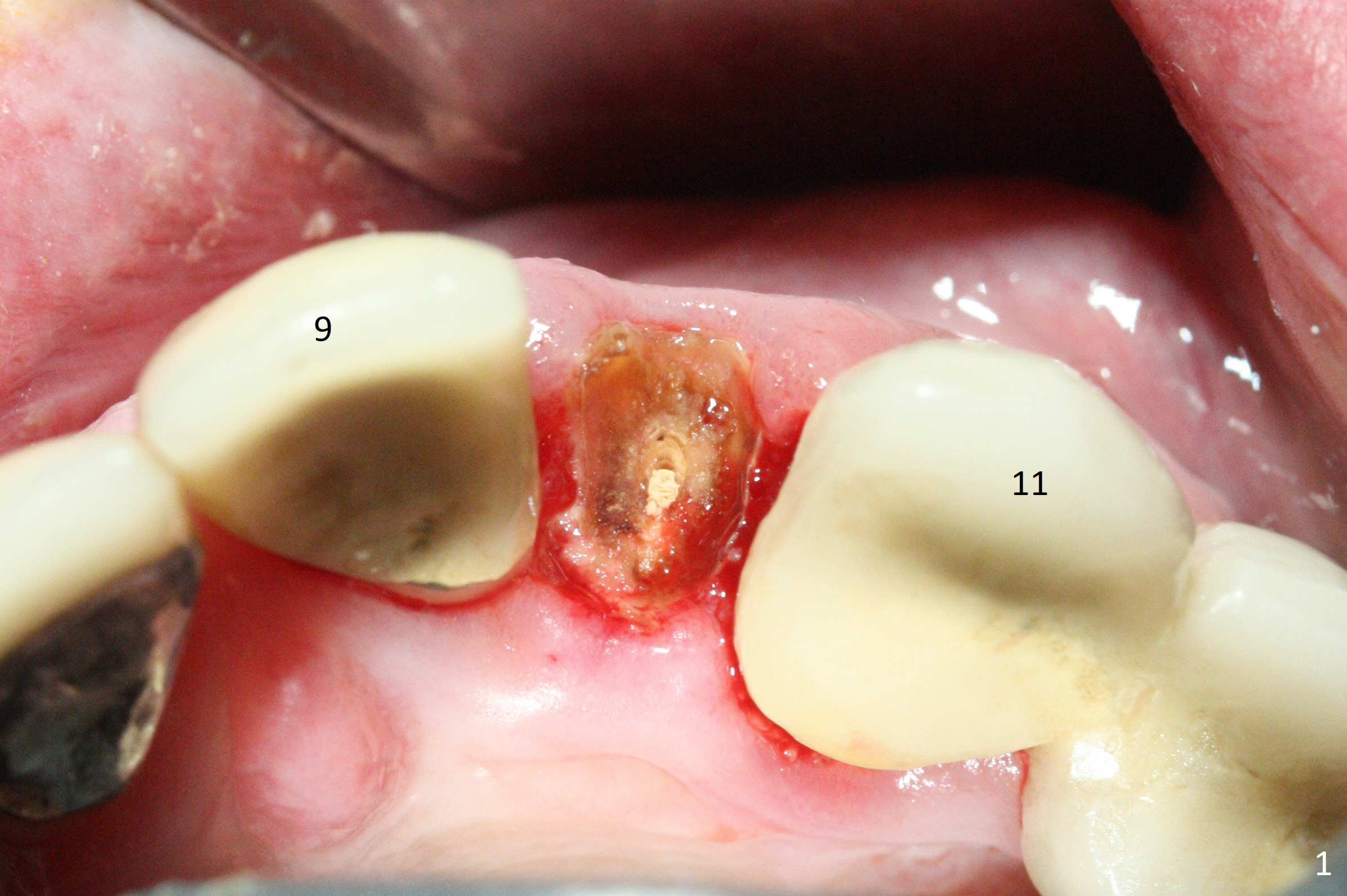
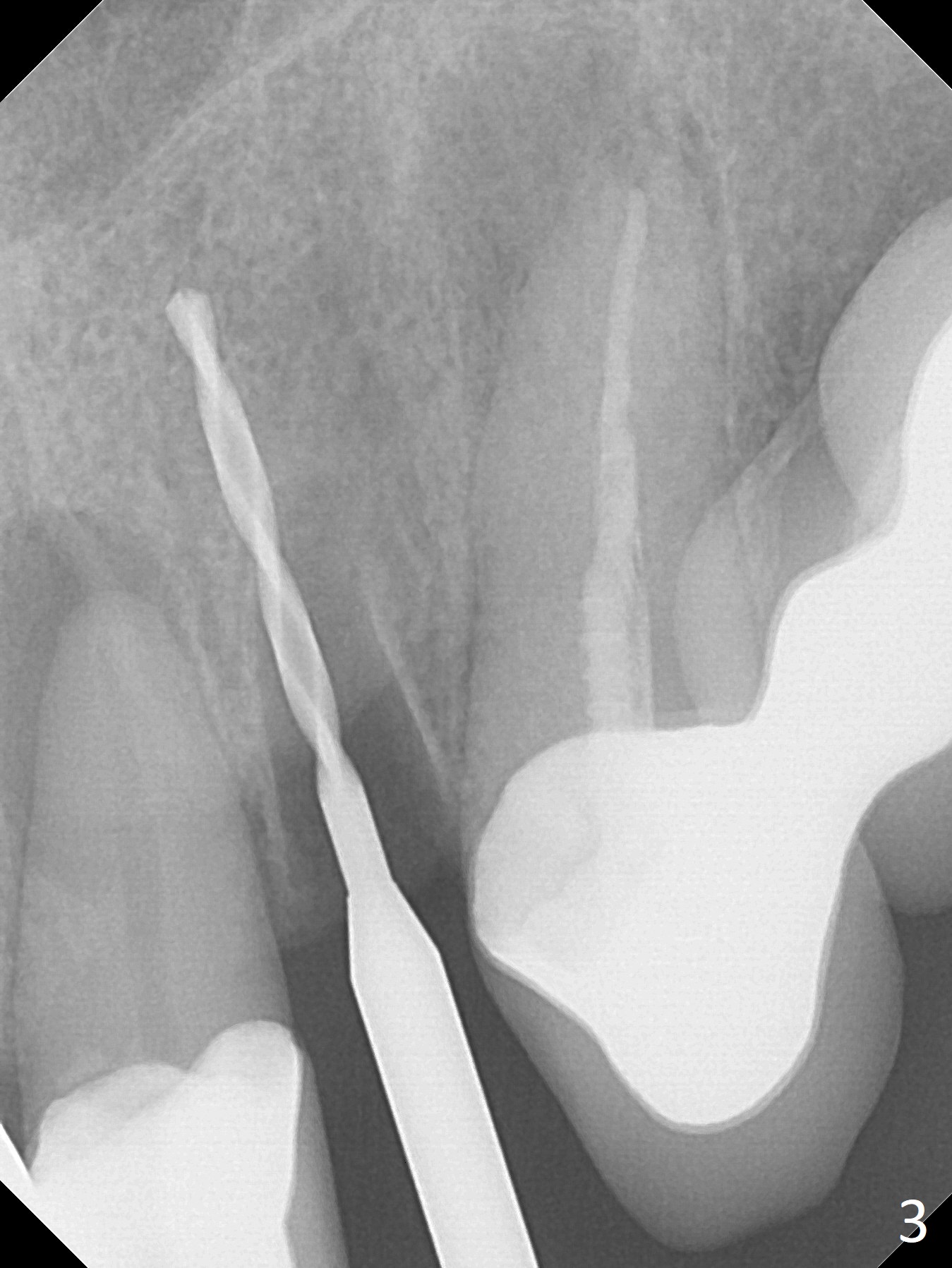
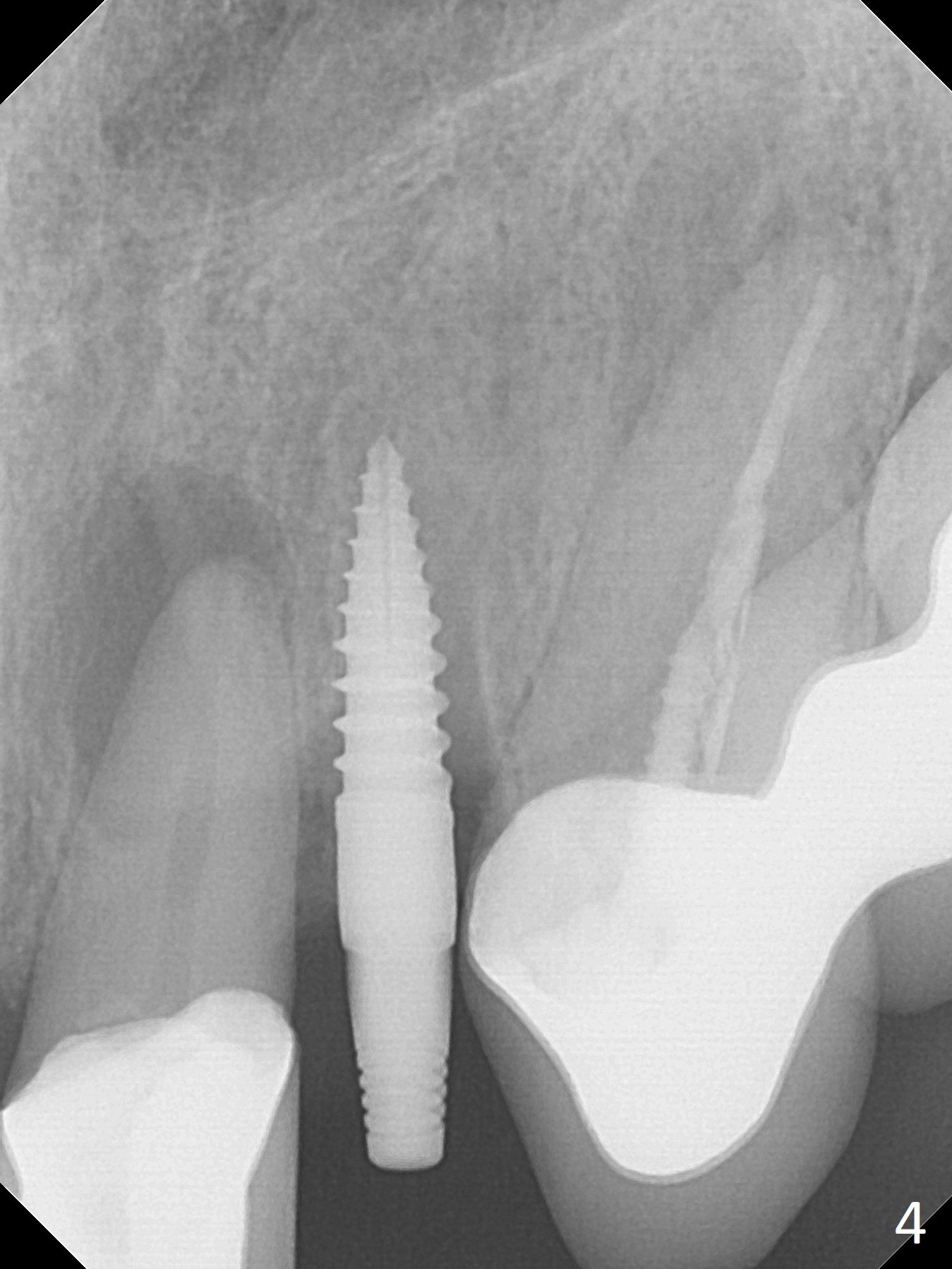
The tooth #10 fractures at the cervix, but is attached to the gingiva. After extraction of the coronal portion of the tooth, the mesiodistal space palatally is found to be narrow (~4.7 mm, Fig.1). It appears that a 1-piece implant is indicated because of the narrow mesiodistal space. In fact the buccal plate of the socket is intact (Fig.2). The initial osteotomy seems to be mesial (Fig.1) and is moved distal using Lindamann bur. After sequential osteotomy, a 3x10 mm dummy implant is still mesial (Fig.4). Following further distalization, a 3x14 mm implant is placed (Fig.5,6; <30 Ncm). Vera Graft is placed in the remaining socket prior to provisional fabrication (Fig.6 *). The socket outline disappears 7 months postop (Fig.7). Panoramic X-ray is taken 1 year 3 month post cementation.
Return to Upper Incisor Immediate Implant, Armaments 7 22 Xin Wei, DDS, PhD, MS 1st edition 12/18/2017, last revision 11/05/2019