

 |
 |
 |
 |
Buccal Gap
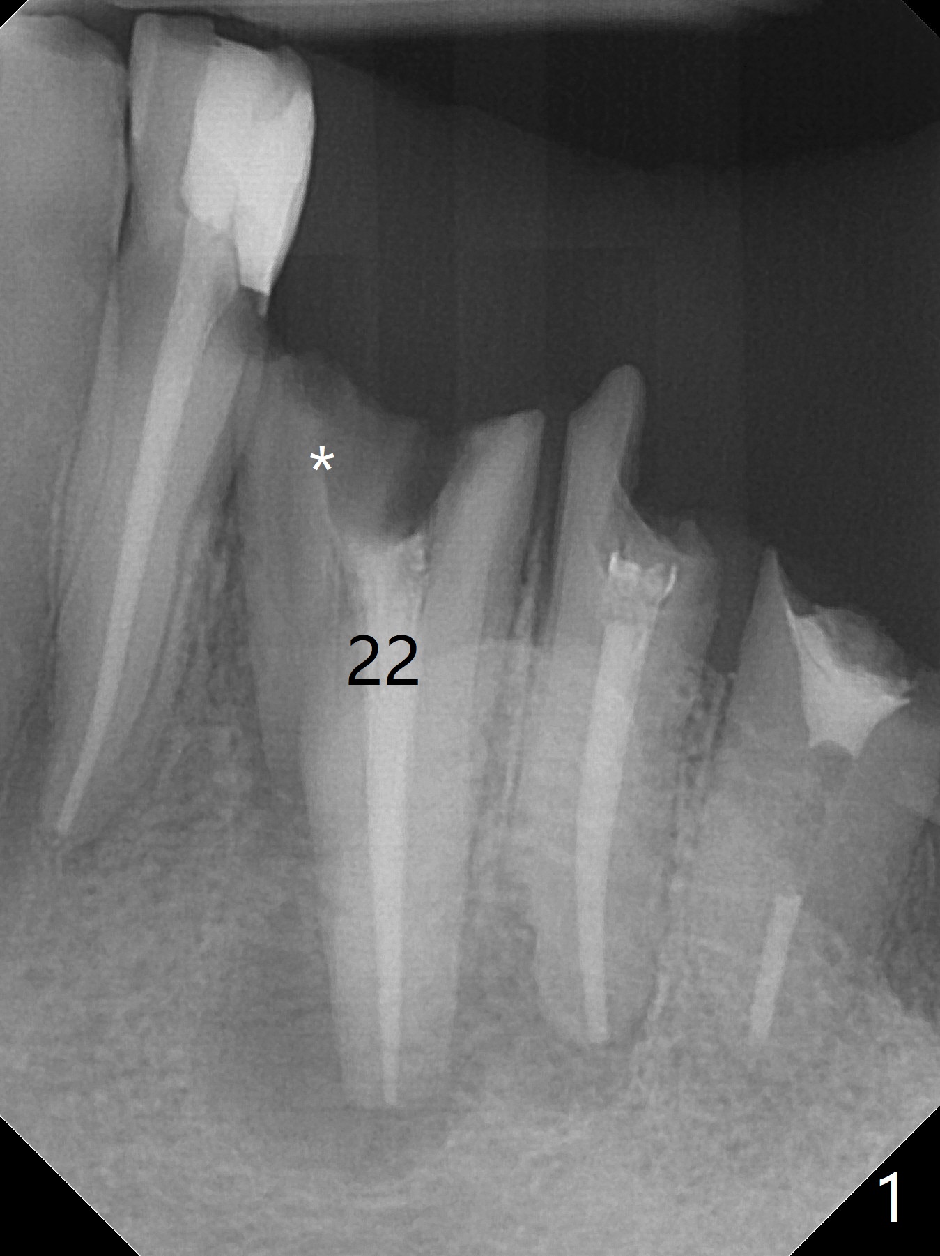
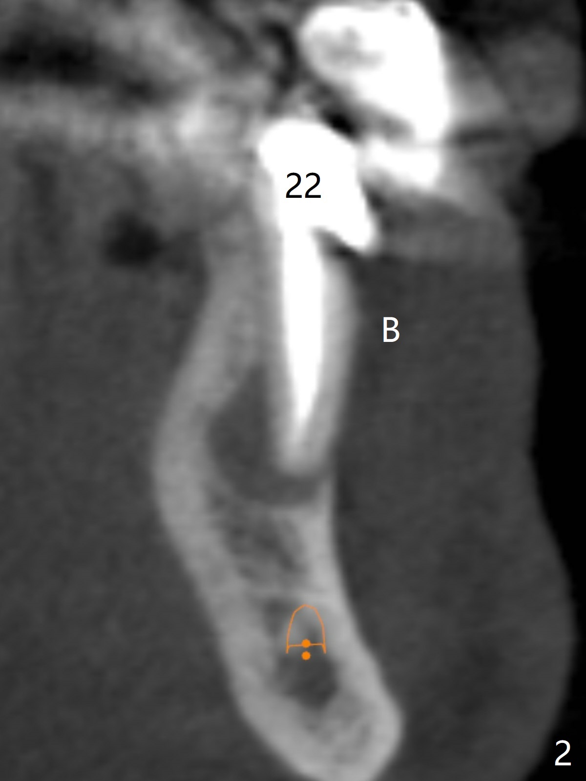
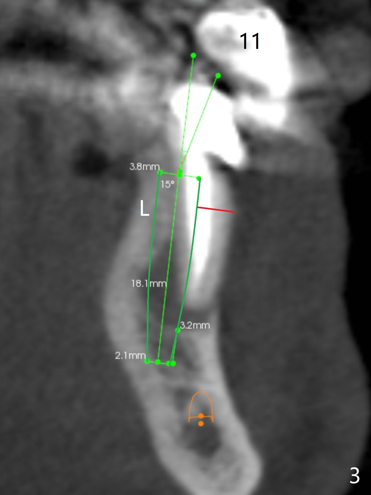
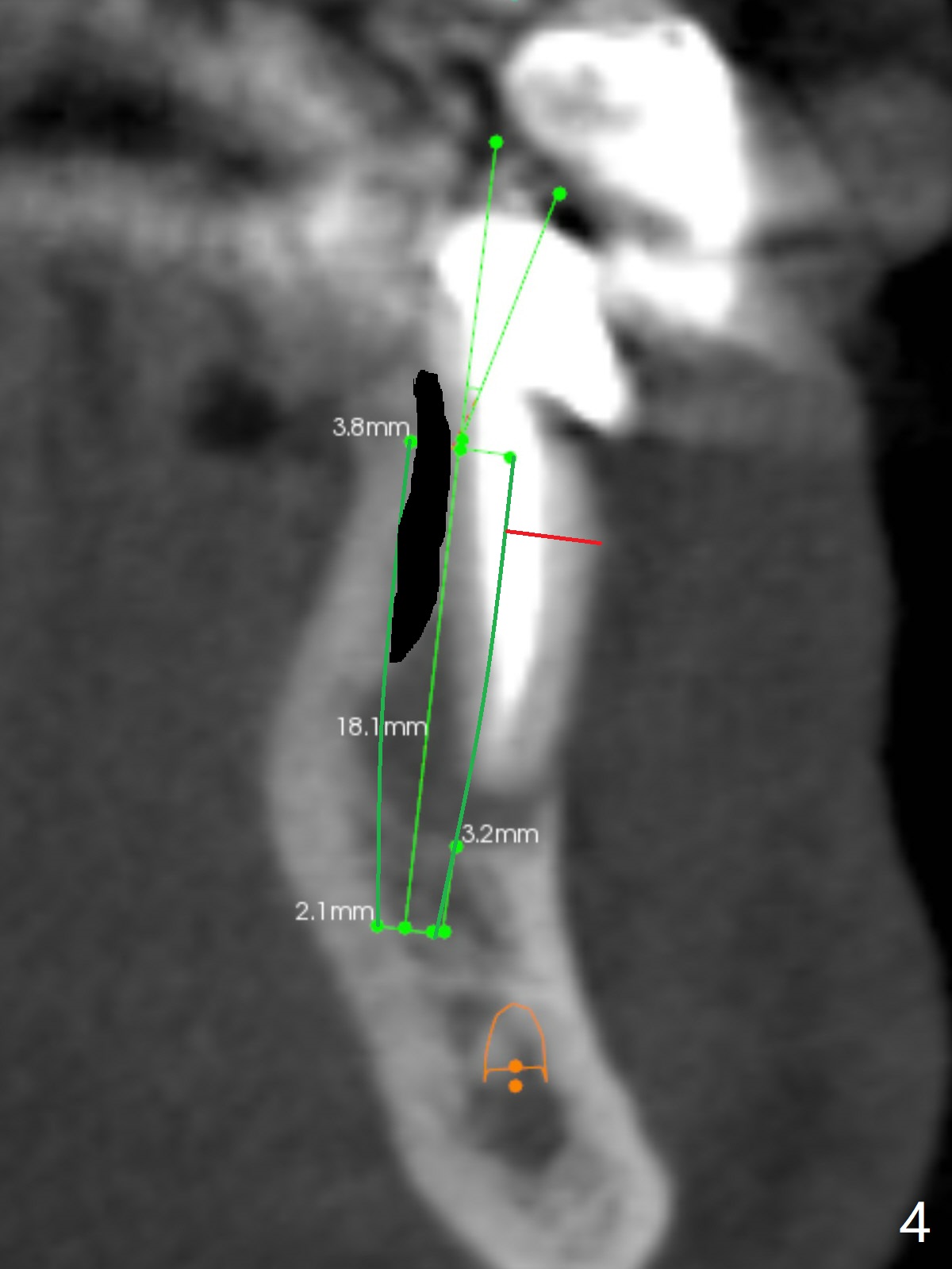
A 61-year-old man has poor dentition (#7 10), requesting extraction of the tooth #22 with root split (Fig.1 *). CT taken 1.5 years earlier (before crown fracture) shows missing buccal plate (Fig.2 B). After extraction, a smallest, longest 2-piece implant (3.8x18 mm) will be chosen to gain ~ 3 mm apical native bone for primary stability; to obtain a 2 mm buccal gap (Fig.3 red line), the implant will be placed as lingual as possible. To achieve the buccal gap, the buccal portion of the lingual plate (Fig.3 L) will be removed (Fig.4 black area) using Lindamann bur. For restoration, a 15 or 25 degree angled abutment may be used (Fig.3,4). If the root is stable, socket shield will be performed.
Return to
Lower Canine
Immediate Implant,
Trajectory,
No Antibiotic,
Socket Shield
Wei, DDS, PhD, MS 1st edition
01/01/2019, last revision
01/10/2019