,%204x8.5,%204.8x7,%20sticky%20bone.jpg)
,%204.5x5.5(4),%2030%20Ncm,%207%20m%20postop.jpg)
|
|
|
|
|
|
|
|
|
|
|
|
|
|
|
Sticky Bone for Large Defect M
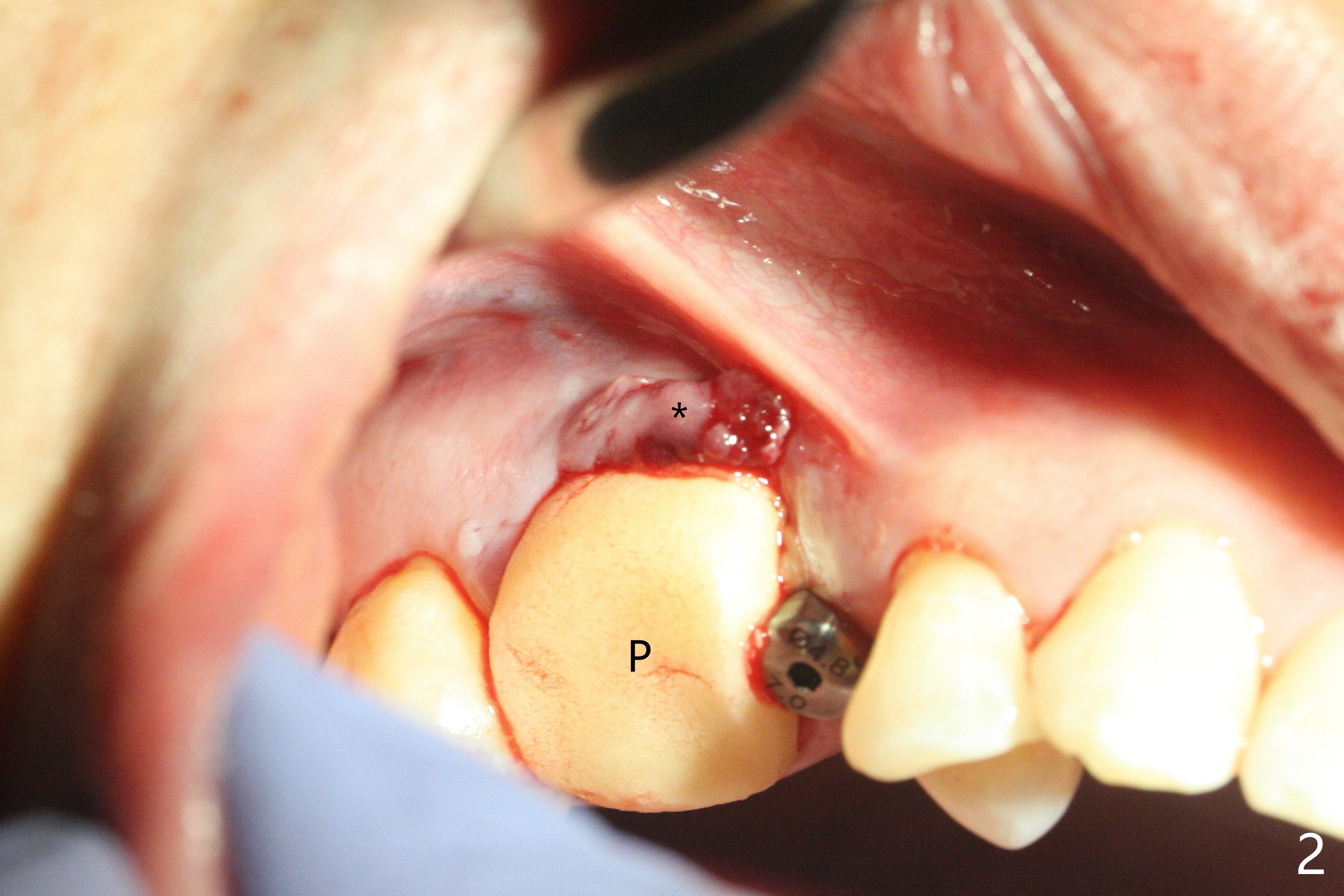
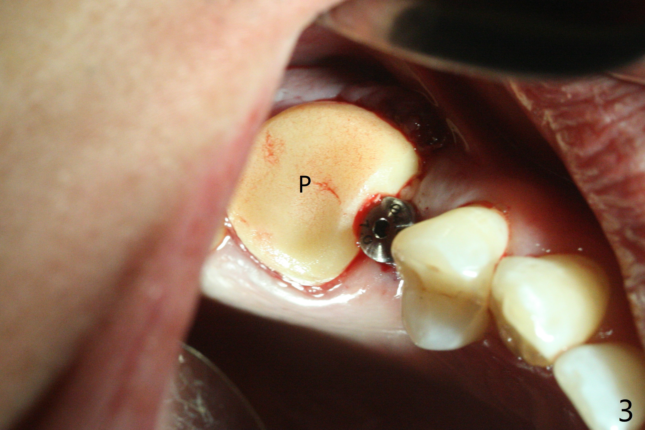
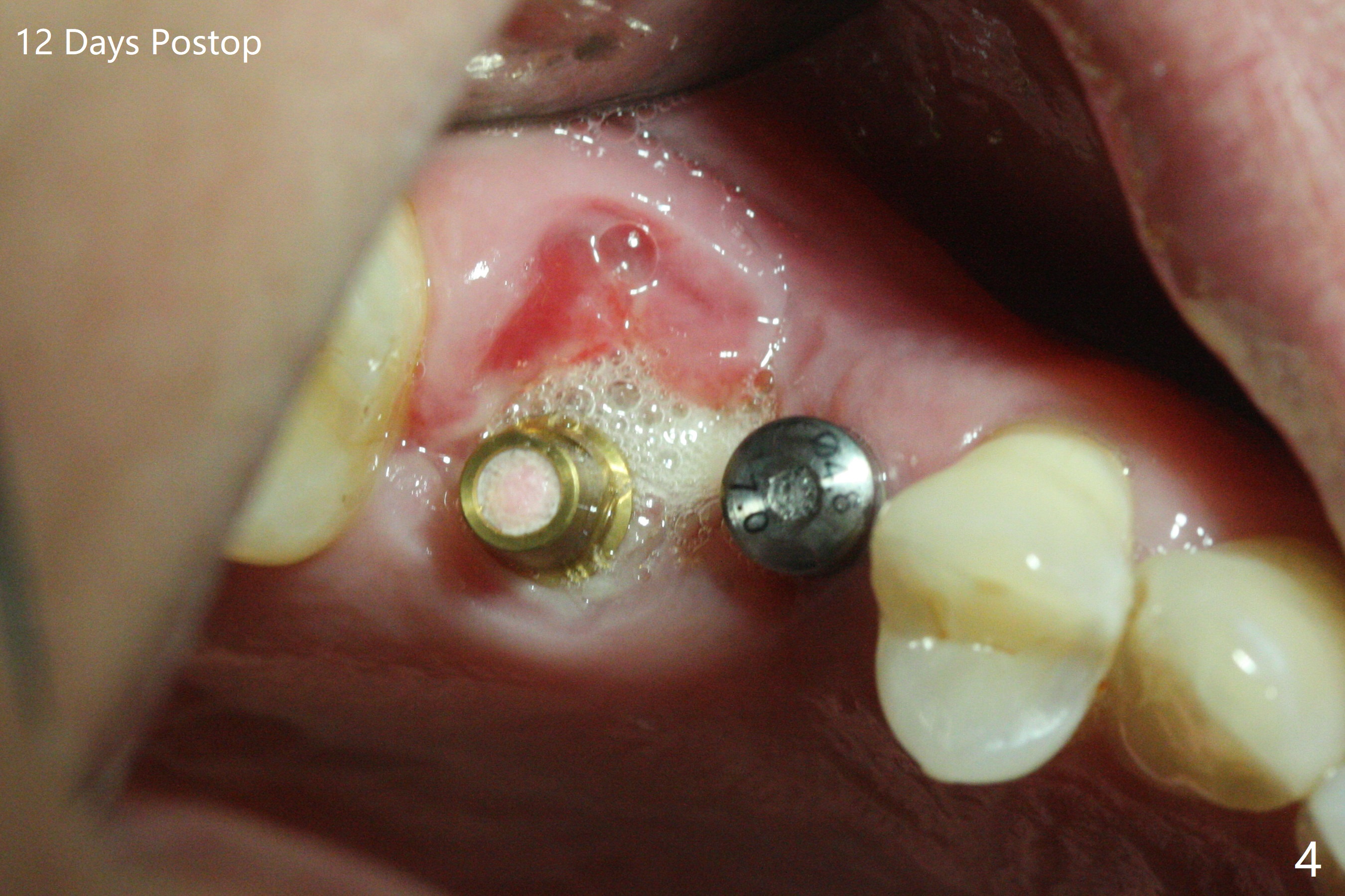
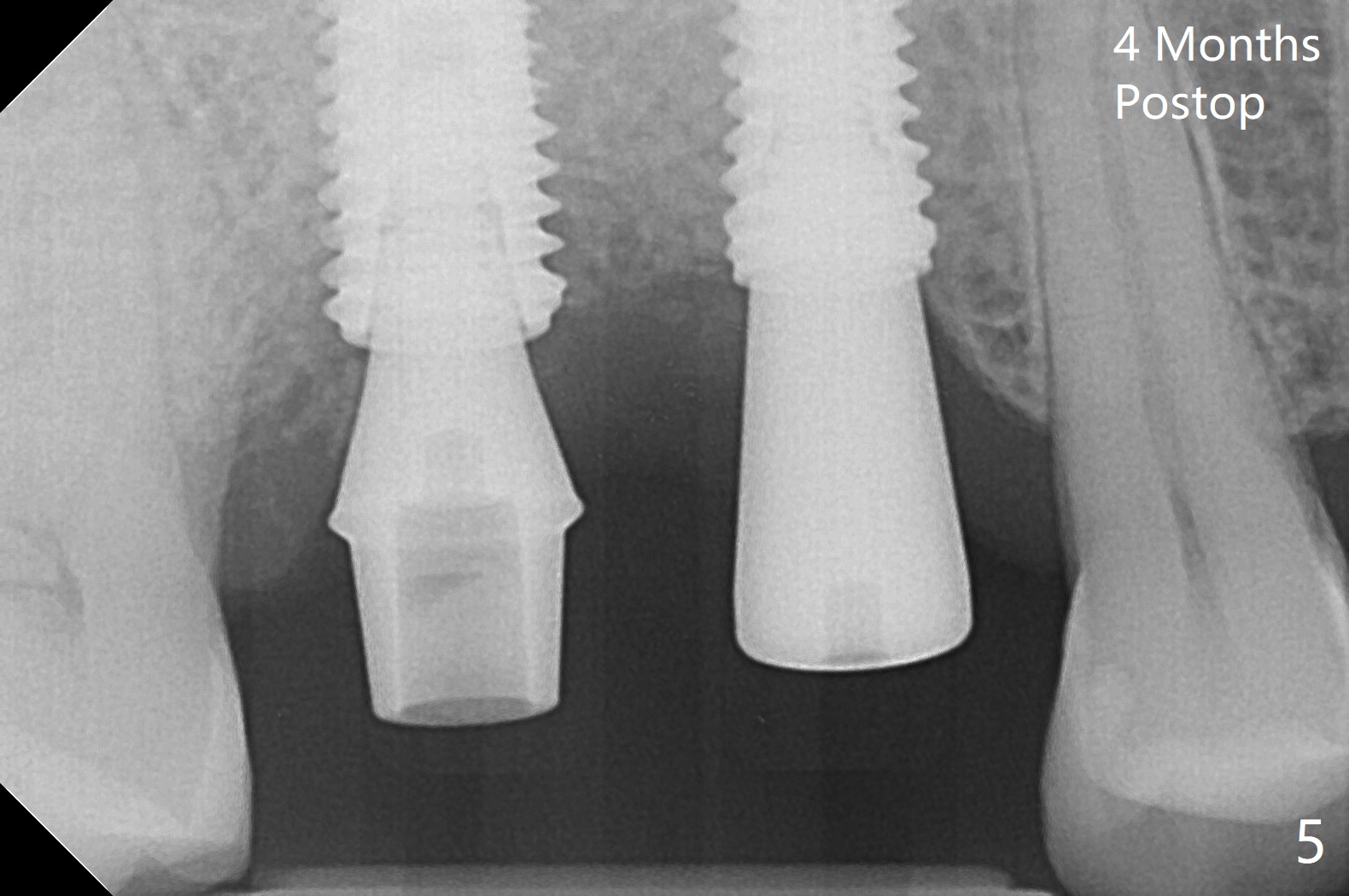
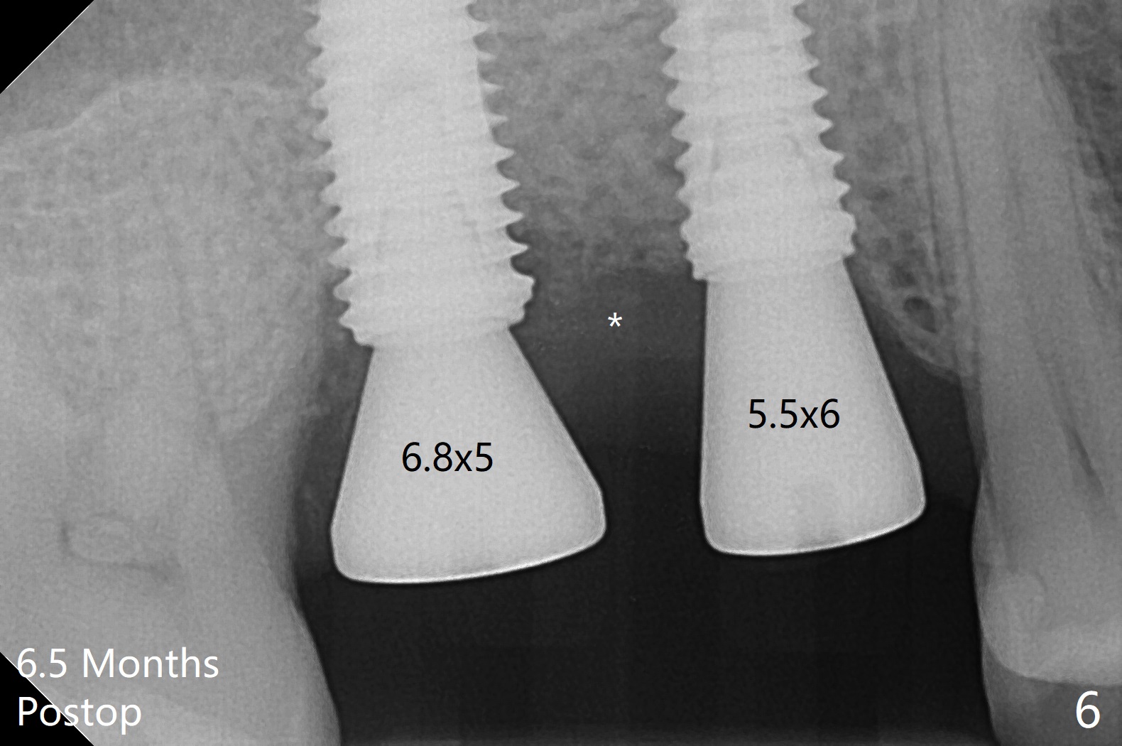
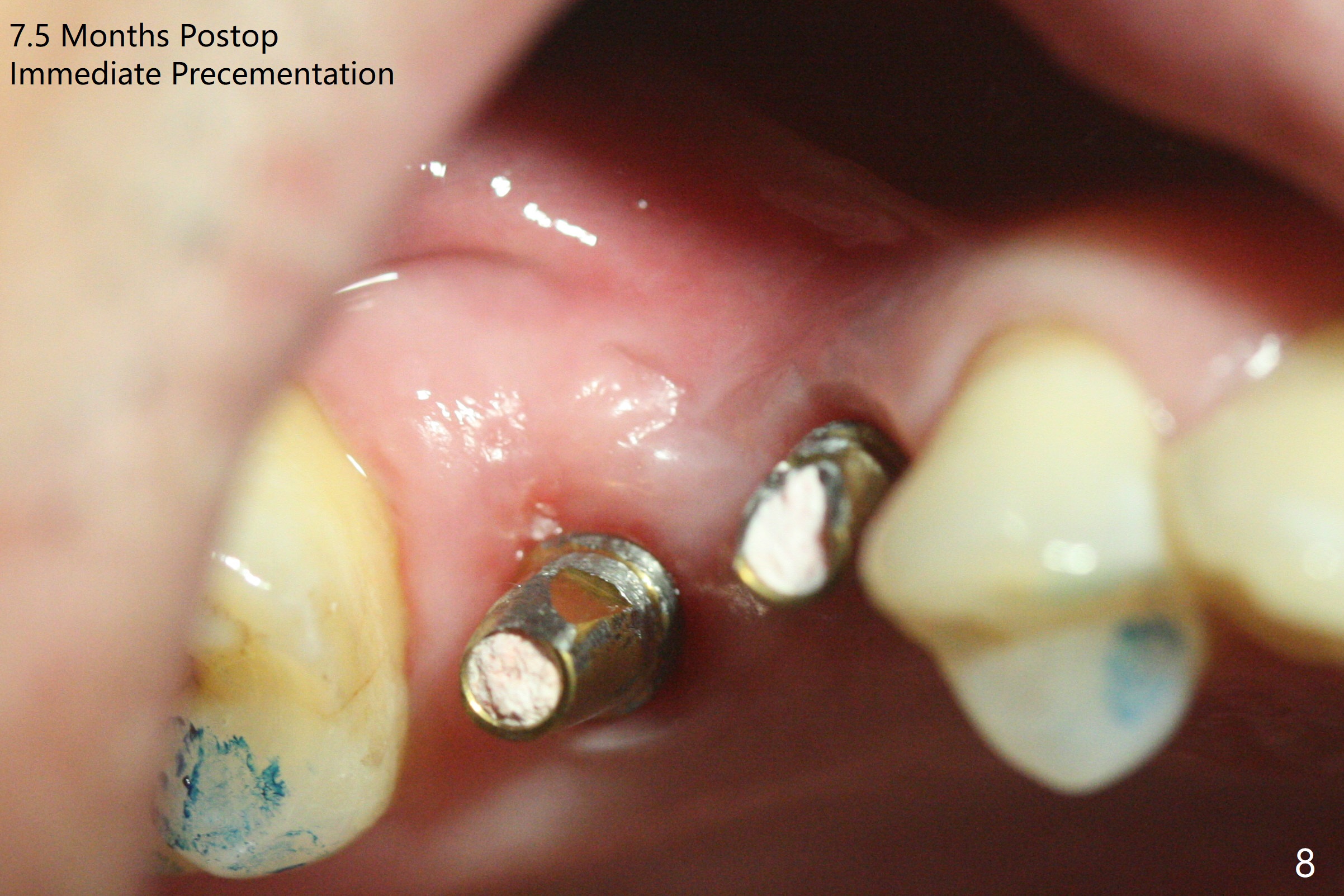
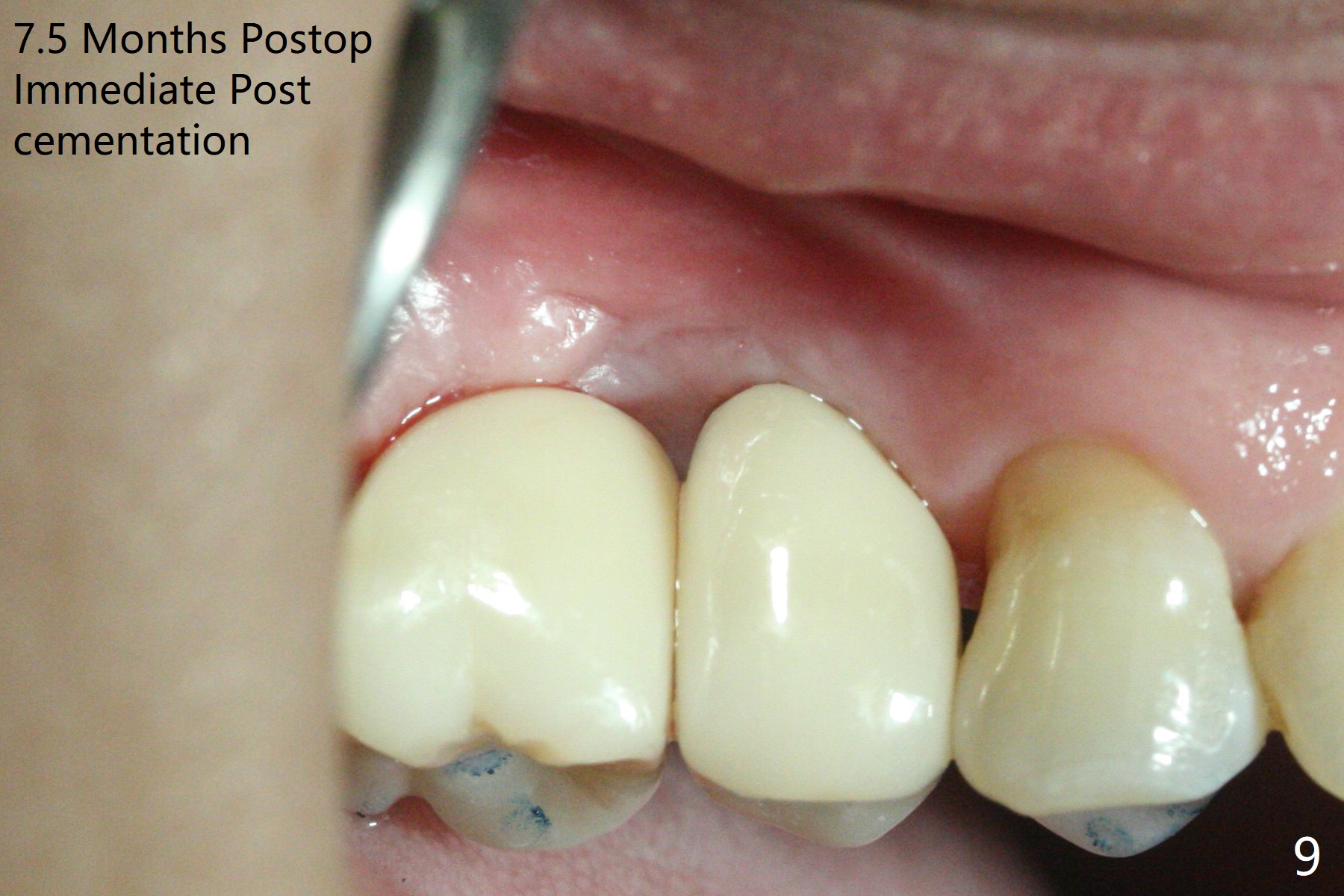
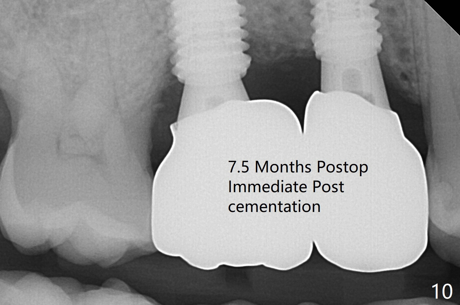
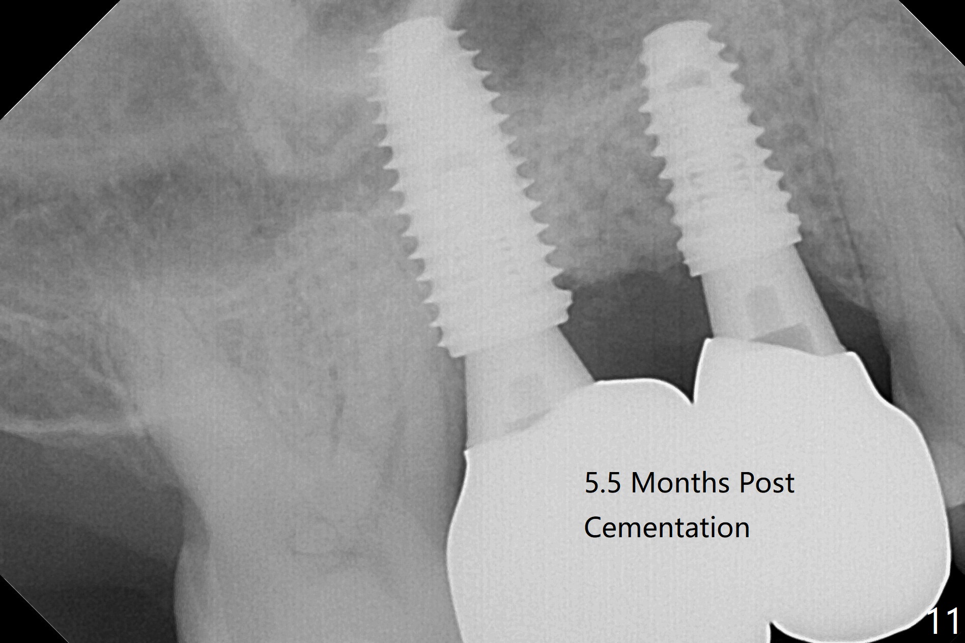
When the tooth #3 with severe buccal gingival recession is extracted and implants are placed at #3 and 4 with guide, sticky bone in 2 large pieces is packed between these implants with a previously large defect in a seemingly secure manner (Fig.1 *). Use of a longer implant at #3 (11.5 mm vs. 10 mm) will reduce the chance of abutment screw loosening in the future. With 2 pieces of PRF membrane coverage, an immediate provisional is fabricated for graft retention (Fig.2,3 P). To stabilize a buccal flap (Fig.2 *, used to be buccal furca gingiva), periodontal dressing is applied later. The buccal socket heals 12 days postop (Fig.4). Apparently new bone forms between the implants 4 months postop (Fig.5). The abutment at #3 may not be completely seated. In fact it is loose, probably related to buccal gingival and gingival cuff erythema 6.5 months postop (Fig.6). Large healing abutments are placed to form the interdental (interimplant) papilla without effect (*). Provisional crowns will be fabricated for the papilla formation. When cemented abutments are placed (Fig.7), papilla formation by manipulation of provisional crowns seems unlikely. Impression is taken. Although there is no implant thread exposure, the buccal plate is concave 7.5 months postop (Fig.8). With special crown design, food impaction should be minimal post cementation (Fig.9,10). There is no bone loss 5.5 months post cementation (Fig.11), while the soft tissue is healthy (data not shown).
Return to Upper Molar Premolar Immediate Implant, Prevent Molar Periimplantitis (Protocols, Table)), Trajectory Augma for Space Webinars 10 开场白
Xin Wei, DDS, PhD, MS 1st edition 07/19/2019, last revision 12/28/2020