|
|
|
|
|
|
|
|
|
|
|
|
|
|
|
|
|
|
|
|
Is This Implant Placed Low Enough?
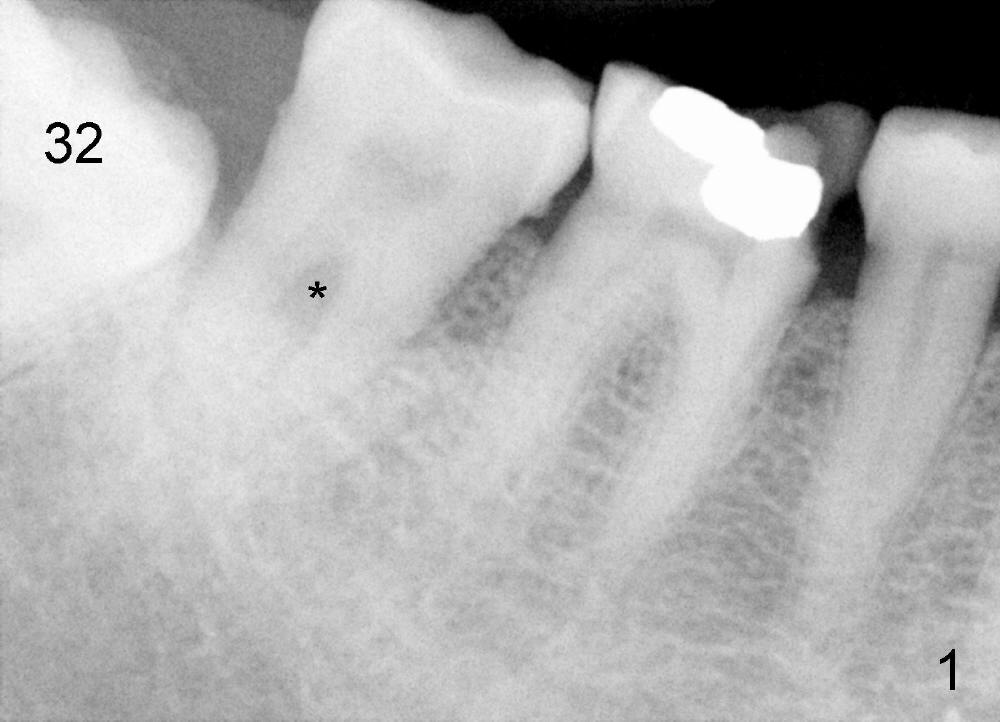
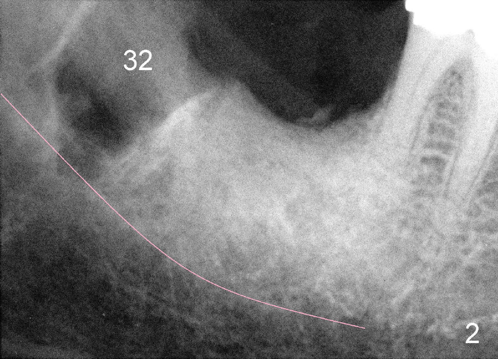
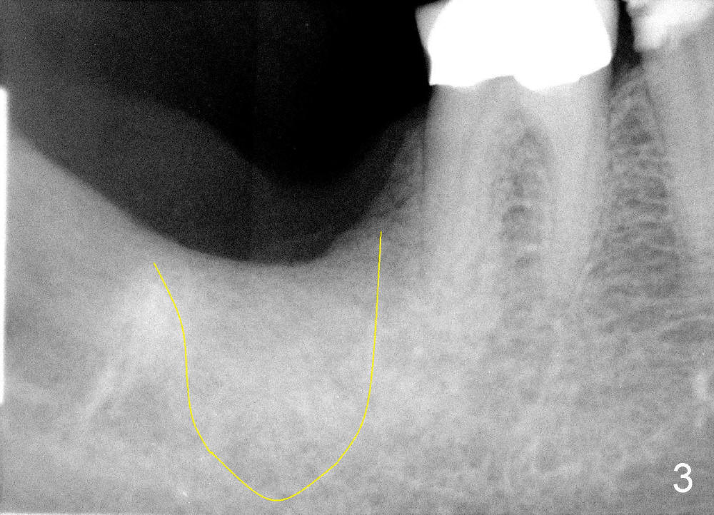
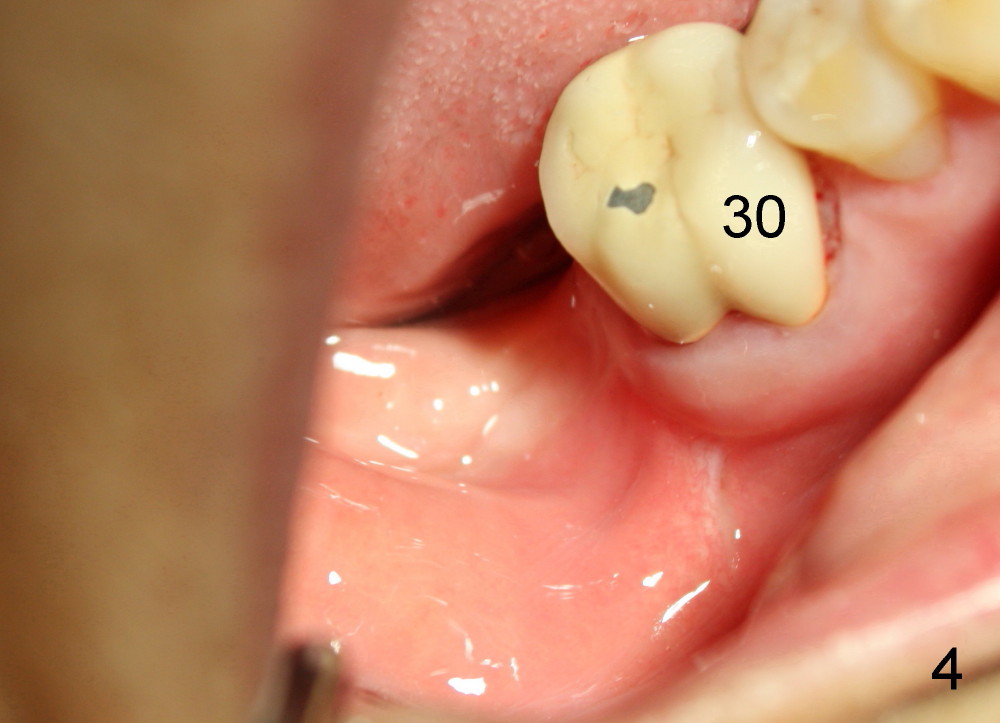
Mrs. Hwang in her sixties has had periodontal disease in her lower right 2nd molar (Fig.1 *). Three years later, both the 2nd and 3rd molars are extracted (Fig.2; pink: the upper border of the inferior alveolar canal). Four months after extraction, the patient returns for implant placement at the site of #31 (Fig.3, 4). The socket of the 2nd molar is outlined in yellow in Fig.3.
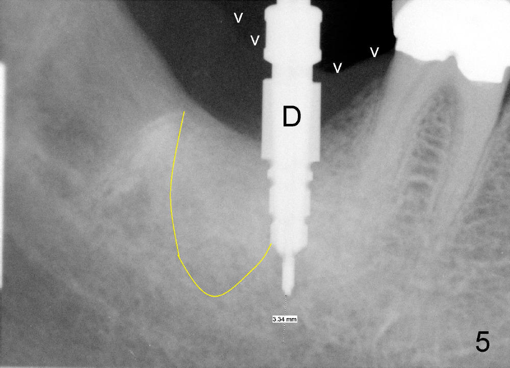
An intraop PA is taken with 4x14mm tapered drill in place (Fig.5 D). It appears that the osteotomy is deviated to the mesial portion of the healed socket (yellow outline). While the top of drill is at the gingival level (arrowheads), the tip of drill is 3.34 mm from the inferior alveolar nerve.
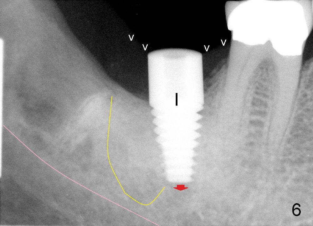
A postop PA is taken immediately after 6x14 mm tapered implant is placed (Fig.6 I). The top of the implant remains at the gingival level (white arrowheads). The implant is later further torqued down about 0.5 mm (red arrow).
The 2nd molar has notoriously short clinical crown (height). Presumably there will be a lot of grinding of the unipost placed upon the implant for this case. Or we will need to cut off the top of the unipost precisely. This patient will return for unipost placement and crown restoration next week, four months after implant placement. We will report what is going to happen. Shorter abutments may have their application in our daily practice, such as narrow vertical dimension (this case), supraeruption and young guys (1,2). We do not have to trim or cut the unipost. Implant dentistry has much more fun and less risk, including wetting my pants!
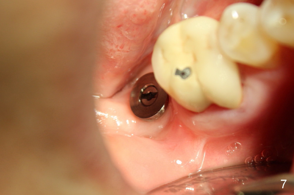
Today the patient returns for implant restoration 4 months after placement. The tissue around the implant is healthy. It appears that there is plenty of clearance (Fig.7). How much I wish no gross reduction!
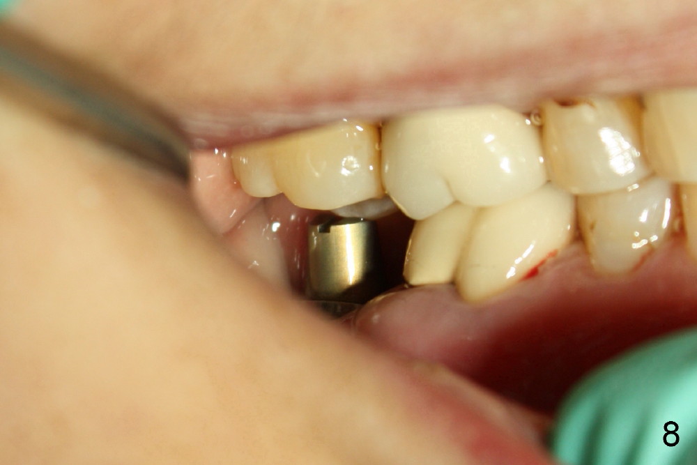
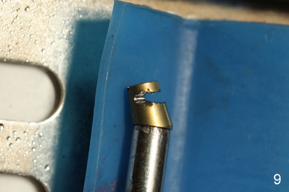
When a 4.5 mm 20 degree short unipost is engaged into the implant, it touches the opposing tooth (Fig.8). The unipost has to be cut (Fig.9). It takes at least 5 minutes to finish. Finally there is enough clearance for crown (Fig.10). This is my first experience to cut the unipost. The good thing is that I learn the shortened unipost does not seem to affect retention of crown.
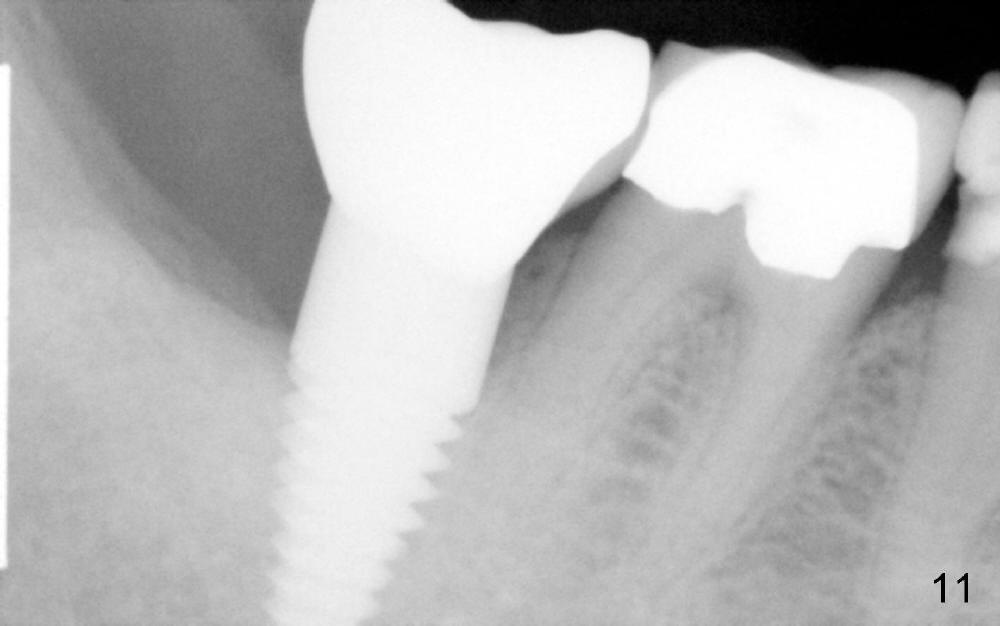
X-ray taken six months after cementation shows that there is no bone loss around the implant (Fig.11).
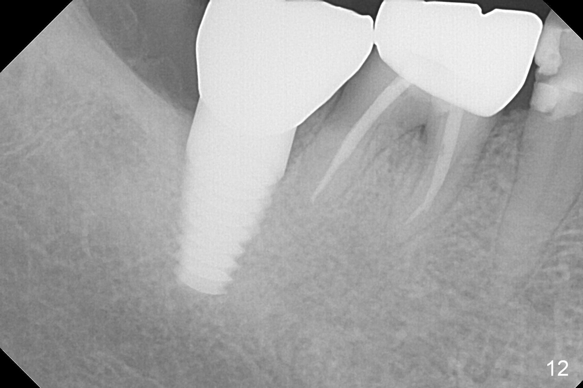
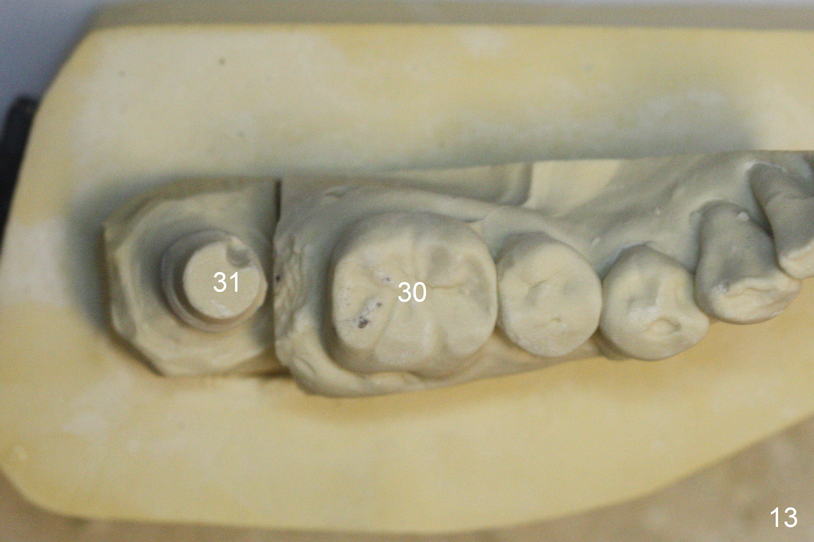
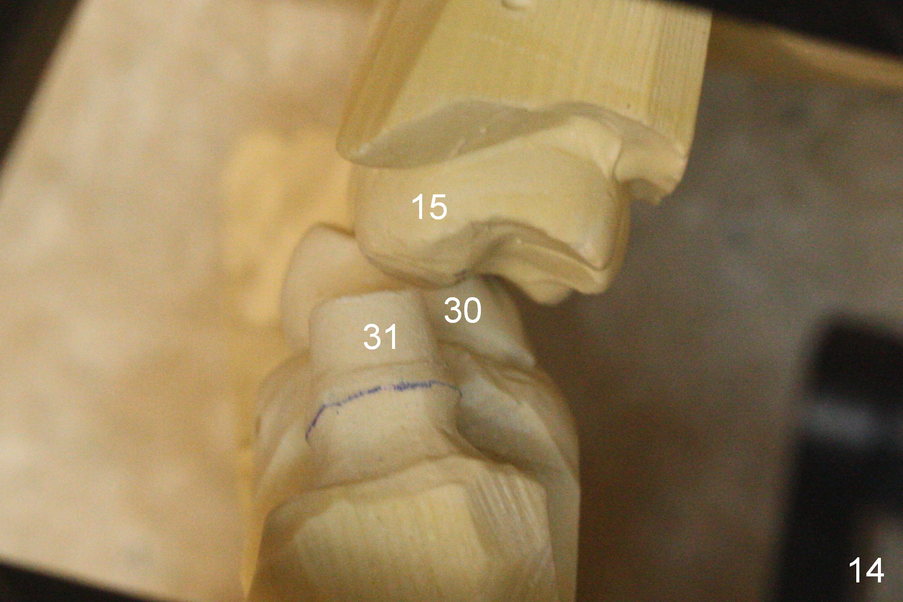
Three years 8 months post cementation, the crown dislodged. A 5x3 mm 0 ° unipost is resin bonded. Retrospectively the angled abutment appears not necessary (Fig.13 (occlusal view), 14 (posterior view)). Fig.12 is taken immediately post cementation of redo crown. There is no bone loss.
Xin Wei, DDS, PhD, MS 1st edition 05/25/2013, last revision 02/02/2017