|
|
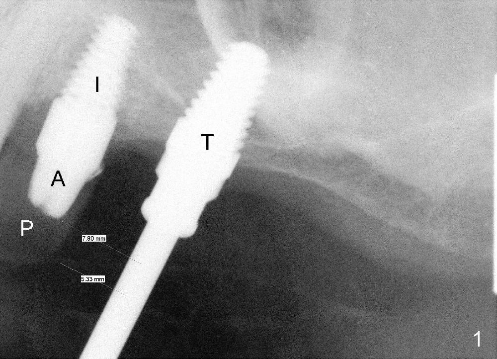
Fig.1: Intraop PA helps determine the position and trajectory of the osteotomy at the site of #14 (tap, T), relative to the neighboring tooth (implant (I), abutment (A) and provisional (P) at the site of #13) mesiodistally. In fact the X-ray is taken while a surgical stent (suction down, transparent radiographically) in place. One of the measurements, 5.33 mm, is from the distal surface of the immediate provisional to the center of the osteotomy so that the future molar tooth will be approximate 10 mm wide mesiodistally, which is ideal restoratively. The 5x11 mm tap is stable in bone. To achieve better primary stability, a 6x11 mm is to be inserted before placing 6x11 mm implant (bone height).
Return to Sinus Lift & Implantation
Xin Wei, DDS, PhD, MS 1st edition 04/19/2013, last revision 09/15/2014