|
|
|
|
|
|
|
|
|
|
|
|
|
|
Simultaneous Sinus Graft and Implantation
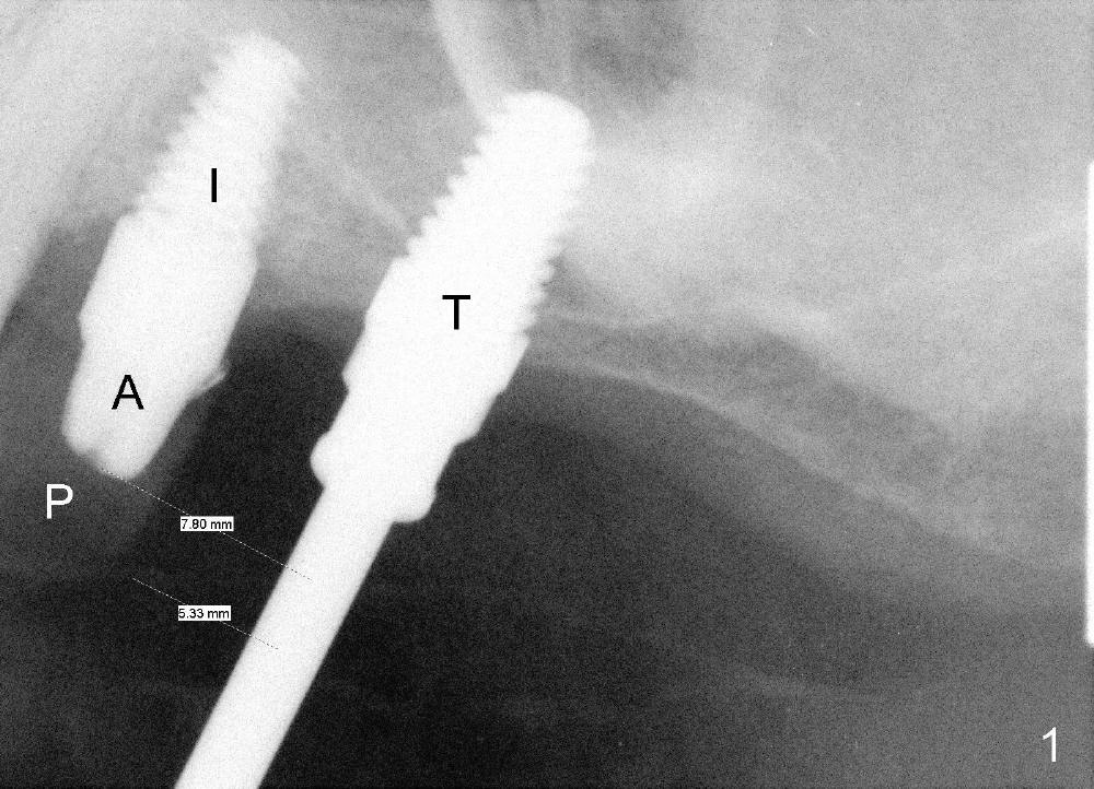
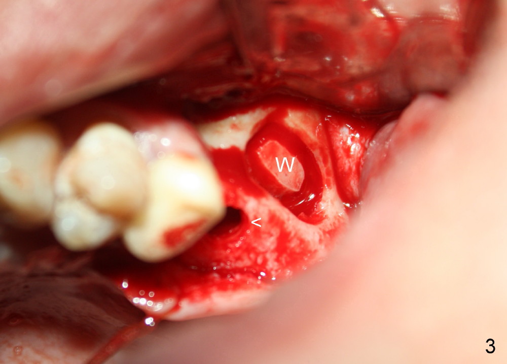
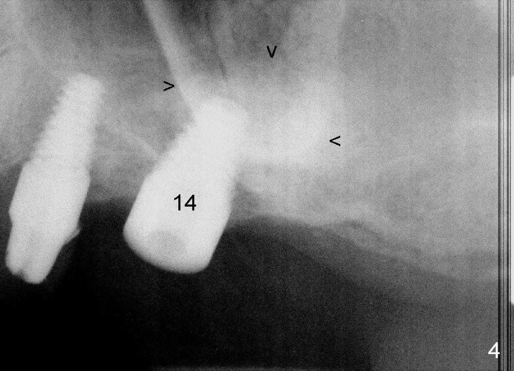
A lateral sinus window is created with a round diamond bur in high speed handpiece with copious irrigation as planned (Fig.3 W). The sinus membrane is lifted without tear. With the membrane protected with 2x2 gauze, an osteotomy (Fig.3 <) is developed at the site of the tooth #14 with 2 mm pilot drill, 2.5-3.5 mm reamers, 5x11 mm tap (Fig.1: T) and 6x11 mm tap. A synthetic bone graft (Osteogen 300-400 micron) is placed in the sinus before placement of 6x11 mm implant (Fig.4: 14). More of the synthetic bone mixed with autogenous bone is added superficial to the apex of the implant inside the sinus. The flaps are sutured.
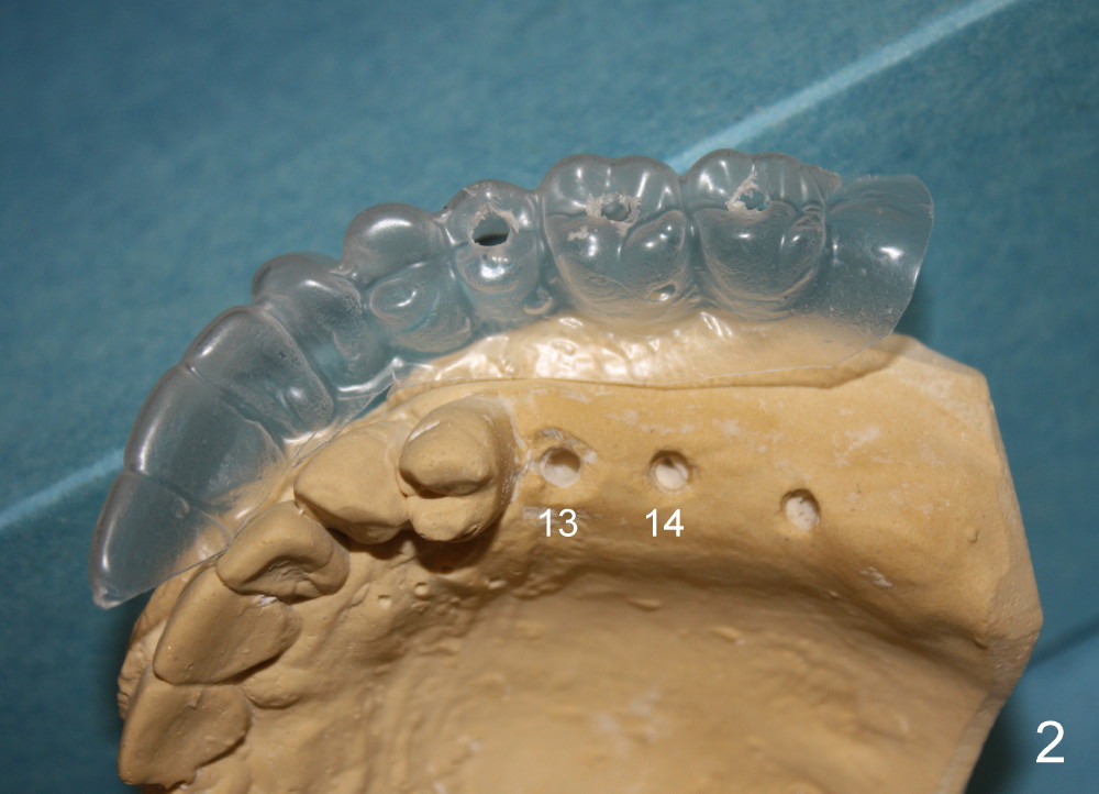
The position and trajectory of the osteotomy (Fig.1 T) and subsequently those of the implant (Fig.4: 14) are aided by a surgical stent, which is fabricated from the model (Fig.2).
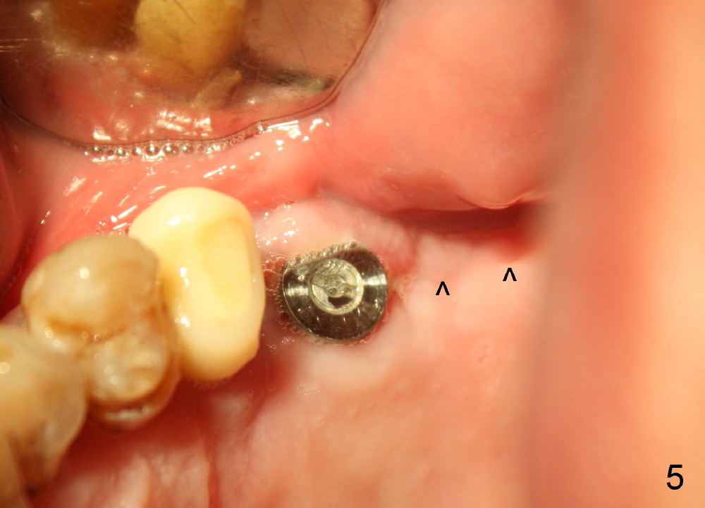
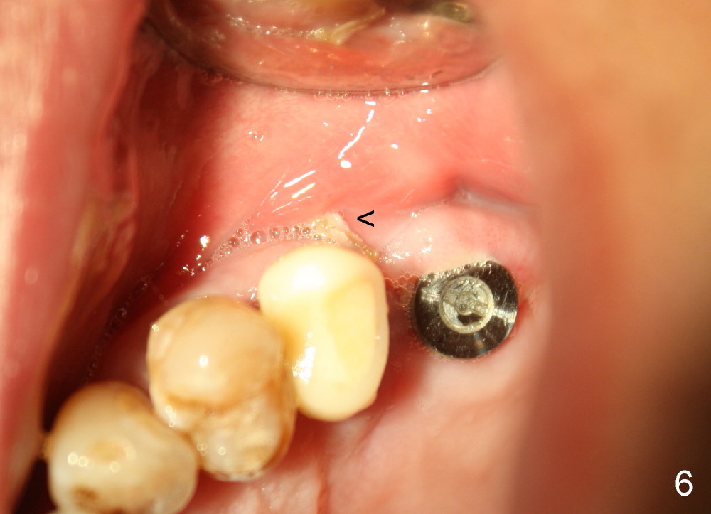
Although there is swelling 1-2 days postop, the wound is healing 8 days postop (Fig.5,6).
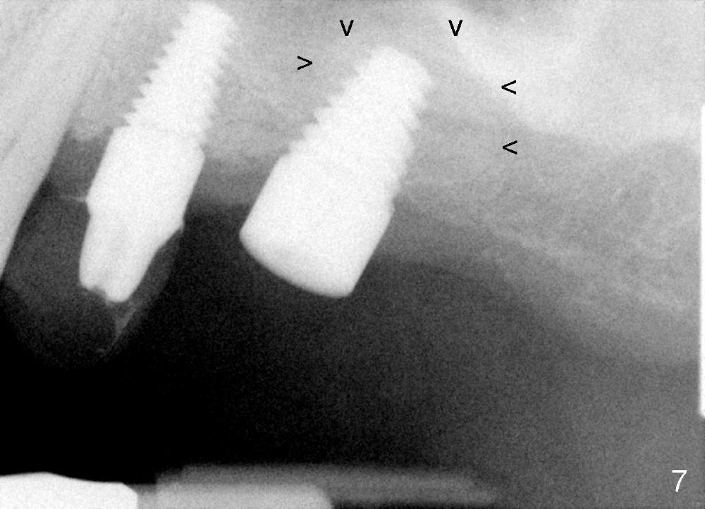
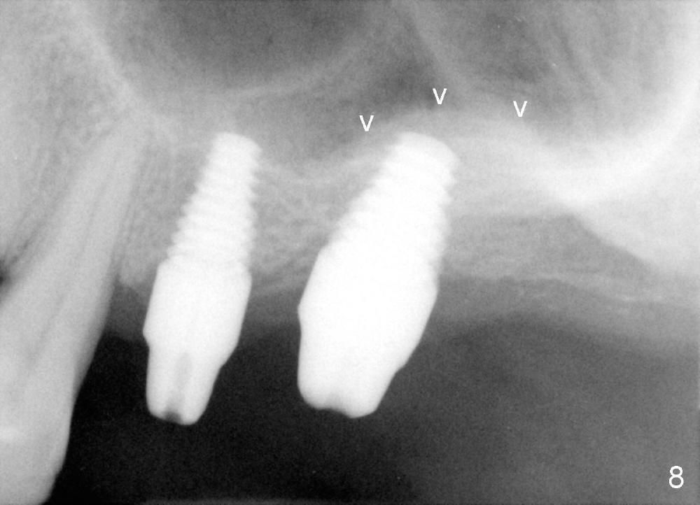
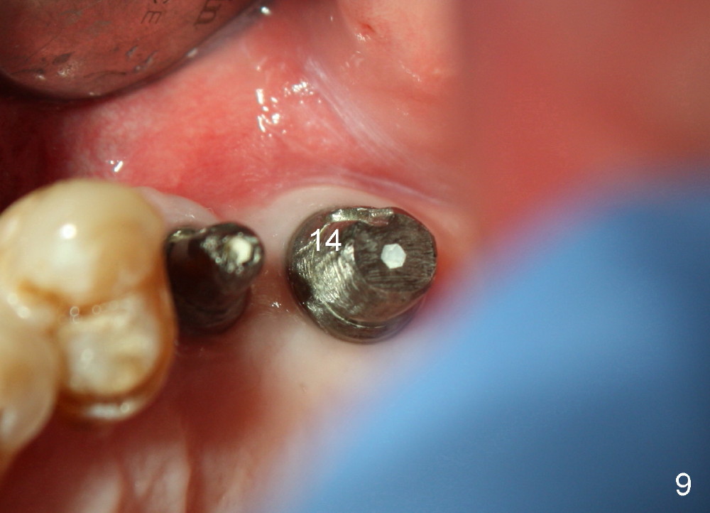
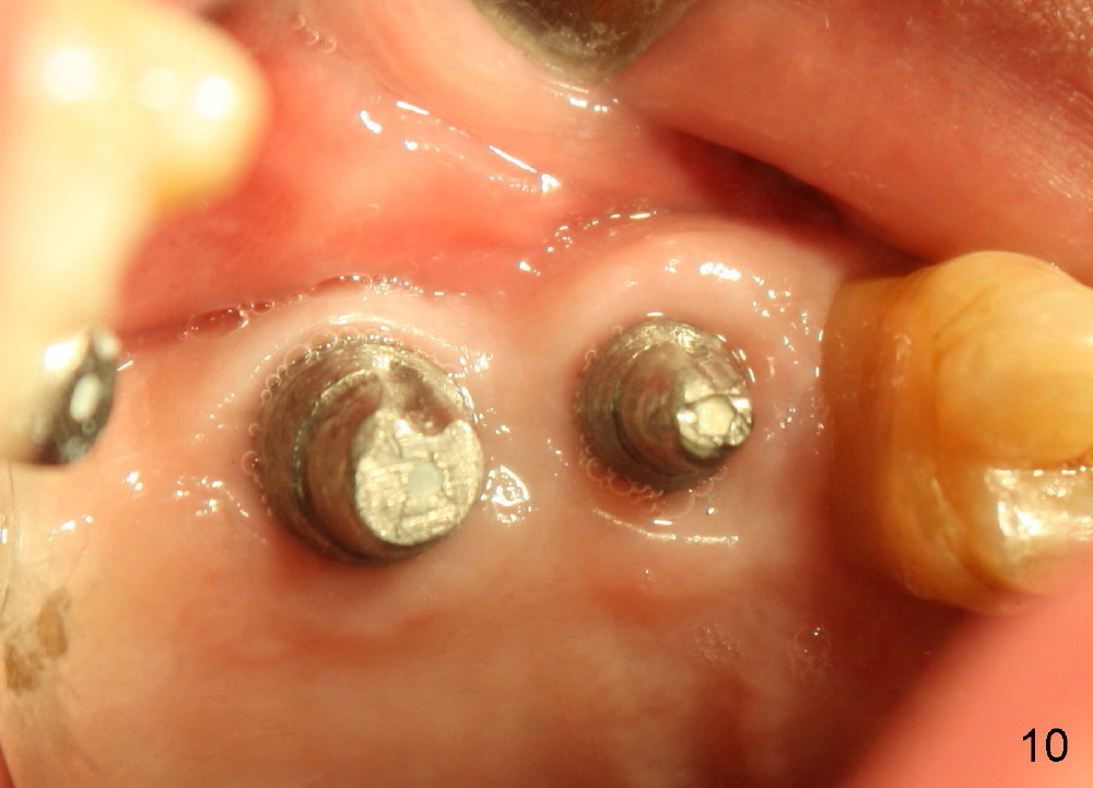
Bone graft remains in place 2 months postop (Fig.7 arrowheads); its density increses 4 months postop (Fig.8). The gingiva around the implant and abutment at the site of #14 is healthy (Fig.9 (4 months postop before final impression);10 (5 months postop immediately before final cementation)).
Return to Sinus Graft Introduction
Xin Wei, DDS, PhD, MS 1st edition 04/19/2014, last revision 09/20/2014