,%20VeraGraft.jpg)
|
|
|
|
|
|
|
|
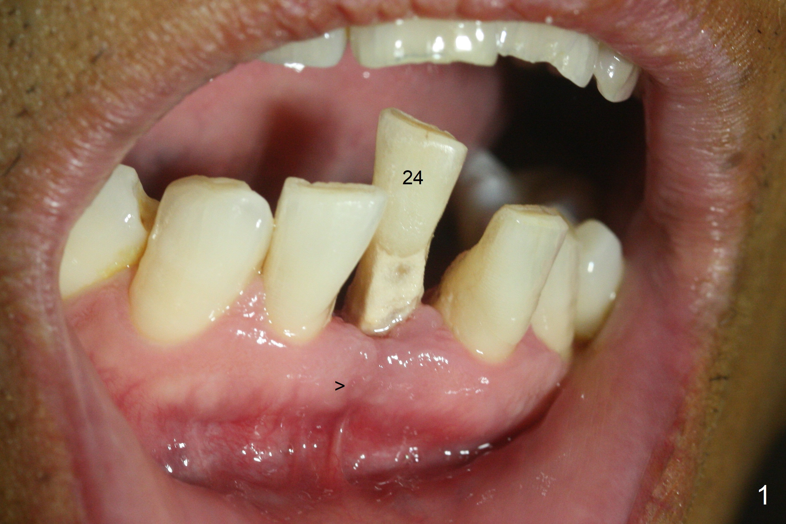
Loss of Buccolingual Plates
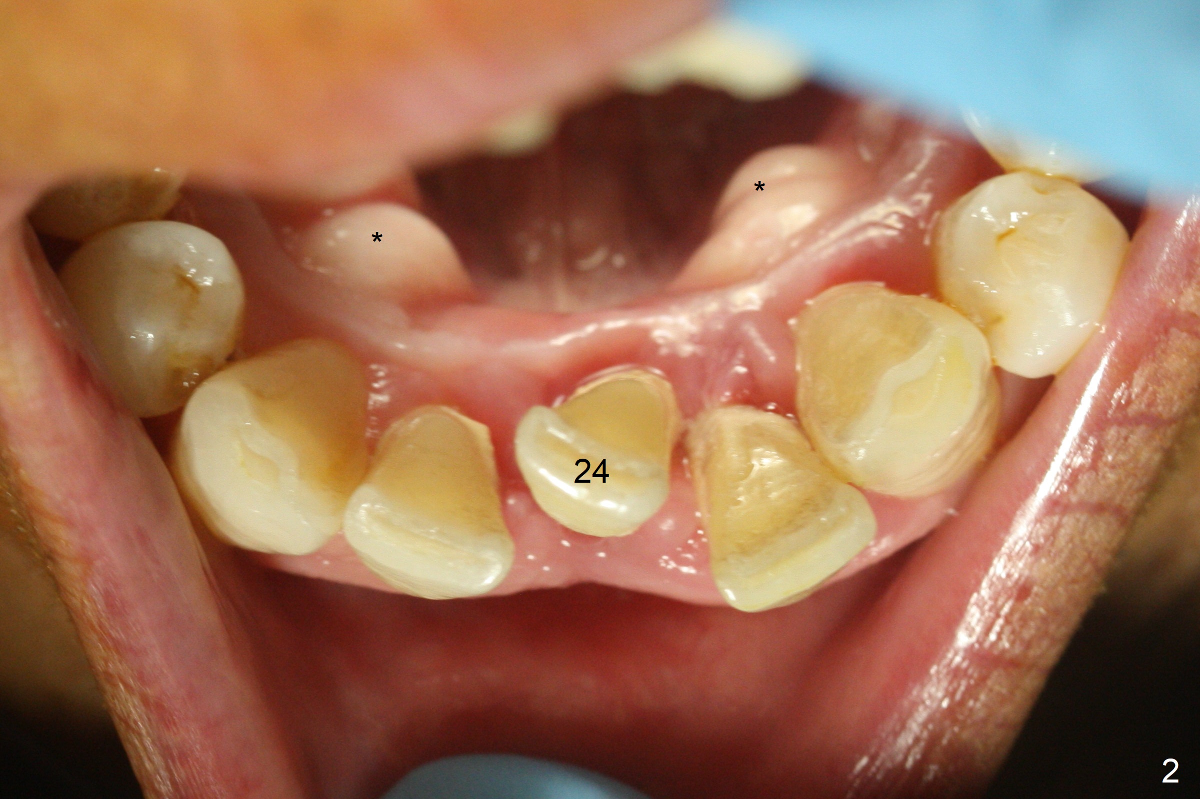
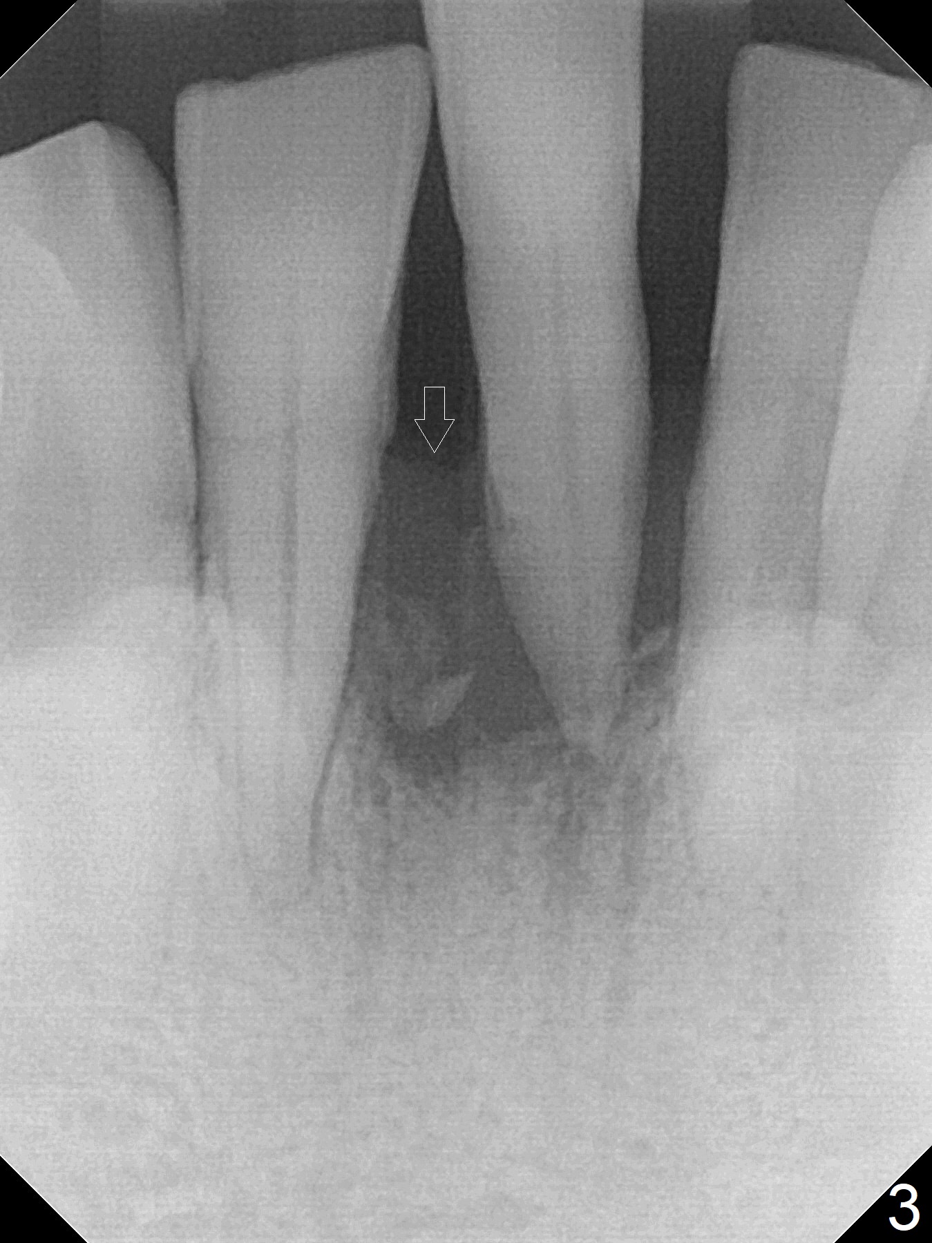
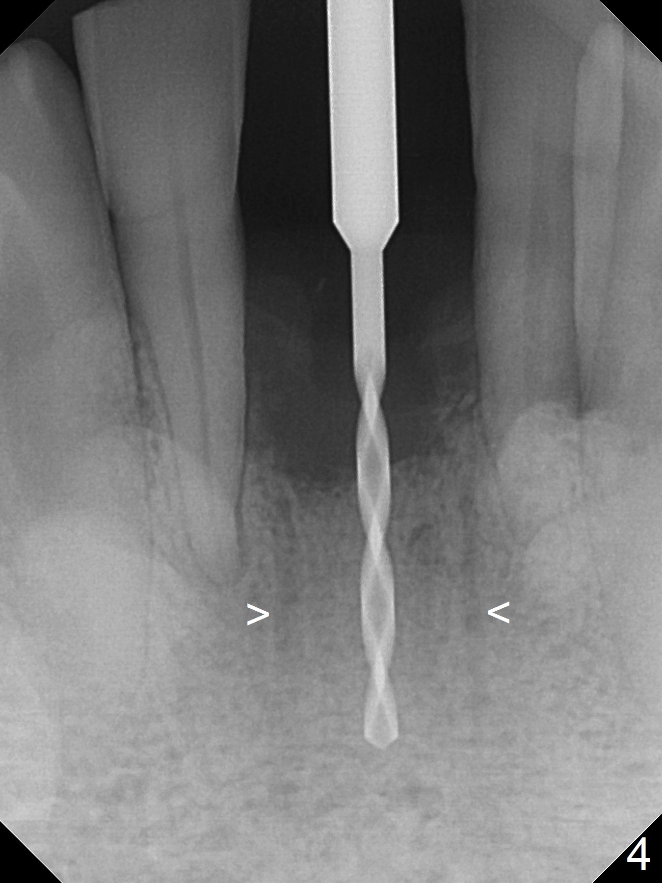
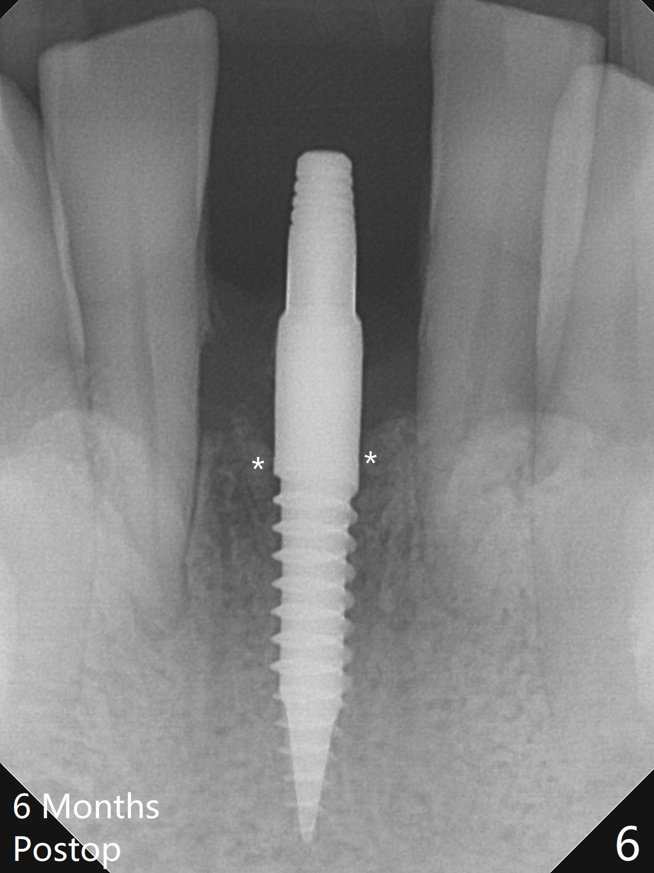
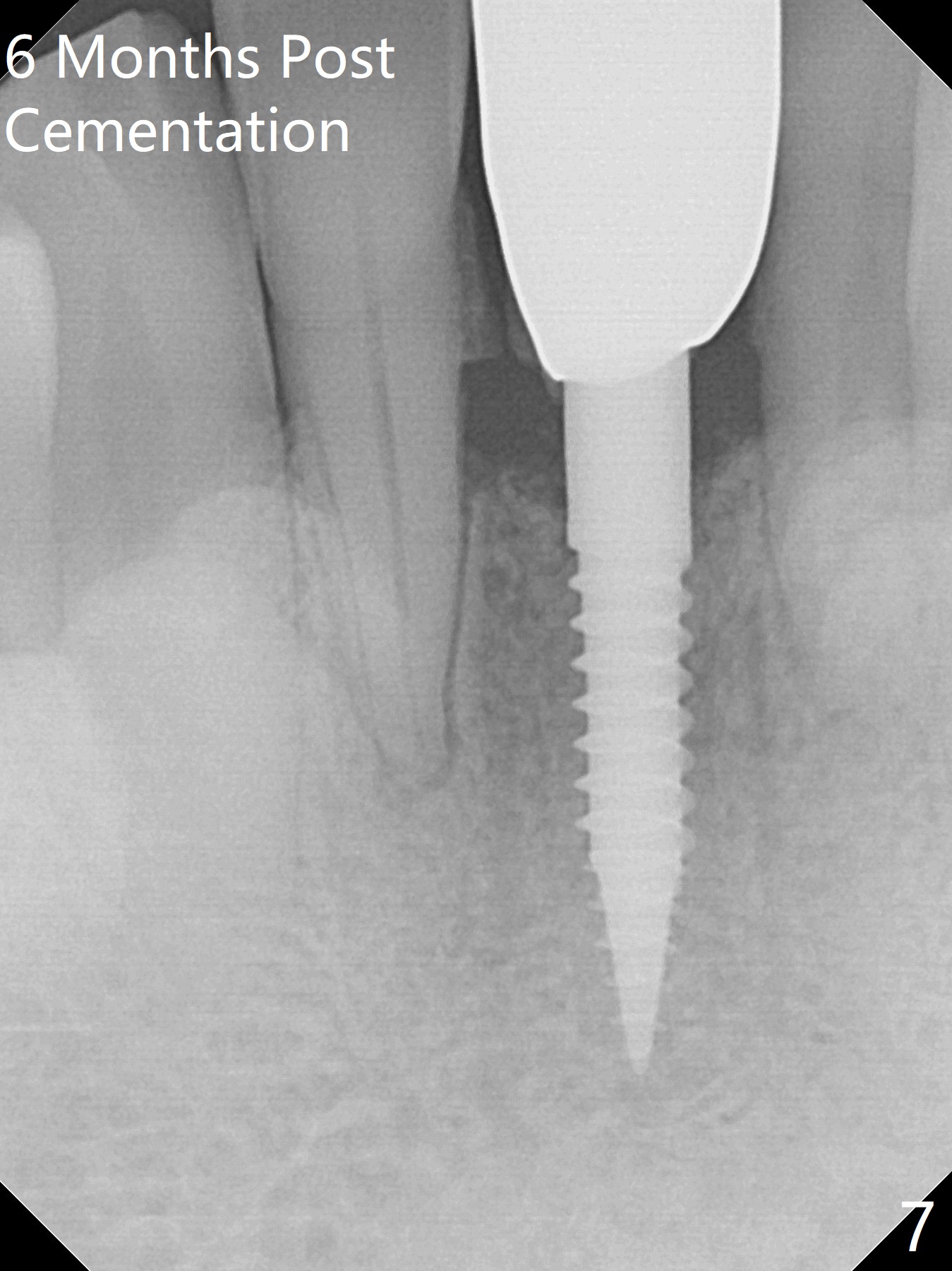
When the patient returns for implant placement 1.5 years after the last treatment (SRP), the tooth #25 has exfoliated, whereas the tooth #24 is severely displaced (Fig.1-3). In fact the buccolingual plates are found to be lost after extraction, corresponding to change in gingival color indicated by an arrowhead in Fig.1. Initial osteotomy with 1.2 mm drill is parallel to the terminal branches of the Incisive Canal (Fig.4 arrowheads). Since the gingiva is as thick as 7.5 mm (Fig.3 arrow), a 3x14 mm 1-piece implant with 4 mm cuff is placed (Fig.5); three implant threads are outside the native bone; with allograft (*) placed and the neighboring crests being coronal to the threads, the chance of periimplantitis should be remote. An immediate provisional is fabricated to contain the graft in place. Meanwhile the tooth #2 is symptomatic with crack. The top 3 threads appear to be contacted by the newly formed crestal bone 6 months postop (Fig.6 *). Bone appears to have grown into the space between implant threads 6 months post cementation (13 months postop).
Return to Lower Incisor Immediate Implant, IBS Xin Wei, DDS, PhD, MS 1st edition 08/05/2017, last revision 09/03/2018