
|
|
|
|
|
|
|
|
|
|
|
|
|
|
|
|
|
|
|
|
|
|
|
BEB for Narrow Molar Ridge
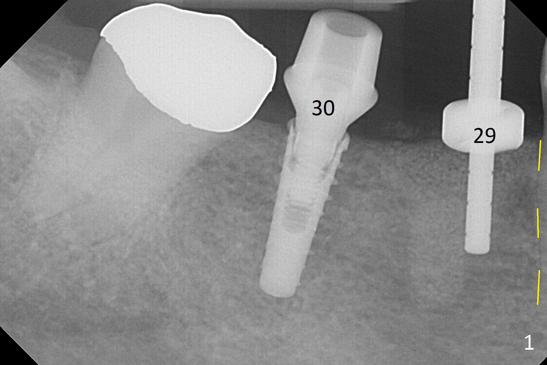
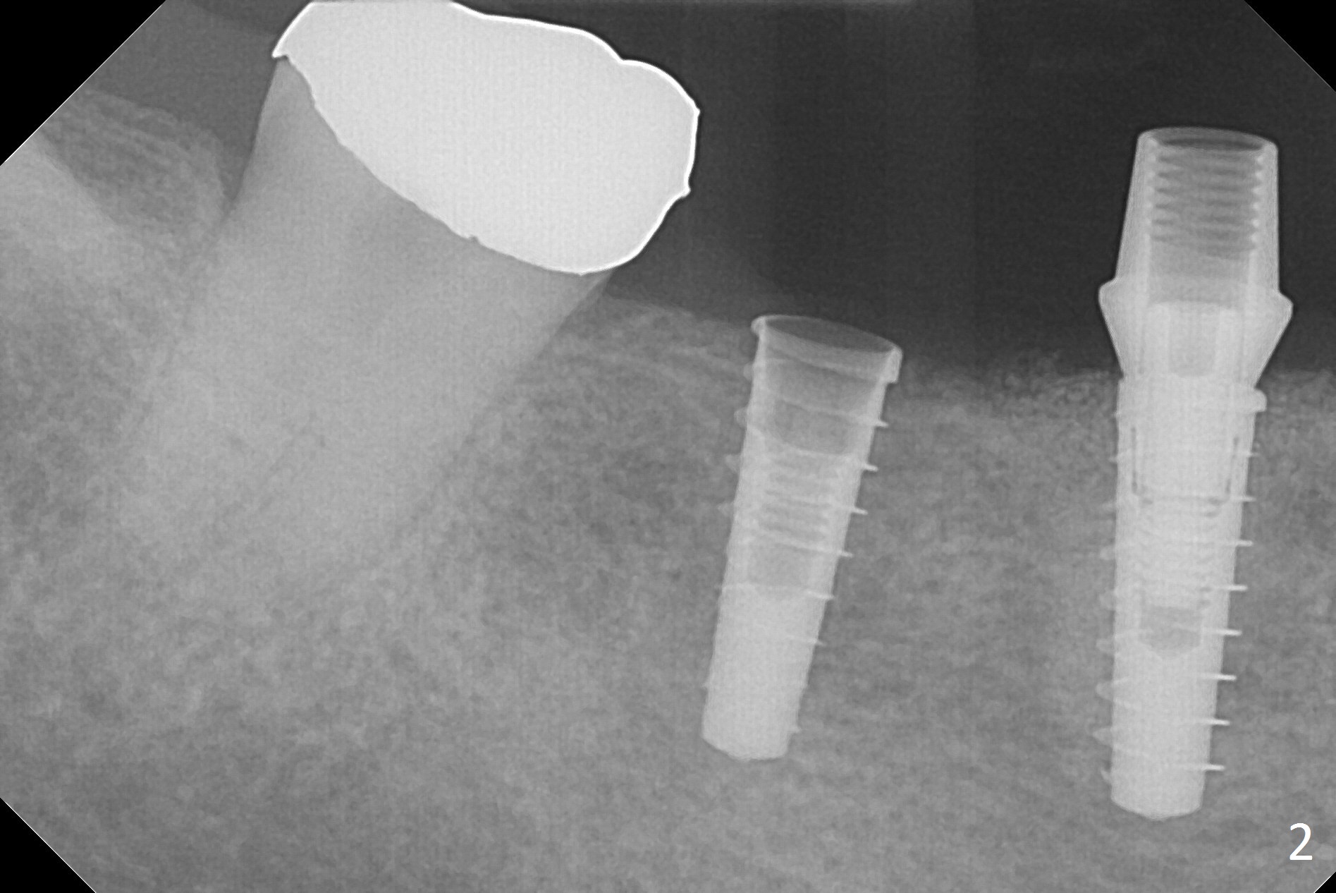
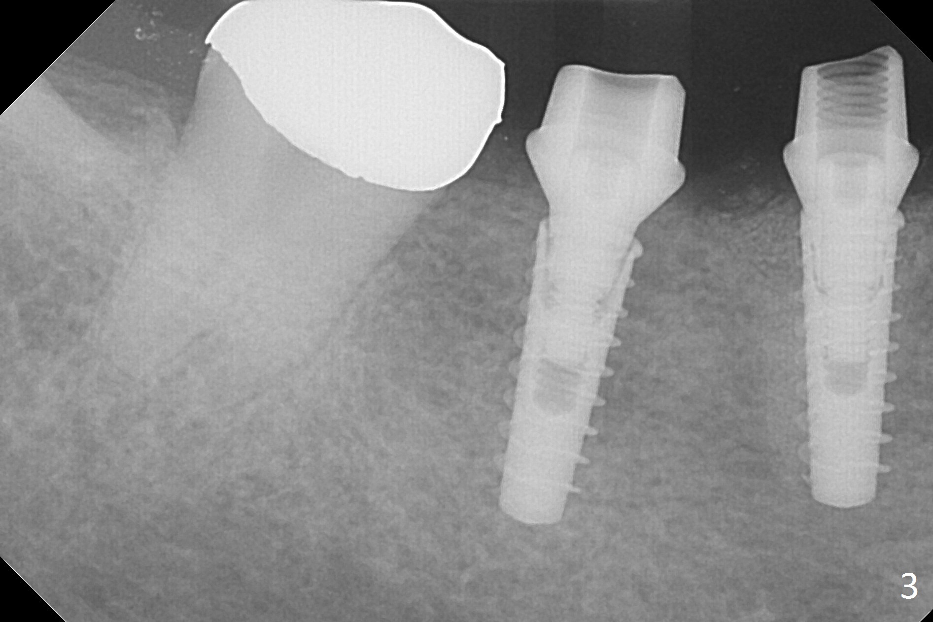
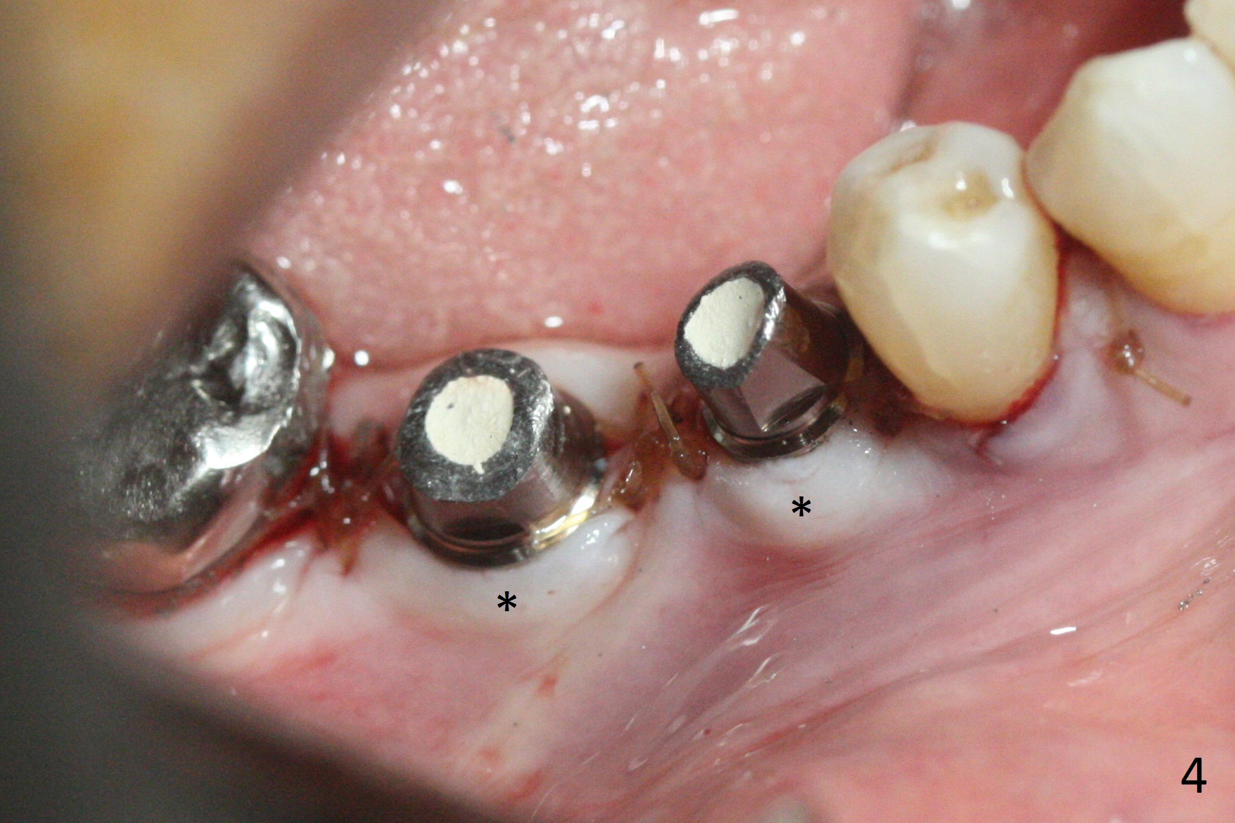
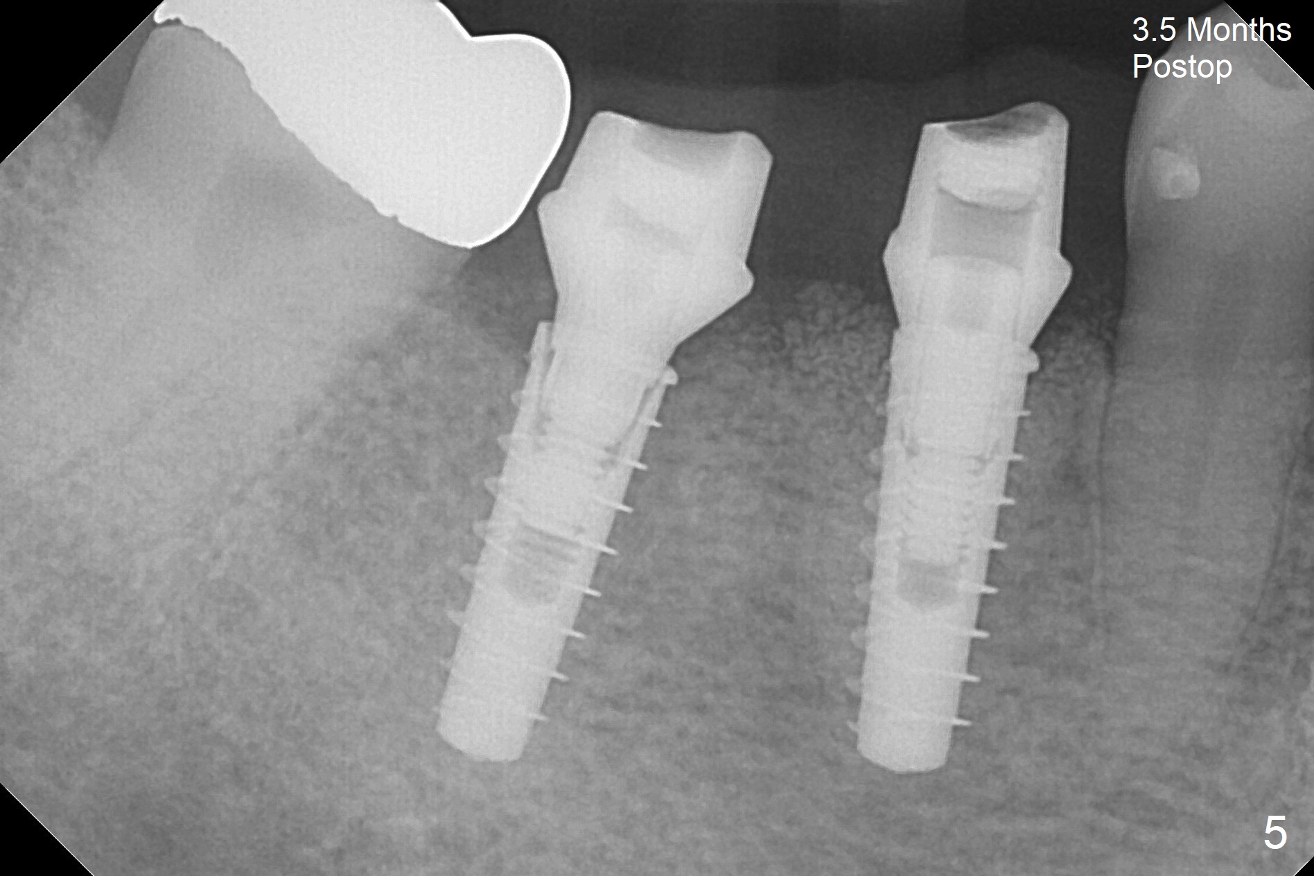
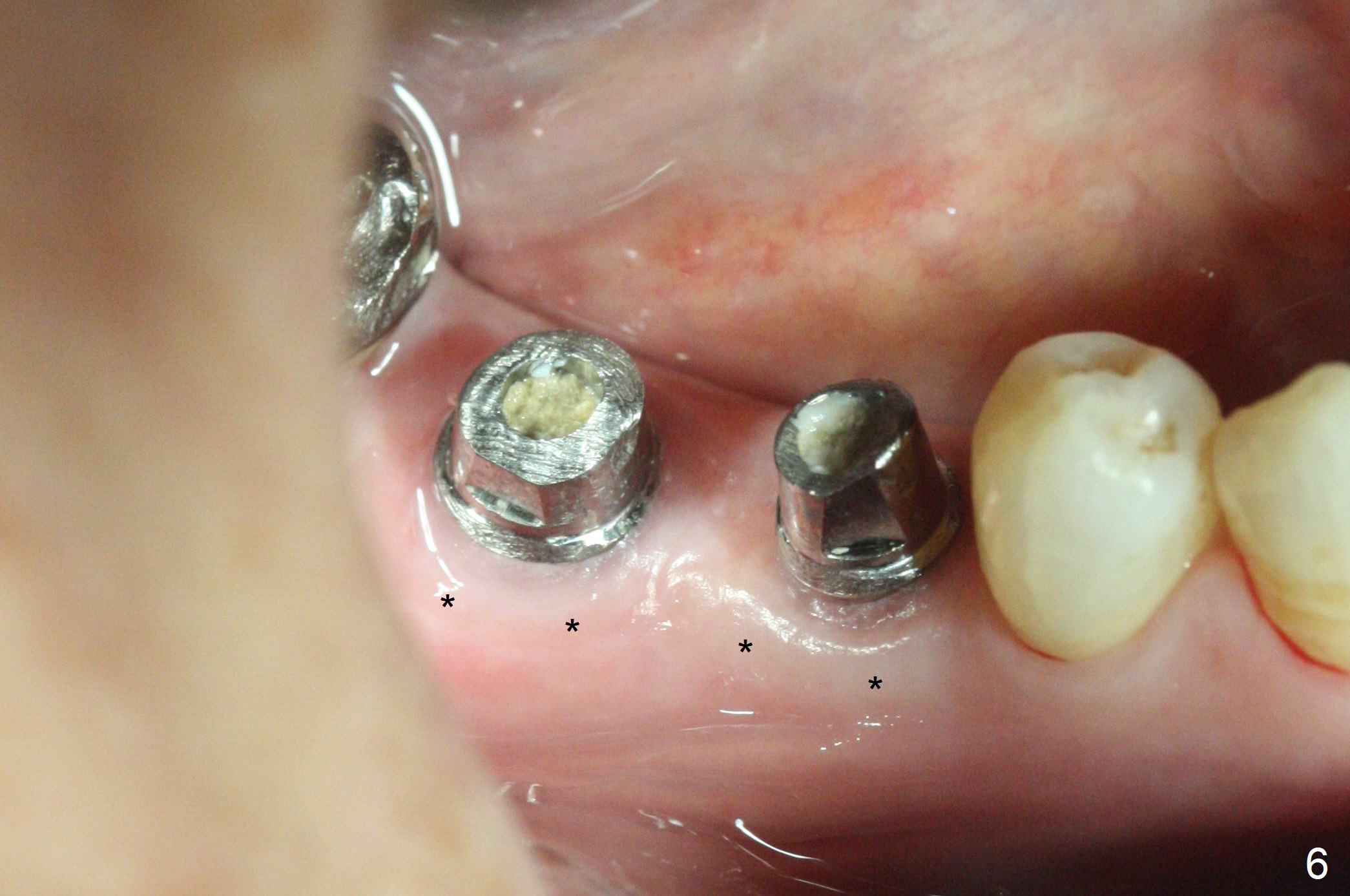
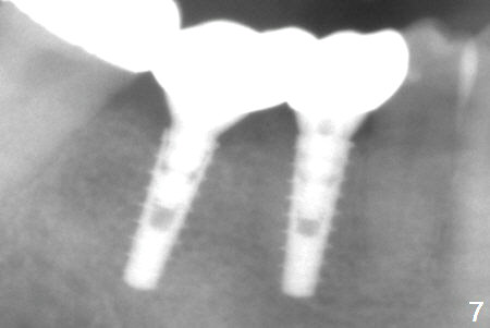
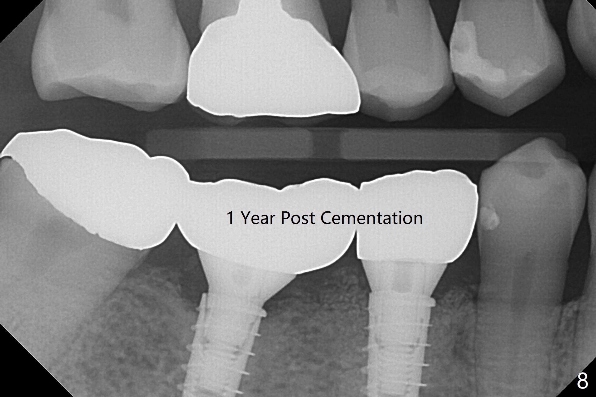
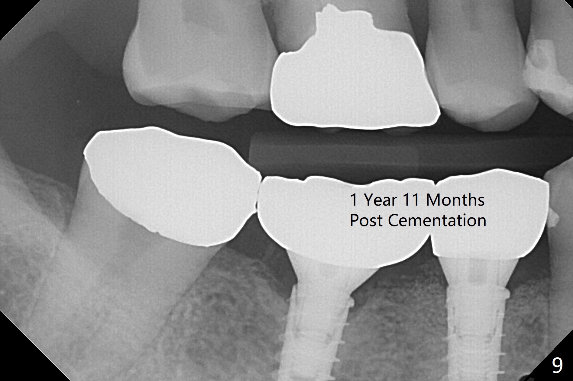
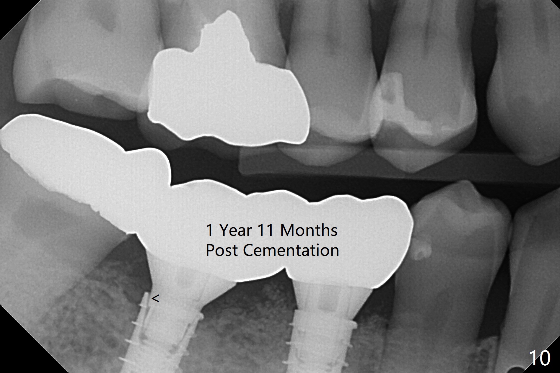
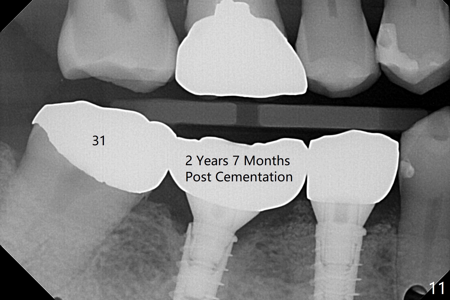
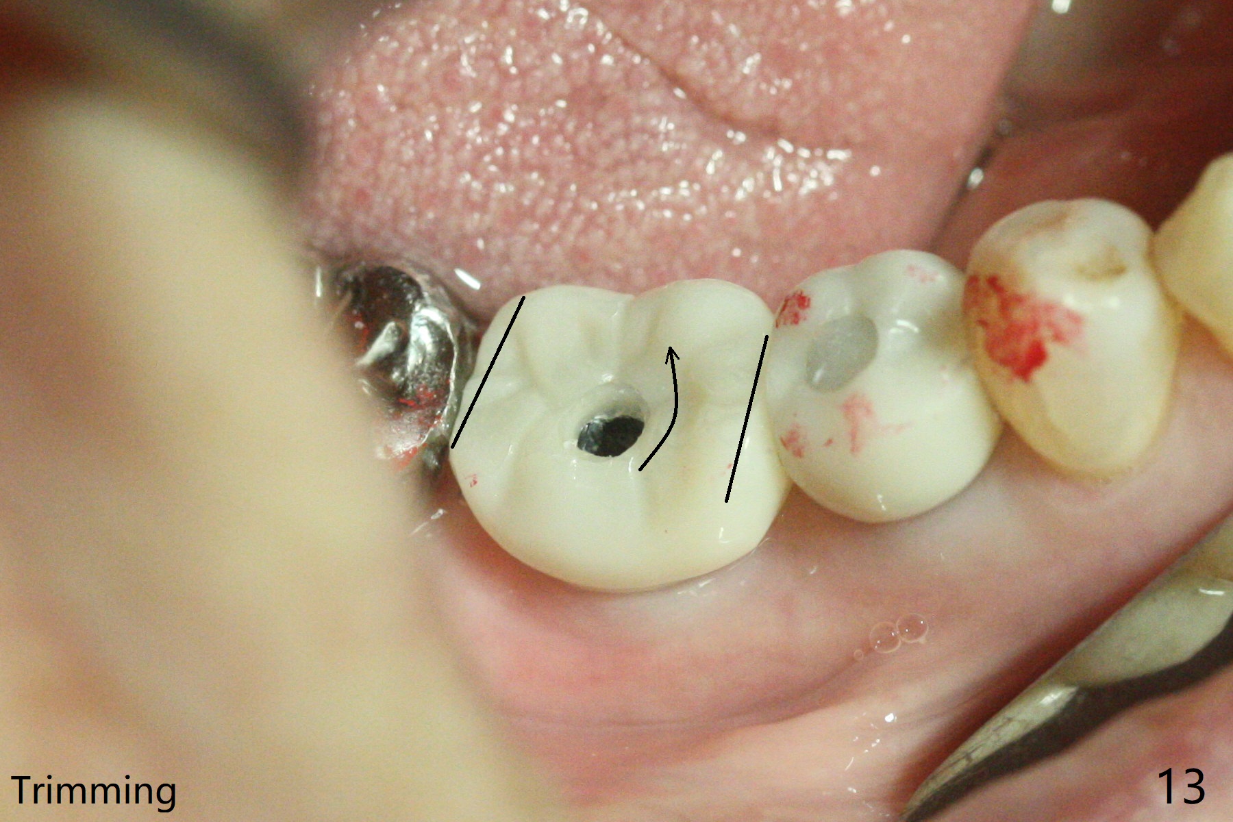
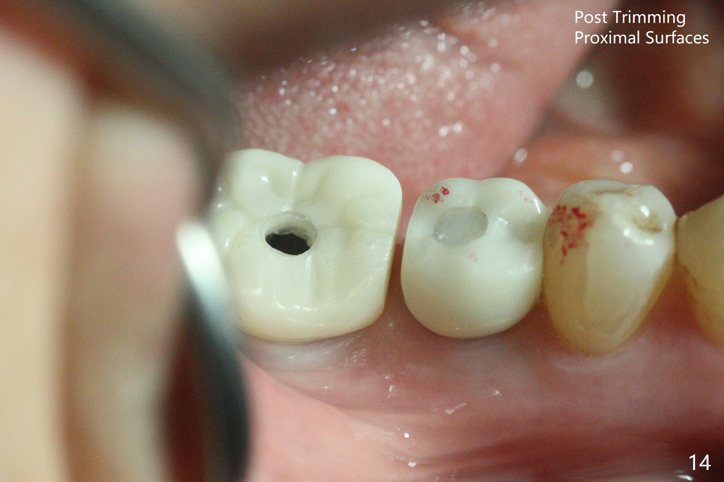
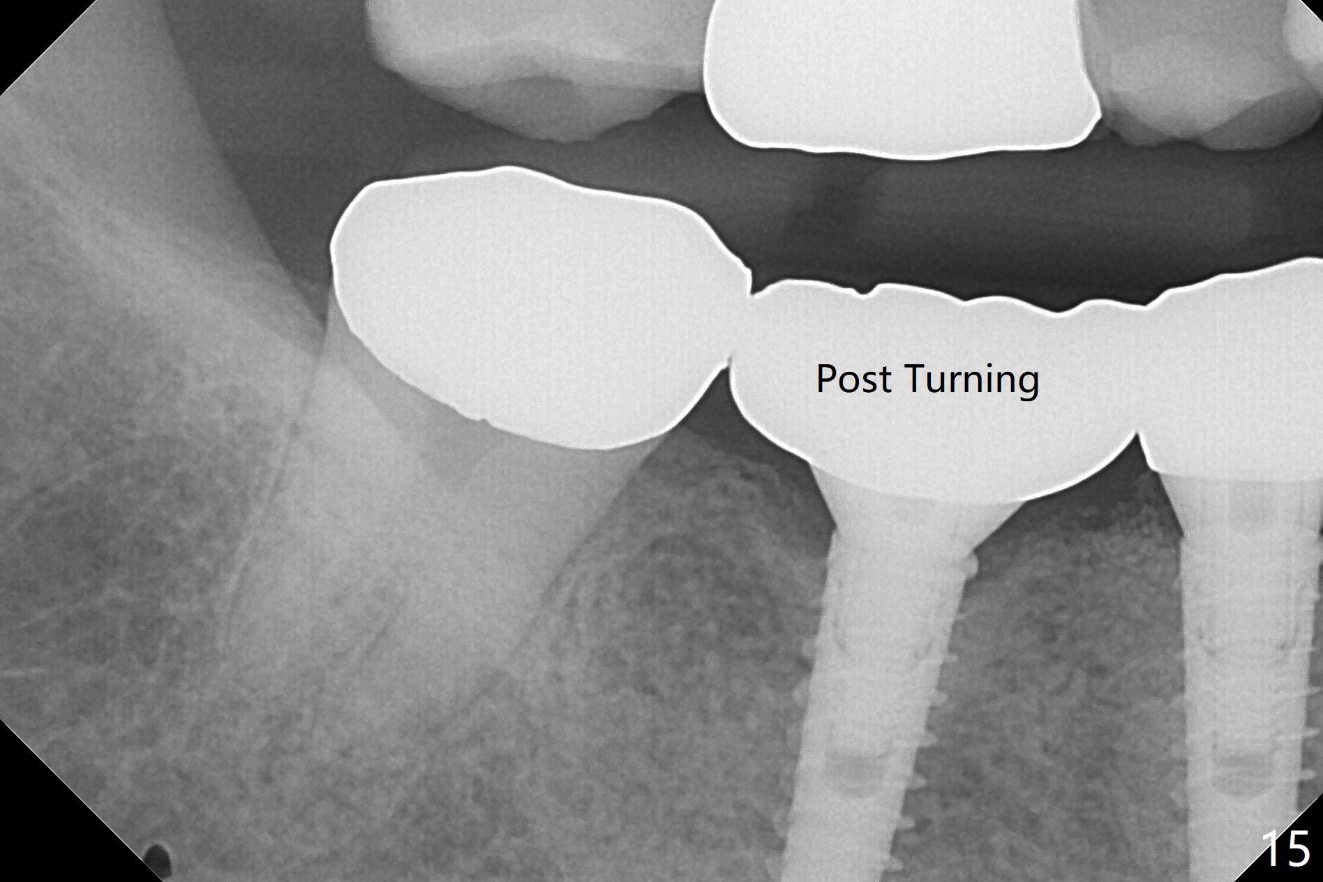
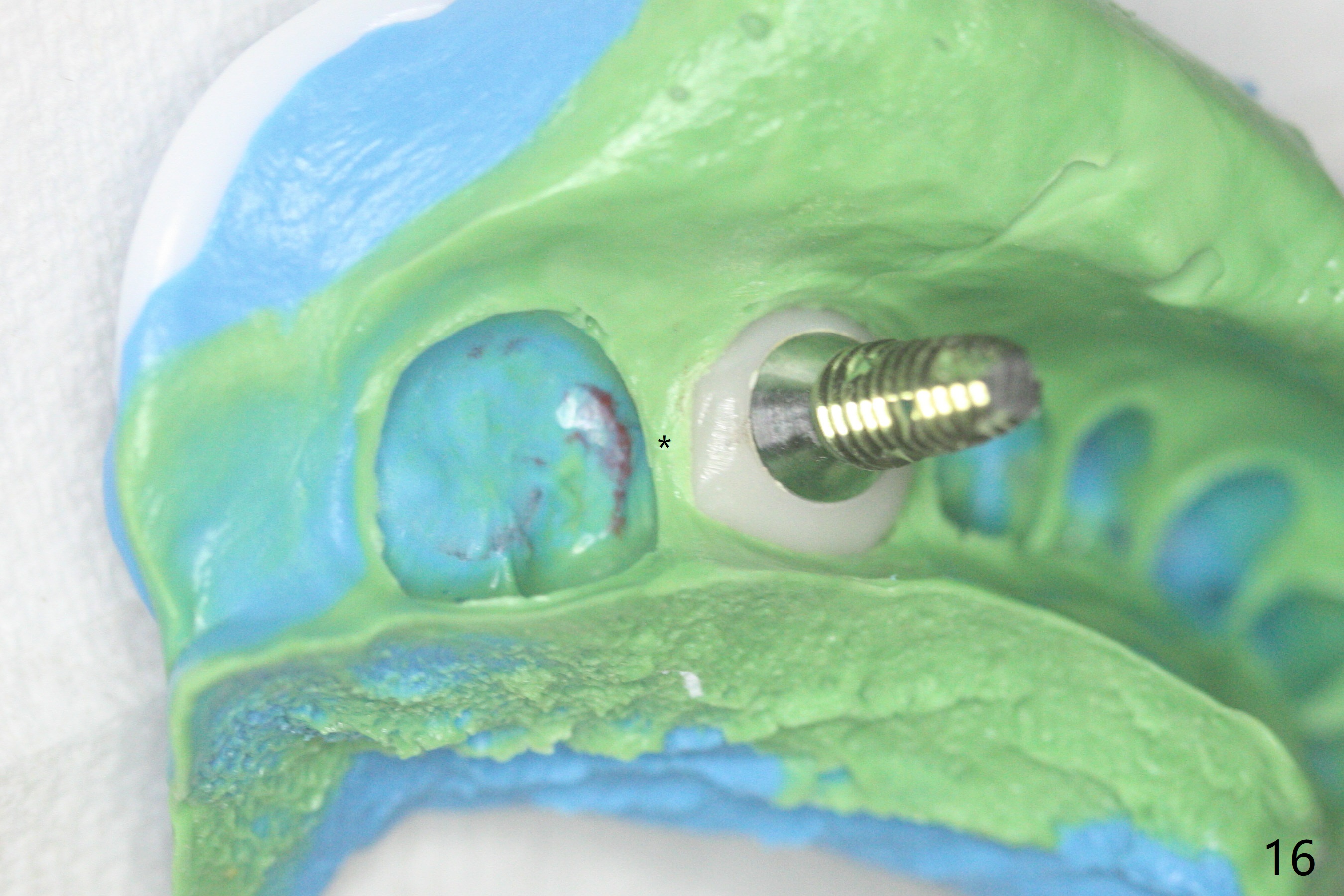
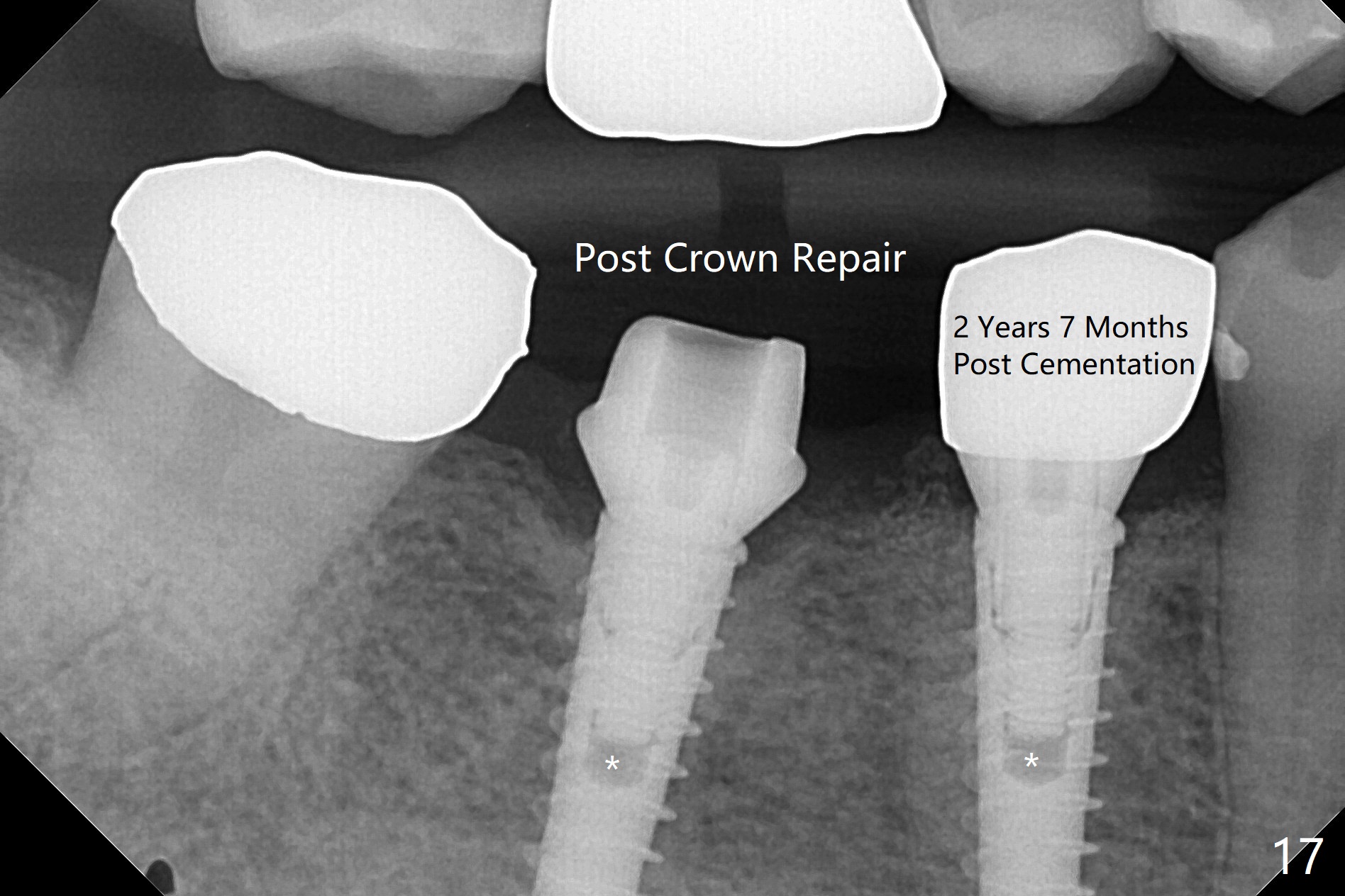
After incision, the ridge at #29 and 30 is found ~ 5 mm wide buccolingually. To place a 4x11 mm IBS implant at #30, the ridge is expanded using BEB technique (bone expansion and bending, Fig.1). It appears that the initial osteotomy at #29 is mesial (Fig.1 yellow dashed line: the distal surface of the root of the tooth #28). After moving the osteotomy distal, the final implant position at #29 (4x11 mm) is within normal limit (Fig.2). The bone at #29 seems to be not so dense that bending (using 1.6 mm drill) is not necessary (using Magic Split and Magic Expanders 3 and 3.8 mm). Later the implant at #30 (4x11 mm) is placed deeper (Fig.3). After placing bone graft around the implants/abutments and suturing, the ridge looks wider with apparent formation of the gingival bands around the abutments (Fig.4 *). Three months and a half postop, bone loss is minimal (Fig.5) and gingival bands forms around the abutments (Fig.6). Fig.7 is taken 1 month post cementation (panoramic X-ray). The patient chews normally 1 year (Fig.8) and nearly 2 years (Fig.9,10) post cementation. The crown at #31 needs recementation 2 years 7 months post #30 cementation; the incomplete seating of the abutment was noted for the first time (Fig.11). Five months later the patient is going to be retired and wants to travel abroad. After approval, the access hole was reopened; articulating paper shows under occlusion of the crown (Fig.12). Since the gap between the abutment and the implant is large, the abutment/crown complex seems to be necessary to be turned (Fig.13 curved arrow). The proximal surfaces of the crown need to be trimmed (straight lines). After turning, the crown sits down with screw tightening; the patient feels pain from the gingival cuff (Fig.14). After turning, the abutment appears to be completely seated (Fig.15). In fact the mesial and distal surfaces of the crown should have clearance from the neighboring teeth (Fig.14) so that pick-up impression is able to hold the crown/abutment complex securely (Fig.16: *). The crown is separated from the abutment after crown repair. They are seated together (loose connection) using the crown as a guide to seat the abutment. BW is taken without the crown. It appears that the abutment remains seated completely (Fig.17). The apical space is equal between #29 and 30 (Fig.17, as compared to Fig.5).
Return to
Lower
Molar,
Premolar Immediate Implant
#15
19/20 Course
1
2
螺丝松动
Xin Wei, DDS, PhD, MS 1st edition 03/27/2017, last revision 03/13/2021