
|
|
|
|
|
|
|
|
|
|
|
|
|
|
|
|
|
|
Magicore
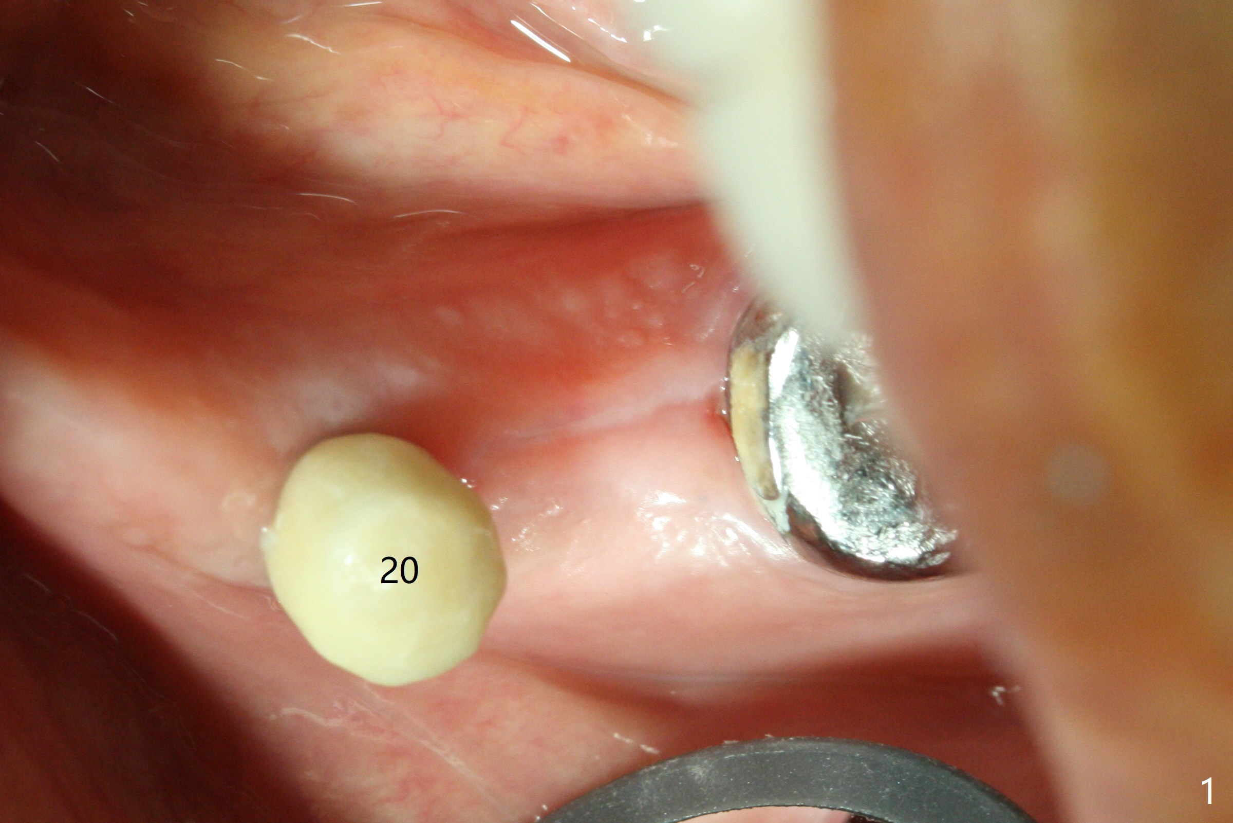
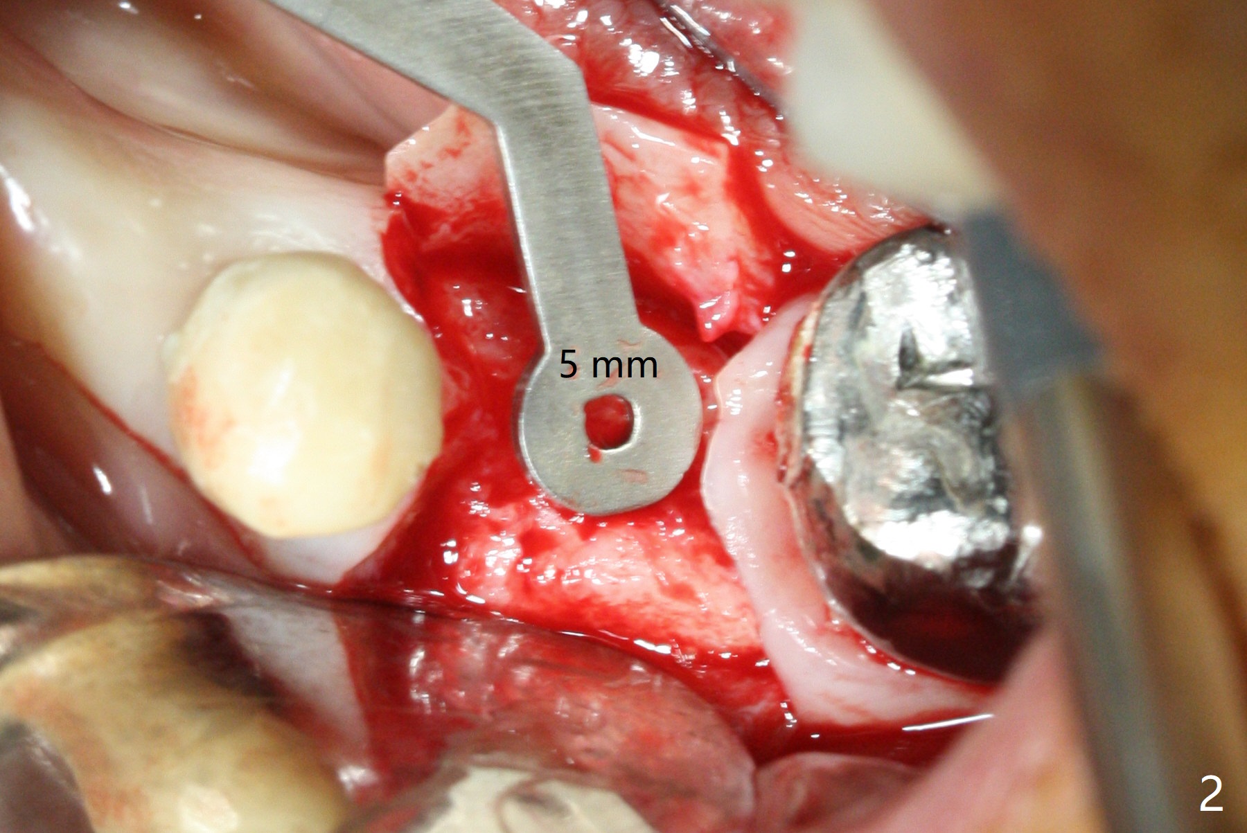
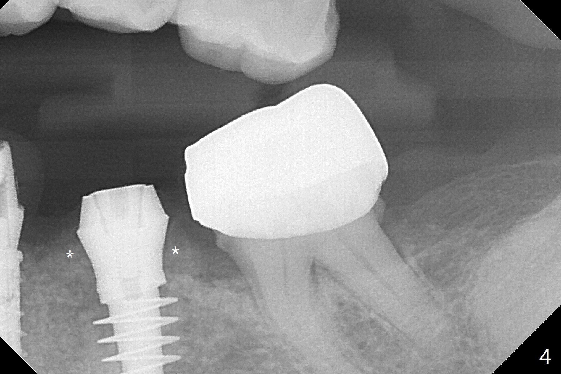
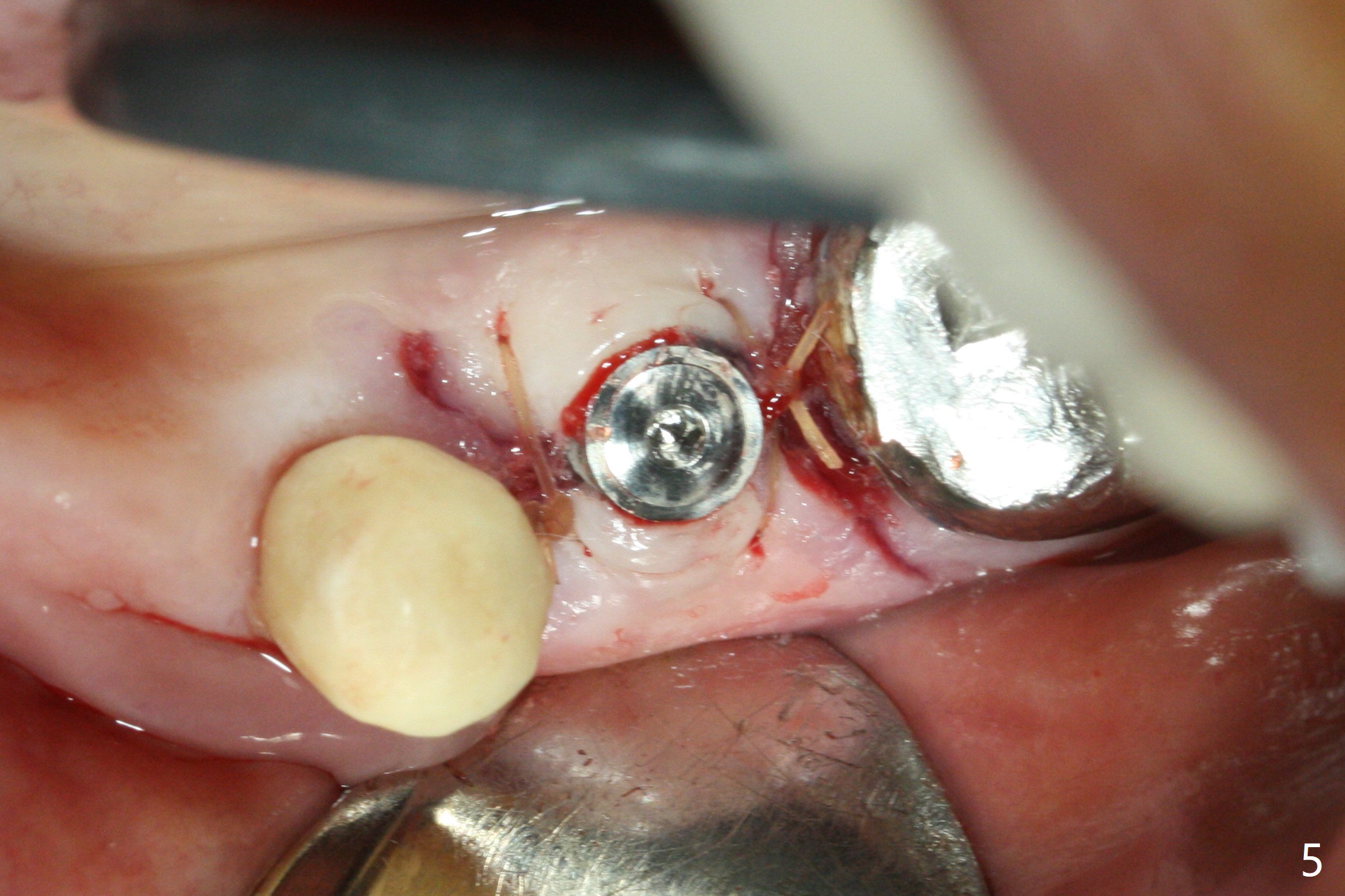
In fact the ridge at #19 is wide (Fig.1); there is enough bone to place a 5 mm implant (Fig.2 (5 mm implant positioner)). The gingiva is 3 mm in thickness. After use of Marking Drill, 5.3 mm Magic Drill (MD) for 9 mm and 4.8 mm MD for ~10 mm, a 5.5x9(3) mm Magicore is placed with primary stability, but too deep. When the implant is reversed, stability loses. After the autogenous bone from bone core is placed in the osteotomy, stability restores to a certain degree (Fig.3); there is no occlusal clearance when a 4.3x3 mm solid abutment is placed. Vera graft is placed around the implant (Fig.4 *) and a healing screw is placed (Fig.4,5).
Dr. Wang thinks that the size of Magicore and placement depth are OK but he is wondering why you did bone grafting. Thursday, October 5, 2017 7:11 AM
Good question. Initially the implant was placed too deep because of lack of experience and concept. When it was backed up, there is a little gap between the osteotomy and implant at the crest. I wanted to fill in the gap with a little bone graft, but it was dispensed a little too much. The latter should not do any harm, although not so necessary. Thanks.
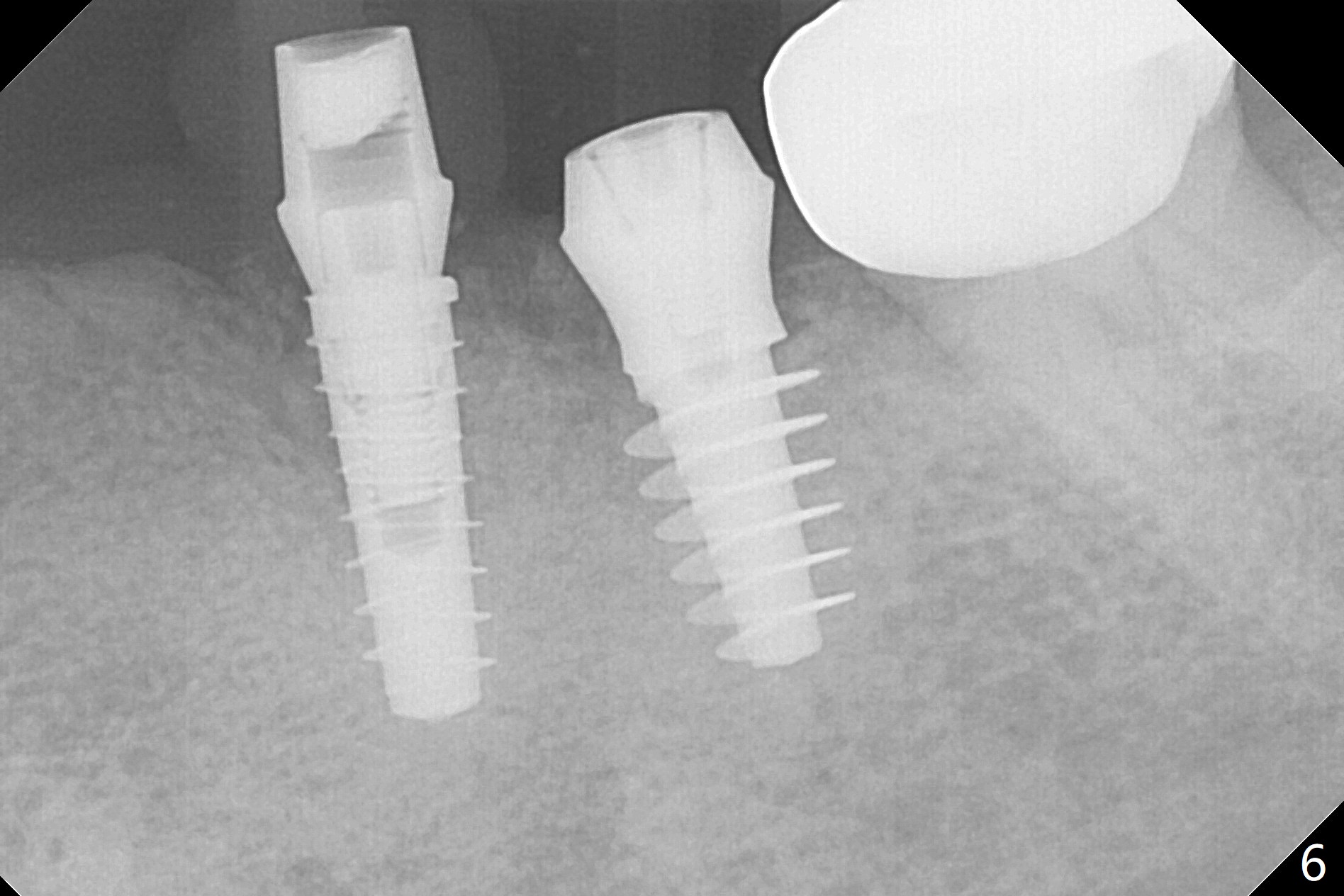
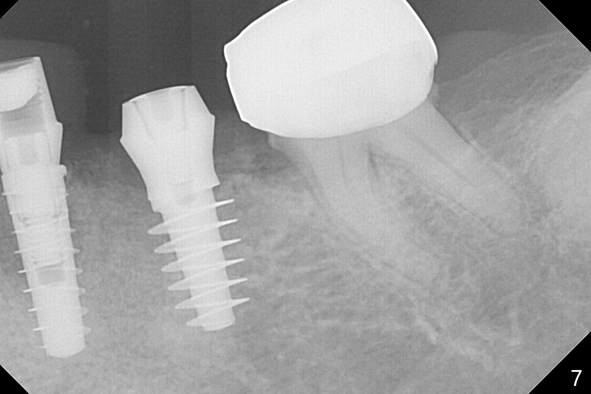
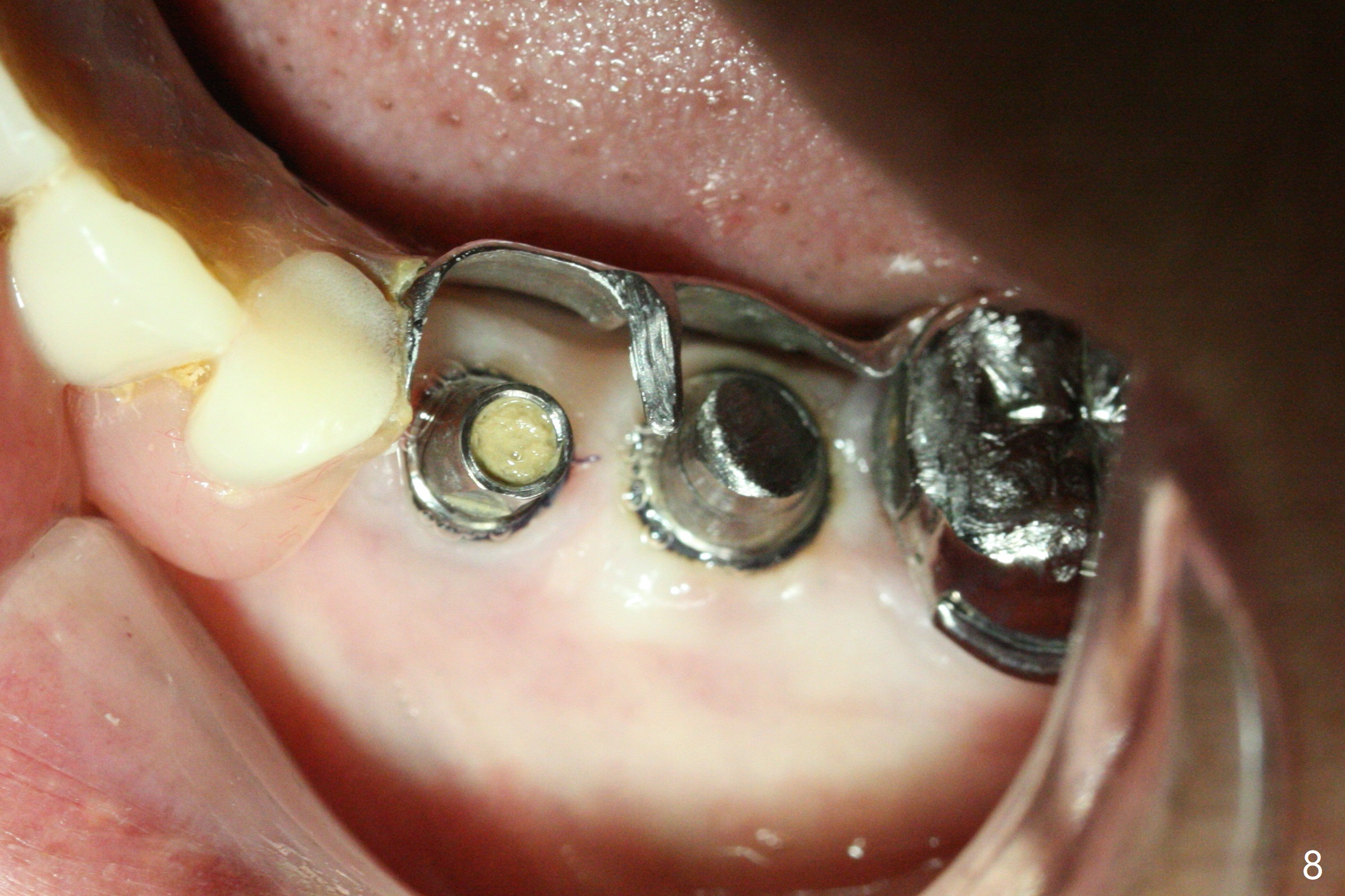
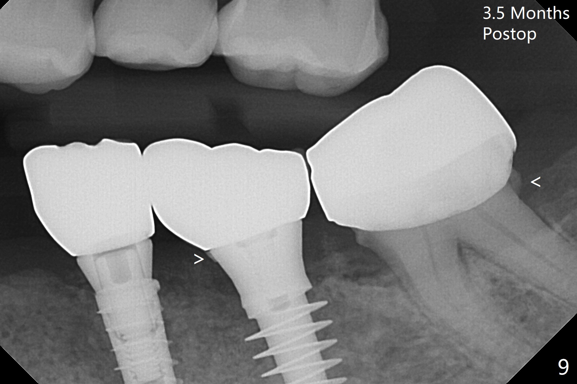
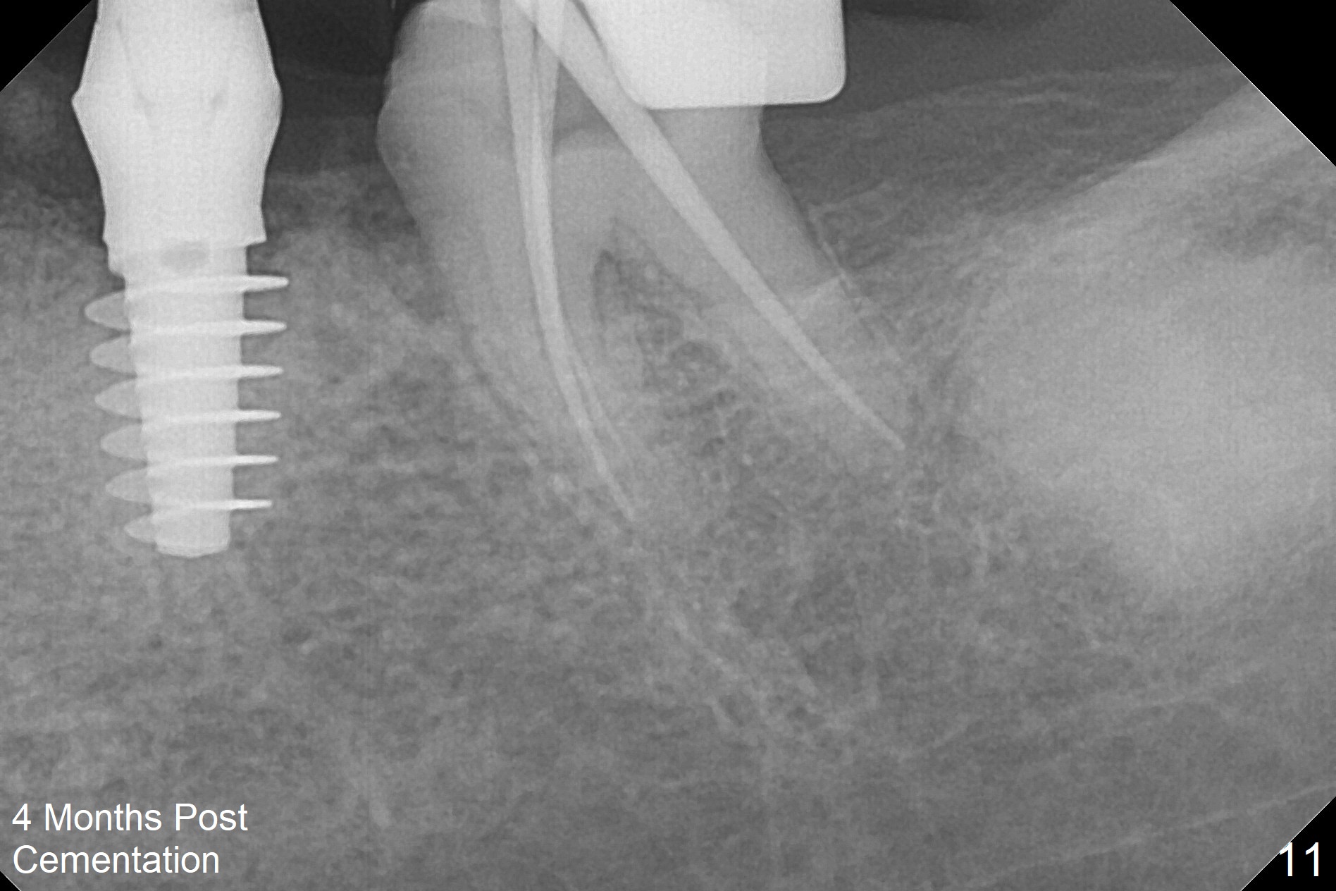
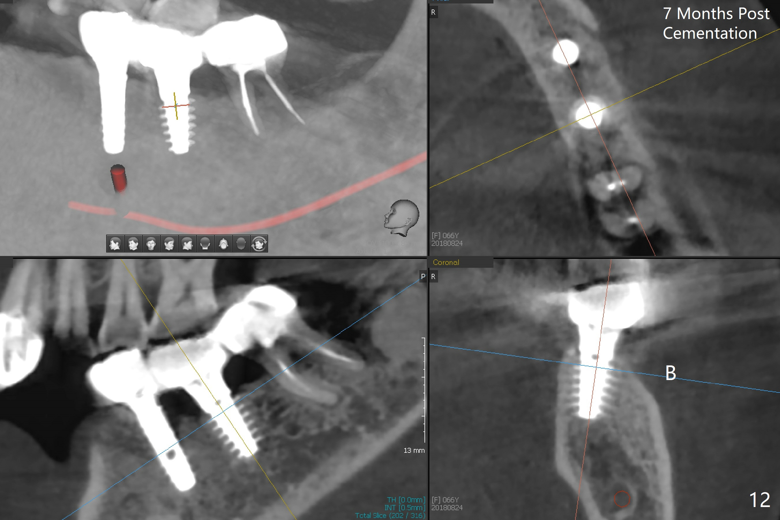
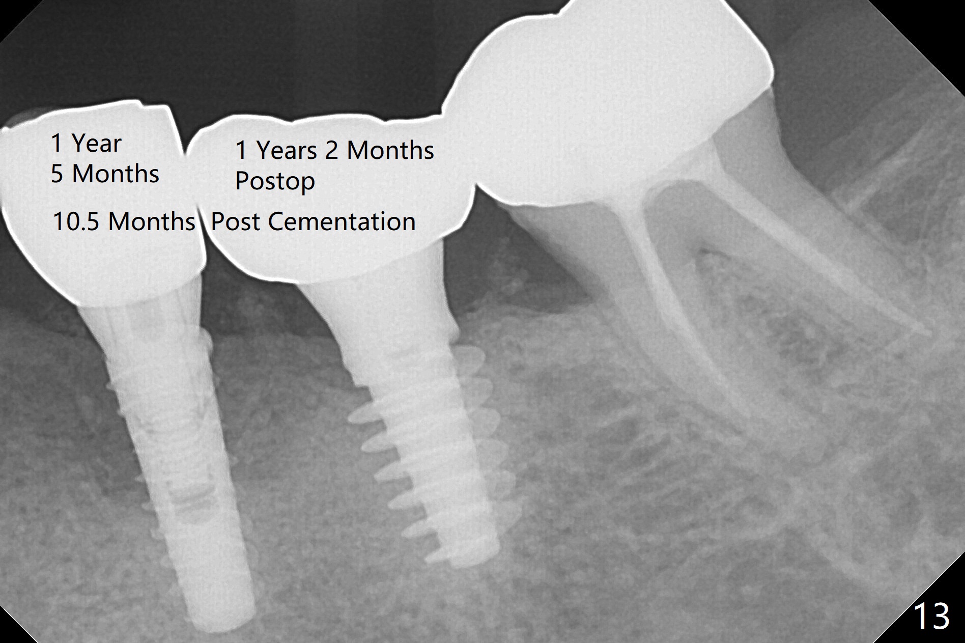
There is no bone loss at #20 or 19 six and 3 months postop, respectively (Fig.6,7). After placing and trimming a 4.3x3 mm Magicore solid abutment, impression is taken (Fig.8). After cementation for #19 and 20 crowns, the crown of #20 is removed for cement removal; attention is paid to cement removal around the crown at #19. In fact, the removal is ineffective with the crown of #20 is reseated and retightened (Fig.9 >). Repeated removal proves to be futile (Fig.10 >). The most effective method will be to take X-ray immediately after #20 crown removal and reseating without torque so that it will be easier to remove the remaining cement if needed. It may be ok in term of hygiene, since proximal brush is used daily. While the crowns at #18 and 19 are being redone because of food impaction, the tooth #18 needs RCT; the Magicore seems to have no bone loss 4 months post cementation (Fig.11). CBCT shows that the Magicore seems to have been placed in the middle of the crest 7 months post cementation (Fig.12 (B: buccal)). The gingiva at #19 is apparently healthy 8 months post cementation. The crown at #19 is recemented 10.5 months post cementation (Fig.13).
Return to Lower Molar Immediate Implant, IBS, #20, 3,4, Magicore Cases
Xin Wei, DDS, PhD, MS 1st edition 10/03/2017, last revision 12/03/2018