,%20no%20graft.jpg)
%20and%20graft%20.5-1%20mm.jpg)
%20abutment.jpg)
|
|
|
|
|
After use of 5x11.5 mm drill against the lingual wall, a 5.5x11.5 mm implant is placed with < 35 Ncm (Fig.3). The low primary stability is in part due to the thin septum (Fig.3 <). The radiolucency apical to the apex of the implant is over preparation of the osteotomy (Fig.3 *). When autogenous bone, .5-1 mm mineralized cancellous & cortical allograft and Osteogen are placed around the implant, the bone density increases (Fig.4 *). An immediate provisional is fabricated without cement to hold bone graft in place. Abutment: 5.5x5(4) mm.
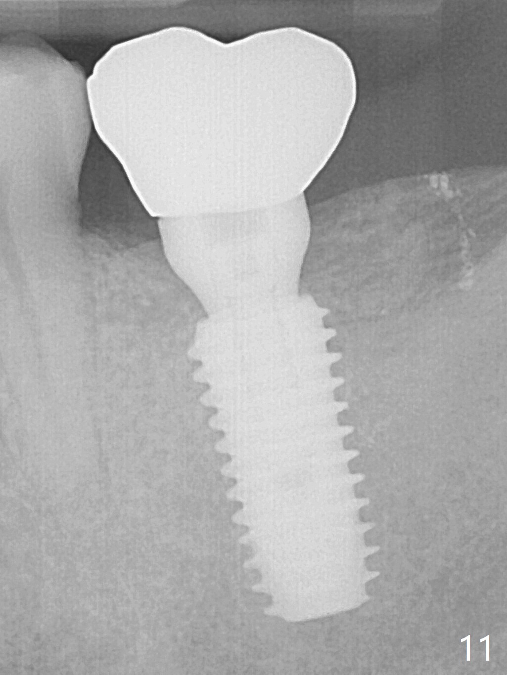
The distal socket and apical space disappear 11 months postop (Fig.5). An angled abutment (5.5x15° B 4 mm) is used before impression to change the mesiobuccal trajectory. There is no bone loss 1 year post cementation (Fig.11).
Implant Position and Trajectory is More Critical
Xin Wei, DDS, PhD, MS 1st edition 12/31/2016, last revision 01/19/2018