|
|
|
|
|
|
|
|
|
|
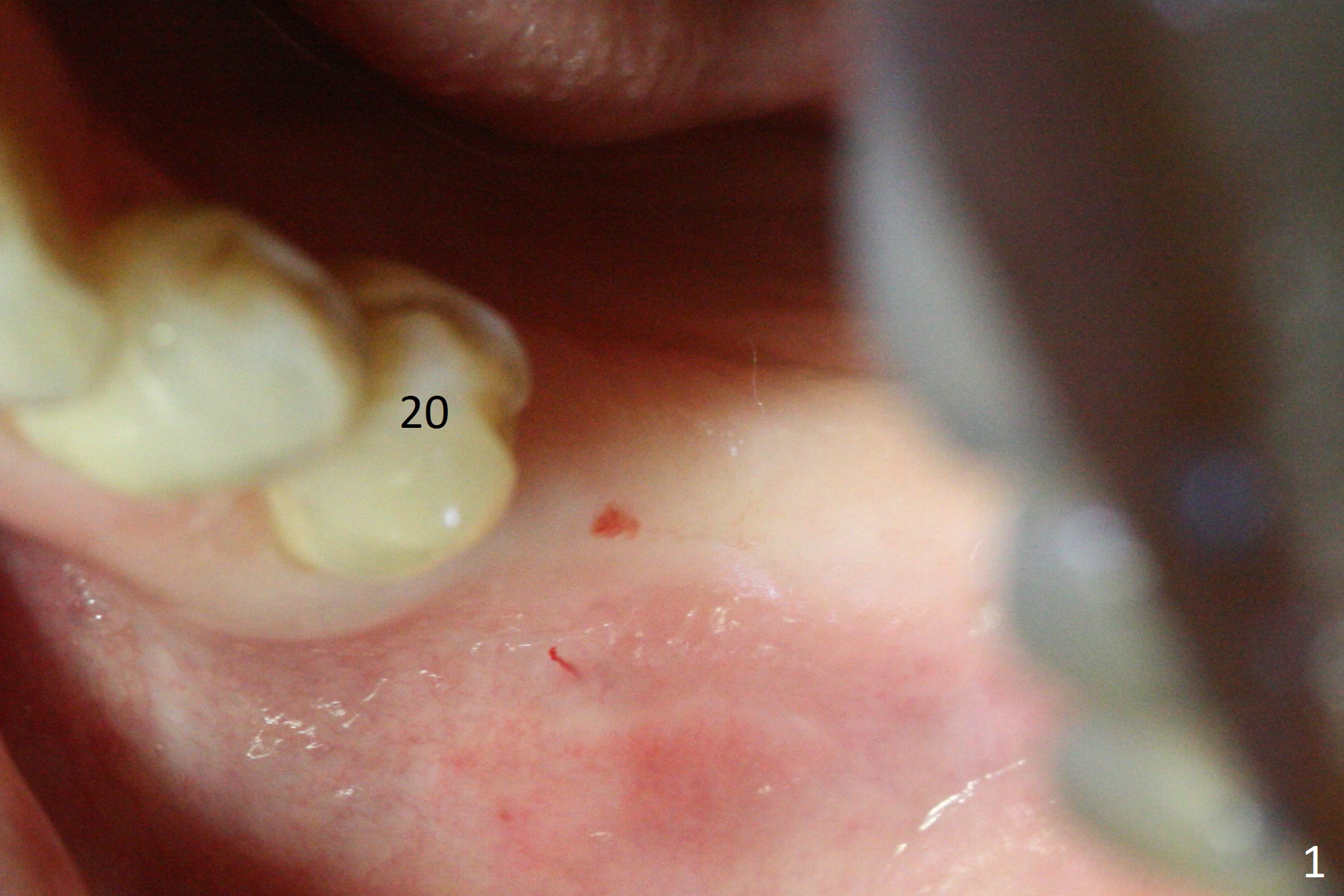
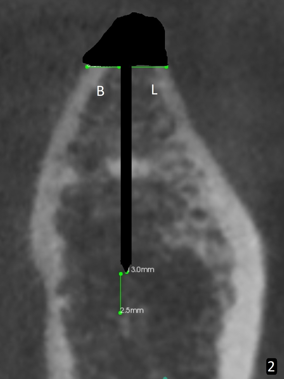
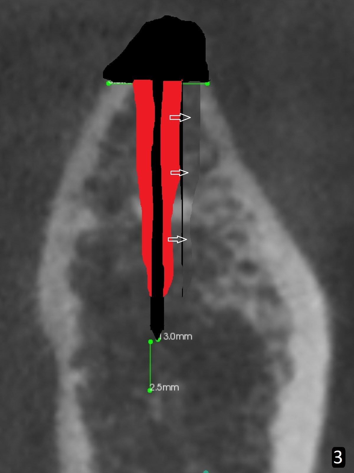
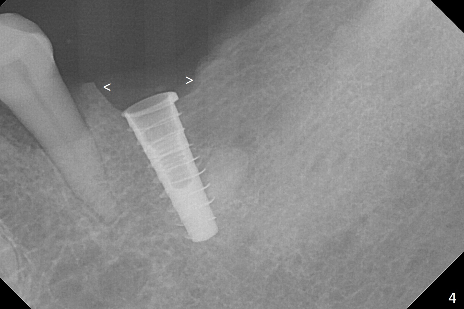
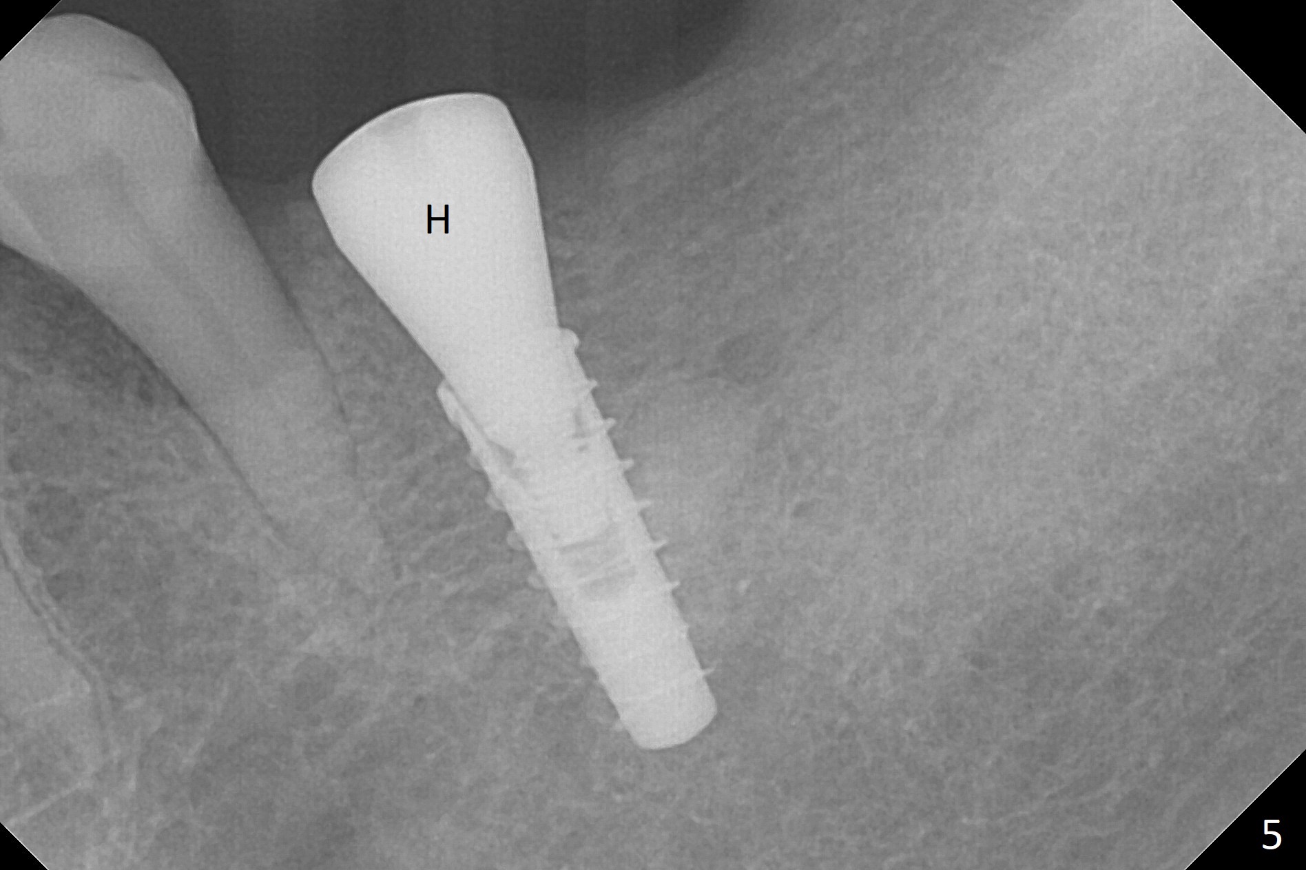
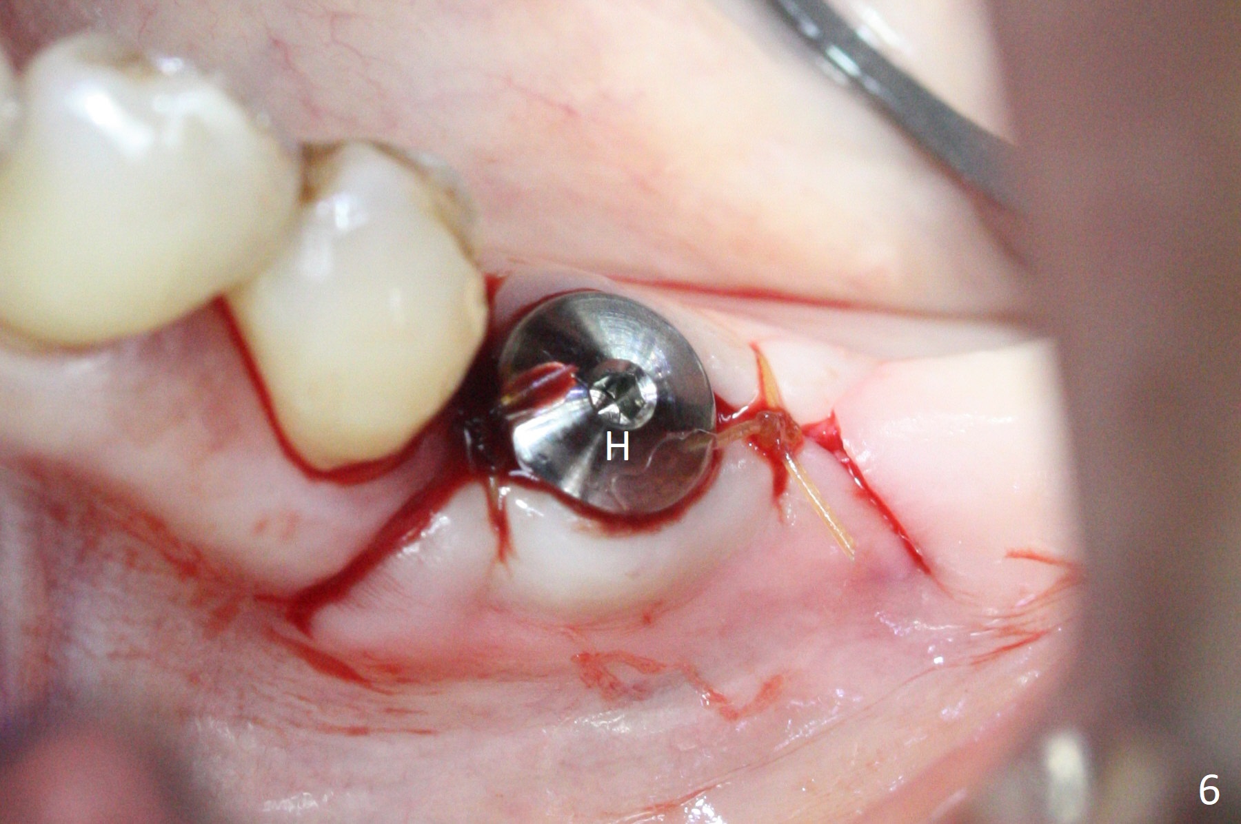
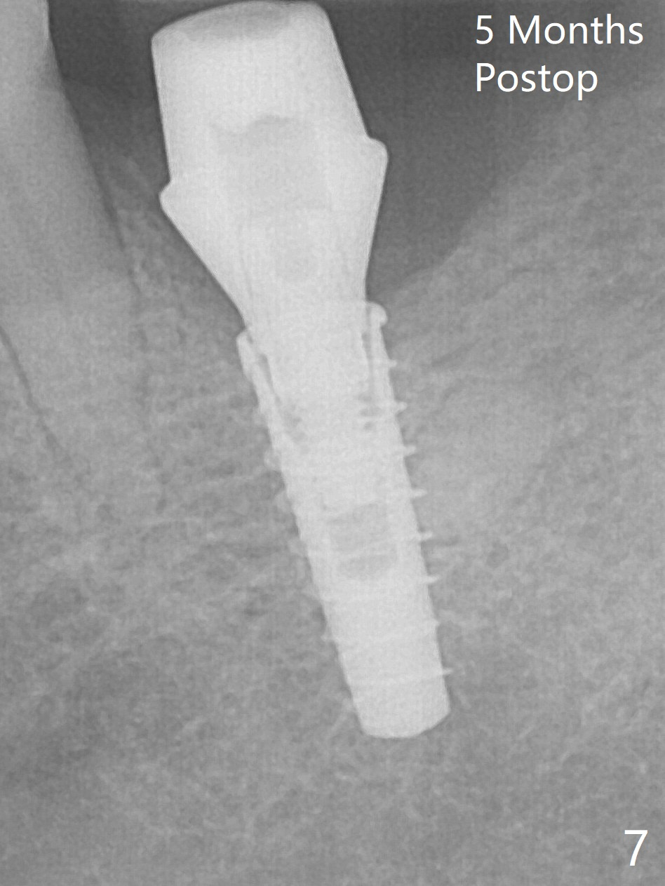
Bone Condensation Instead of BEB in the Mandible
After incision over the narrow ridge at #30 (Fig.1) and ridge reduction (Fig.2 black area on the top; Fig.4 arrowheads), a 1.6 mm drill is used (Fig.2 (CBCT coronal section) long vertical black area) to start BEB (bone expansion and Bending). It is expected that the cortical and cancellous bone of the buccal (B) and lingual (L) plates can be expanded after use of Magic Expanders (ME) and 1.6 mm drill alternatively. In fact it appears that only the cancellous bone is pushed after MEs from 3 to 4.3 mm (Fig.3 arrows). There is no apparent cortical bone bending. There may be cancellous bone condensation. Dummy (Fig.4) and definitive (Fig.5) implants (4x11 mm) are placed. Since torque is 20 Ncm, a 5.5x4 mm healing abutment is placed (Fig.5,6 H). The opposing tooth is supraerupted; intrusion is pending. A 6x4(2) mm abutment is placed with a provisional 2 months postop. There is enough occlusal clearance. Impression is taken 5 months postop with difficulty because of subgingival margin (Fig.7). After cementation, the patient feels discomofort, which is relieved when the mesial embrasure is enlarged. Return to Lower Molar Immediate Implant, Armaments Xin Wei, DDS, PhD, MS 1st edition 12/14/2017, last revision 09/16/2018