
|
|
|
|
|
|
|
|
Septal Placement
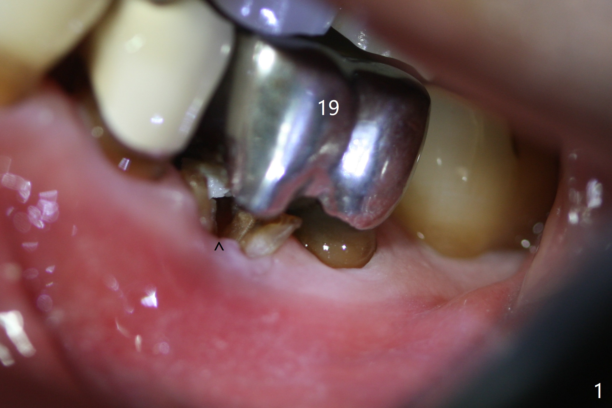
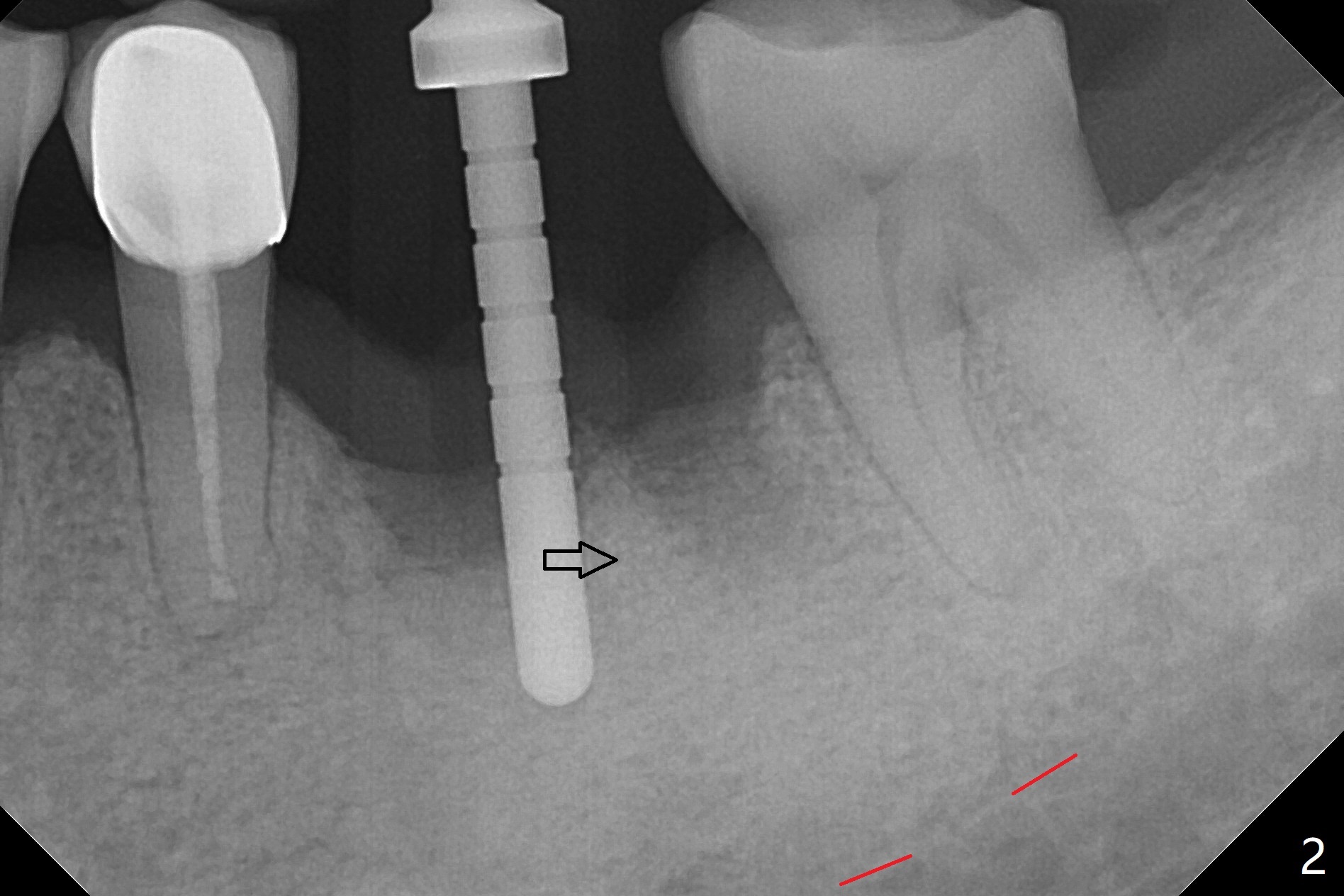
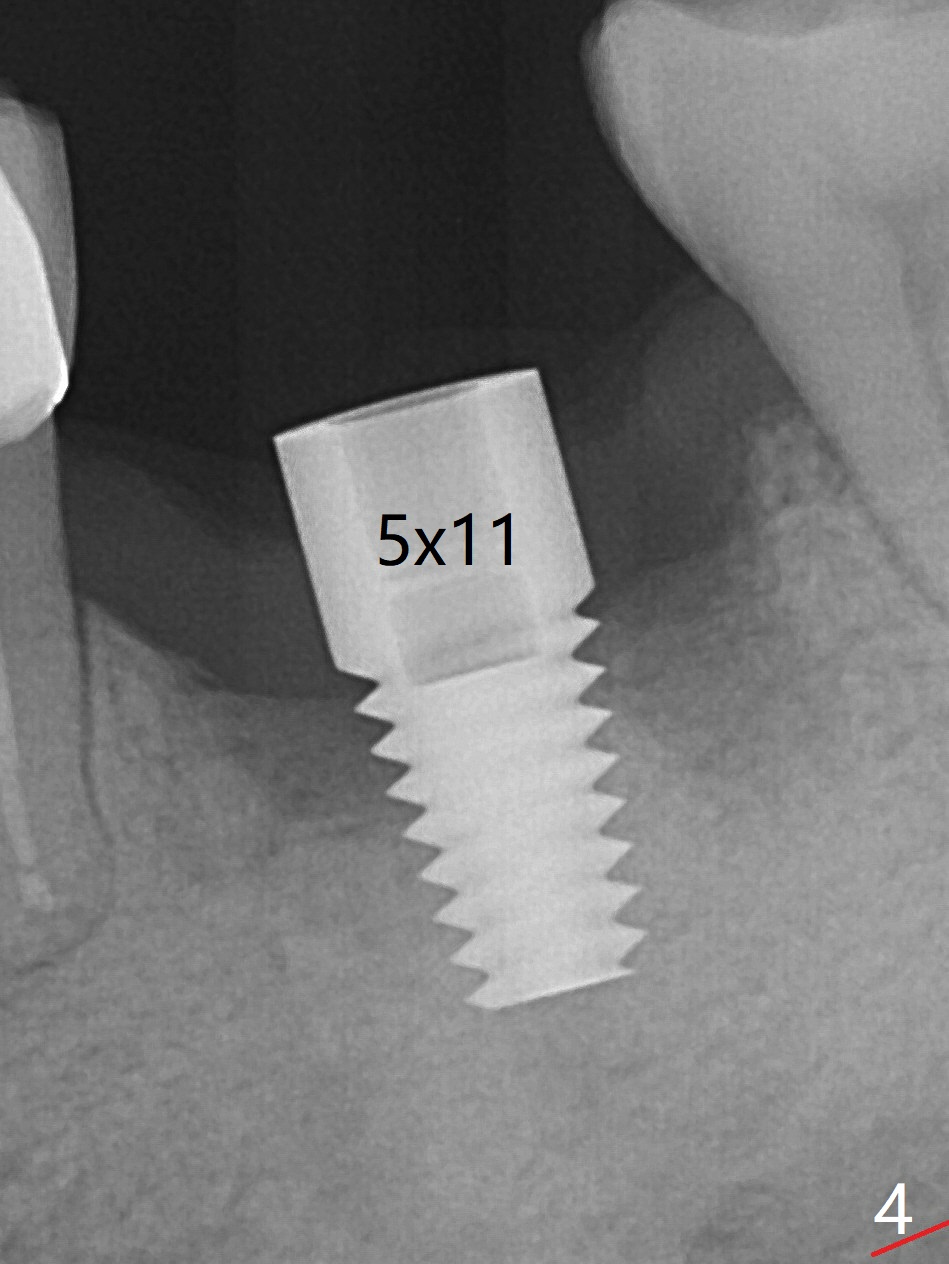
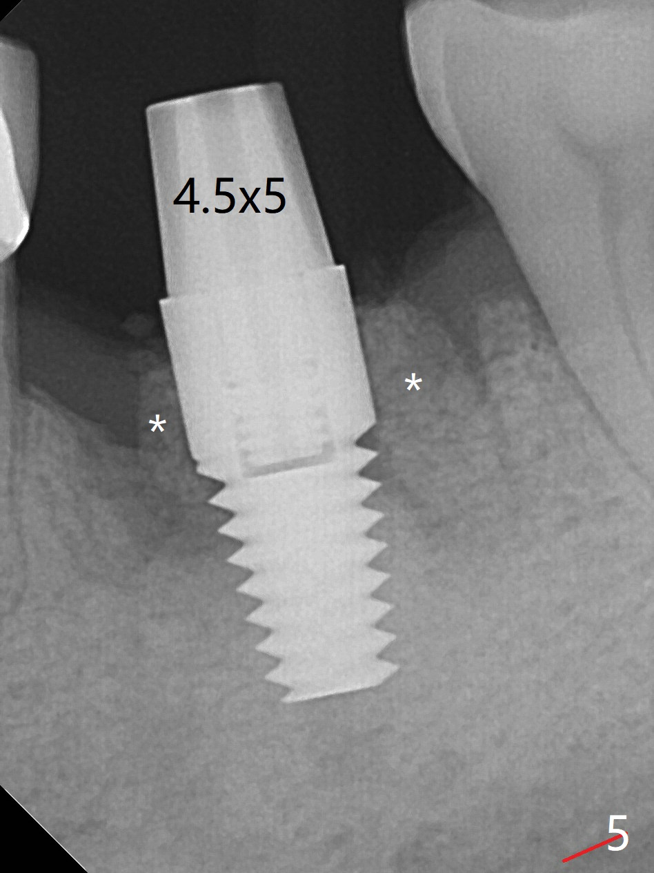
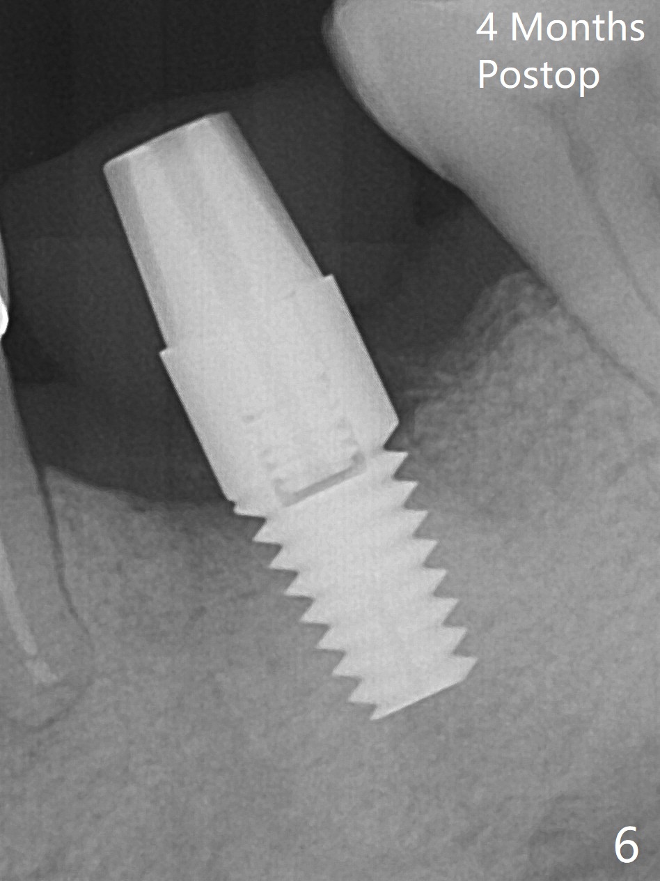
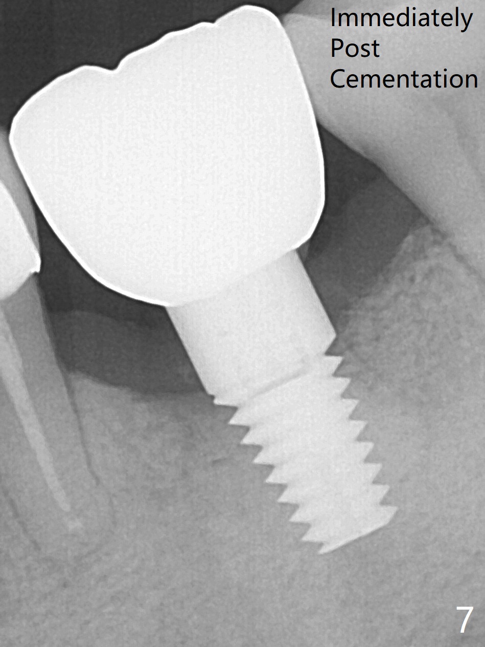
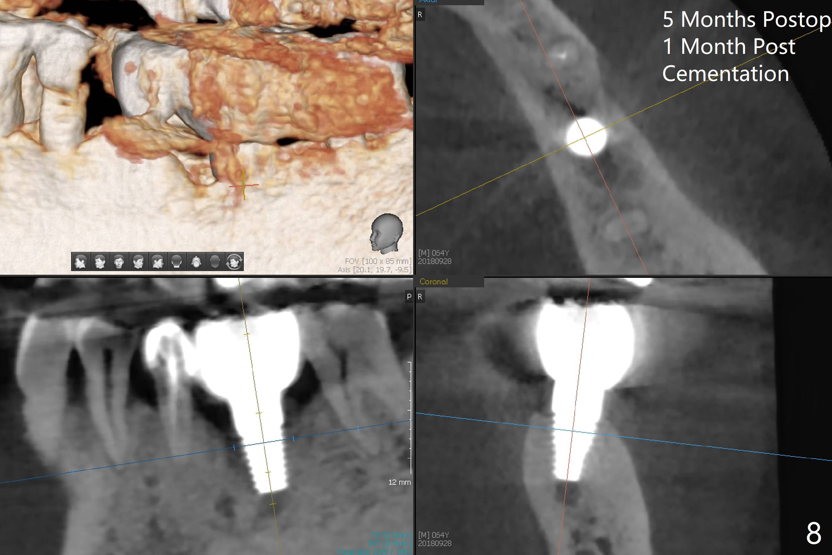
After extraction of the tooth #19 with mesial root fracture (Fig.1 ^) and curettage, the initial osteotomy in the septum is found to need to be distalized (Fig.2 arrow). Following sequential osteotomy, a 5x17 mm tap cannot reach the expected depth (Fig.3 yellow dashed line) because of the dense bone. A shorter implant is placed with >60 Ncm (Fig.4). An immediate provisional is fabricated after placement of a 4.5x5 mm abutment and Vanilla/Osteogen graft (Fig.5 *). The mesial and distal sockets heal 4 months postop (Fig.6). The bone density in the mesial and distal sockets continues to increase when a crown is cemented (~4.5 months postop, Fig.7). The implant at #19 was placed in the middle of the bone (Fig.8).
Return to
Lower
Molar Immediate Implant, Prevent
Molar Periimplantitis (Protocols,
Table),
Armaments,
3
8/9
10
14,15,30
Xin Wei, DDS, PhD, MS 1st edition 04/23/2018, last revision 10/01/2018