
|
|
|
|
|
|
|
|
|
|
|
|
|
|
|
LECTURE: Two Extra Teeth
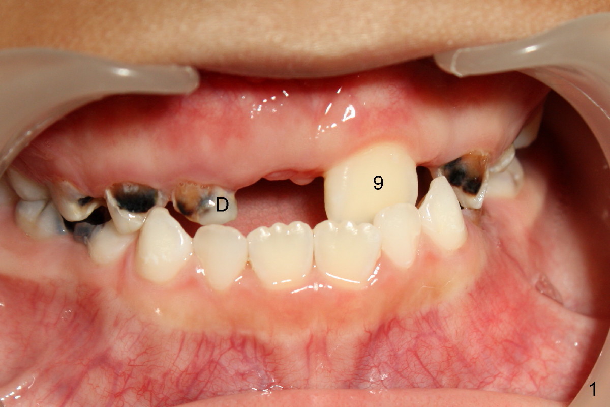
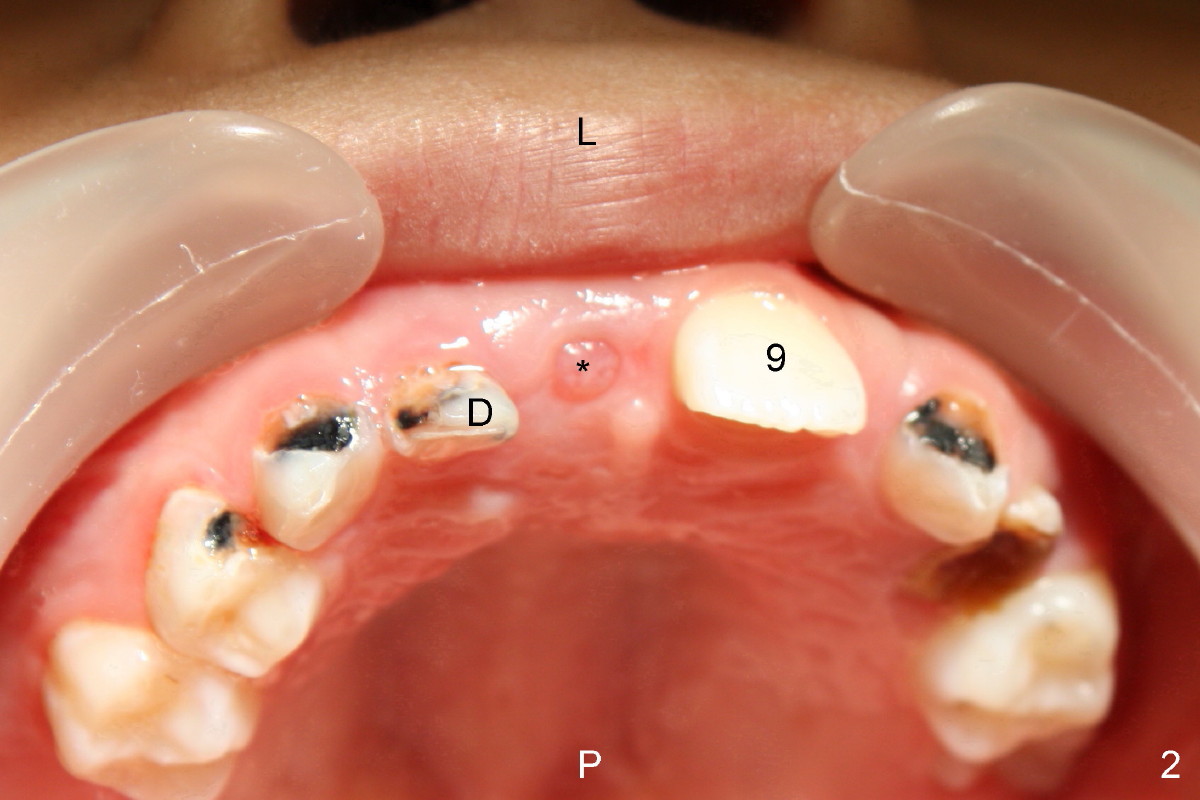
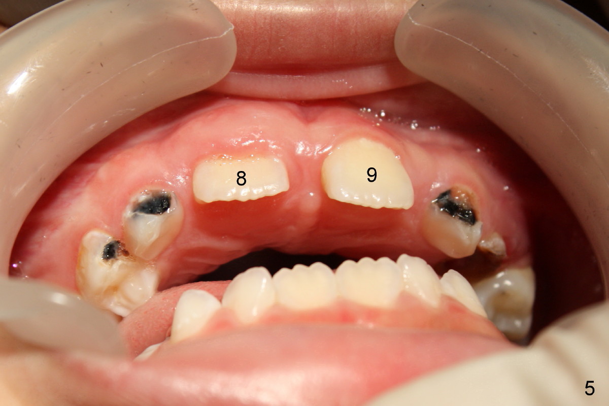
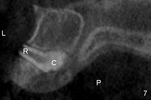
When Leon is at the age of 7, one of the top central incisors (Fig.1,2: #9) erupts, while the other (#8) does not. Initial X-ray shows that there are two extra teeth which block or interfere with eruption of the tooth #8 (1 2 3 4 5). To remove these extra teeth, we need to know where they are: on the lip (Fig. 2 L) or palate (P) side of the central incisors. This information decides surgical approach. Neither clinical or regular X-ray exam can provide this information. X-ray is two dimensional (without information of depth), whereas CT is three-dimensional. Nowadays, CT is available readily, affordable and reasonably safe (without too much radiation).
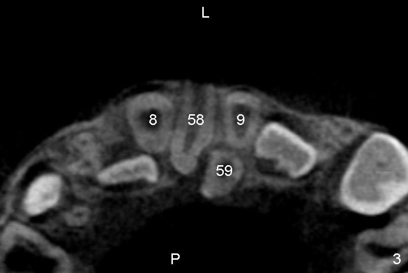
Fig.3 is a CT cross section through the roots of the central incisors with the lip (L) on the top, while the palate (P) on the bottom of the figure. It shows Leon's two extra teeth (#58,59) between the central incisors (8,9). The tooth #59 appears to be completely on the palate side. So we should approach this extra tooth from the palate. How about the tooth #58? The root is on the lip side, whereas the crown on the palate side. The size of the crown and the root is almost the same from this image. We do not know whether it is easy to approach this extra tooth from the palate or not. Let us make a section along the long axis of #58, i.e., through letters L and P in Fig.3.
The section shows that the crown of the extra tooth #58 (Fig.7 C) is larger than the root (R). It appears to be easier to remove this extra tooth from the palate approach.
Let us go back to Fig.3 and make a section through the teeth #9 and #59. The new section (Fig.8) confirms that the extra tooth #59 is completely located palatally with the root (R) on the top and crown (C) on the bottom. In all, CT images help us decide the surgical approach.
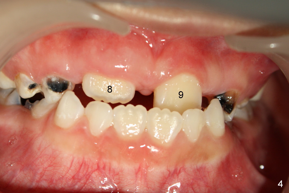
On the day of surgery (two months after initial photographs (Fig.1,2)), the other central incisor, #8, is erupting (Fig.4,5), but there is a big gap between #8 and 9, presumably due to the presence of these two extra teeth. The baby lateral incisor is gone (as compared to D in Fig.1,2), most likely due to being pushed out from the erupting #8.
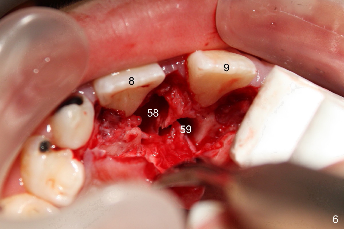
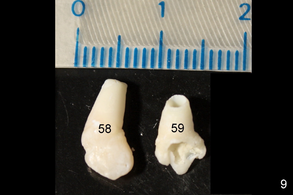
With the guidance of CT information, palatal incision is made with local anesthesia and the two extra teeth are removed smoothly (Fig.6,9). The boy is doing great during and after operation. Braces are needed to close the gap between the central incisors (Fig.4,5) and fix cross bite (the top incisors are inside the lower incisors, which is also not normal).
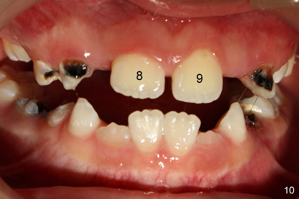
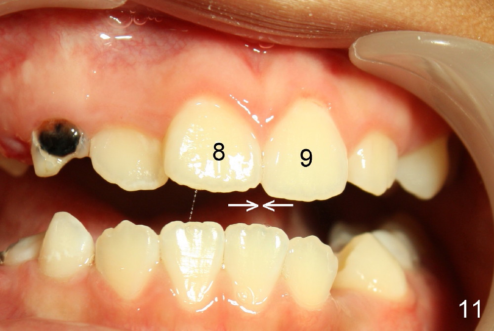
Nine months after extraction of the two extra teeth, the two central incisors are close to each other (Fig.10: 8,9, as compared to Fig.4). Two years after surgery, there is no gap between the two central incisors (Fig.11). No early treatment may lead to unpleasant appearance later on (4 5).
Xin Wei, DDS, PhD, MS 1st edition 07/27/2012, last revision 10/22/2014