|
|
|
|
|
|
|
|
|
|
|
|
|
|
|
|
|
|
||
Immediate Provisional Covers Extraction Socket and Bone Graft I
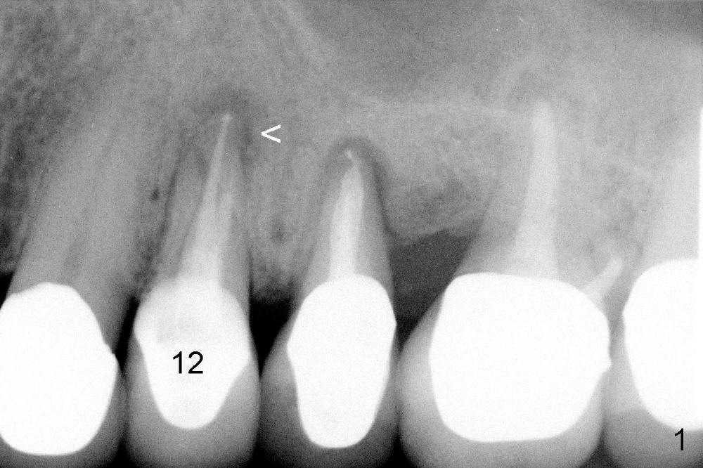
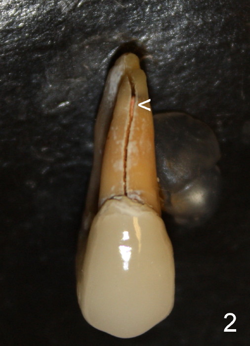
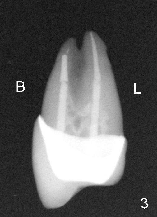
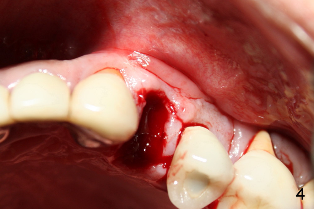
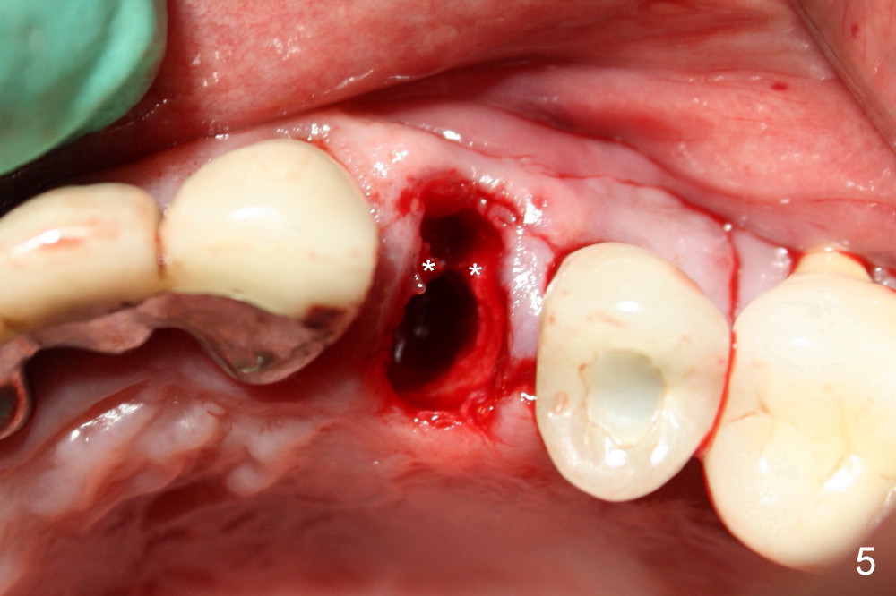
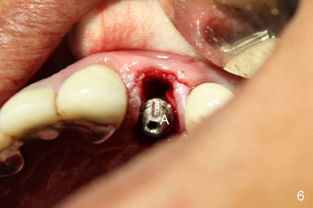
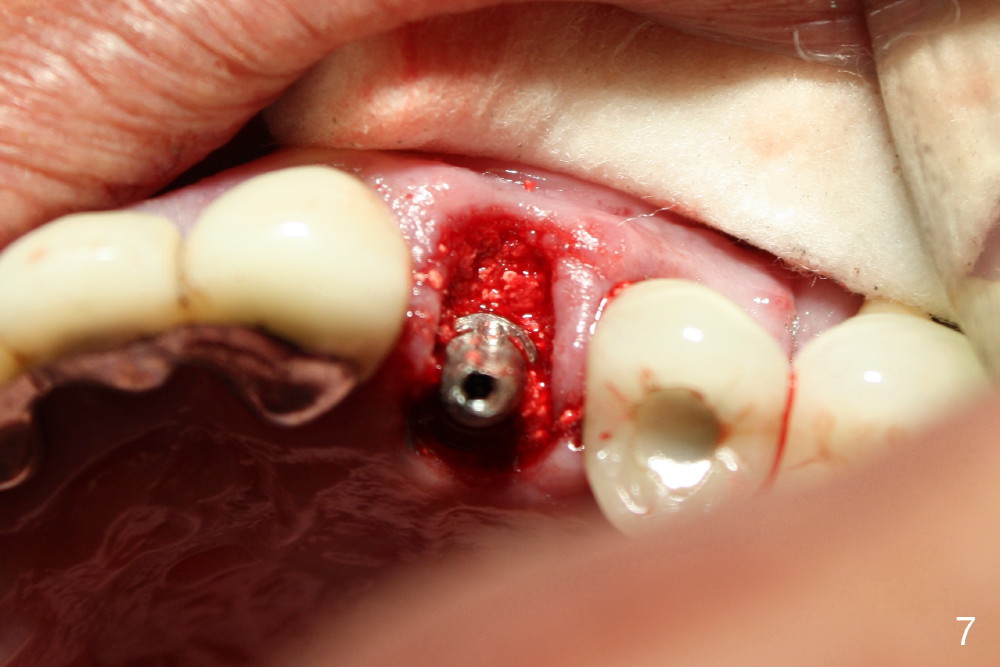
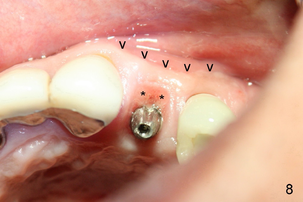
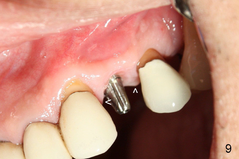
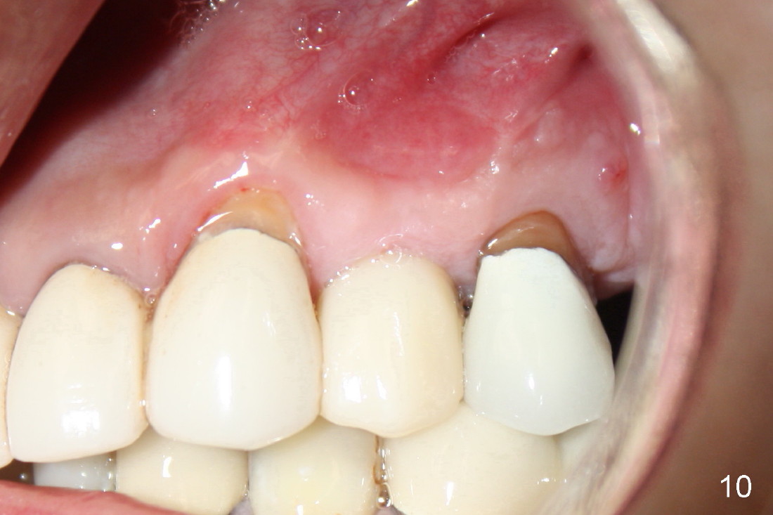
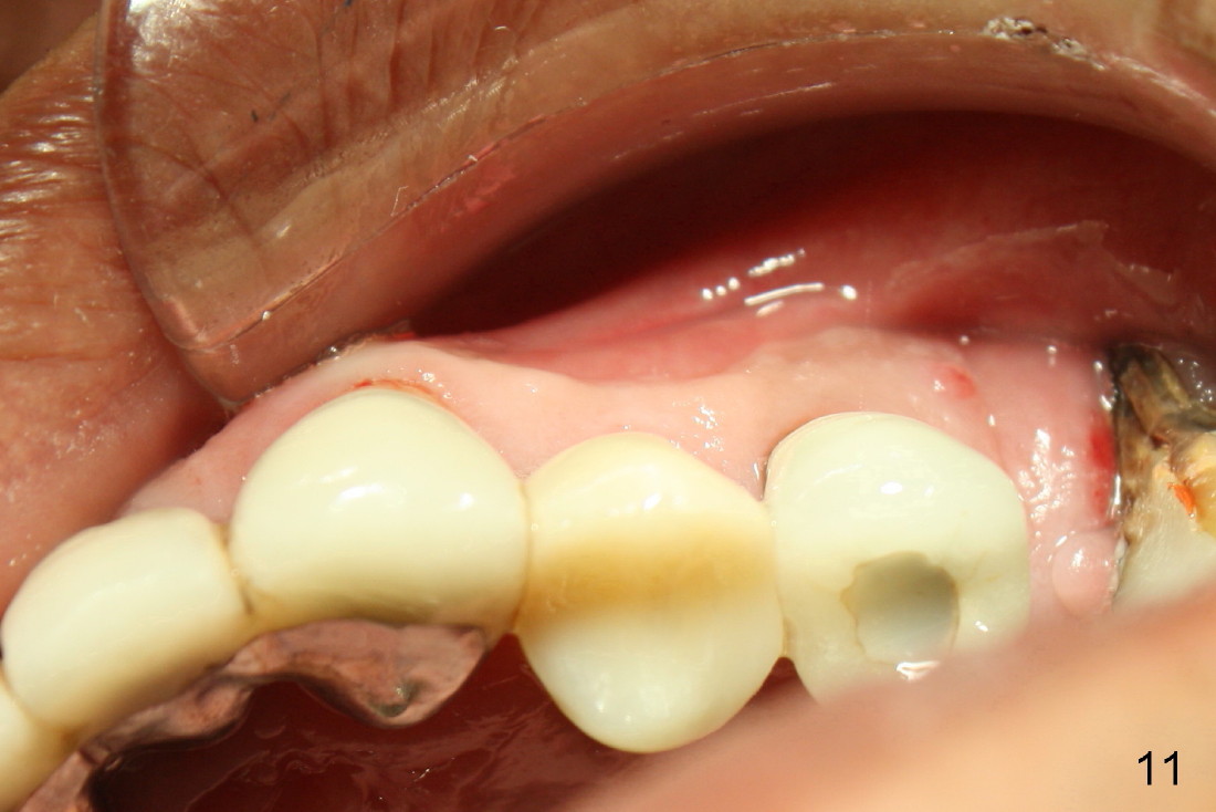
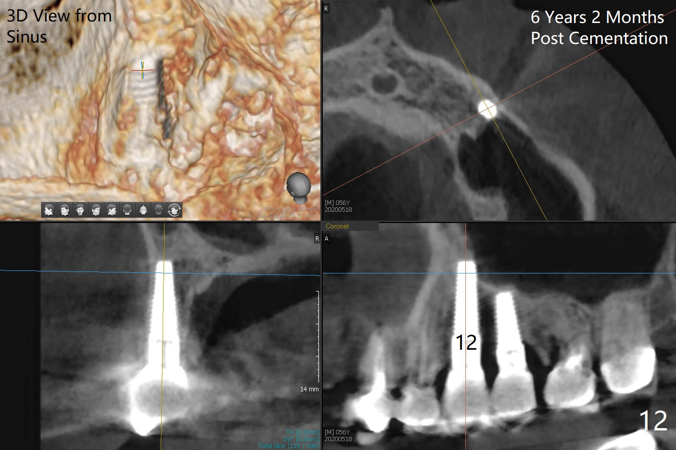
A 50-year-old man has mild pain in the upper left 1st bicuspid 3 years after root canal retreatment with placement of two posts (Fig.1). Findings of clinical exam are consistent with root fracture (Fig.2). Extraction reveals two fused roots (Fig.3,4). Probing indicates that the buccal plate is defective. Osteotomy is initiated in the palatal socket with a 2 mm pilot drill, followed by 2.5 and 3.0 mm reamers and 4.5x20 mm tap. The septum appears to have been pushed buccally (Fig.5 *) and form a new buccal wall (partially, strengthened by bone graft mentioned below) for the implant to be placed. The implant (4.5x20 mm) is placed in the palatal socket (Fig.6 *) with insertion of an abutment (A: 3.5x5 mm 0º), while a mixture of autogenous bone (harvested from reamers) and Synthograft (Bicon) is placed in the shrunken buccal socket (Fig.7 using allograft may decrease postop bony shrinkage). The bone graft is then contained by an immediate provisional without collagen membrane or flaps. The patient is doing well postop. The gingiva is healthy (Fig.8*) when the provisional is removed 3 months postop with normal papillae (Fig.9 arrowheads). It remains the same 1 month post cementation (Fig.10,11). For further follow up, see immediate implant of the tooth #13.
Return to
Upper Bicuspid Immediate Implant
3/4
Xin Wei, DDS, PhD, MS 1st edition 02/23/2014, last revision 05/19/2020