|
|
|
|
|
|
|
Diabetes
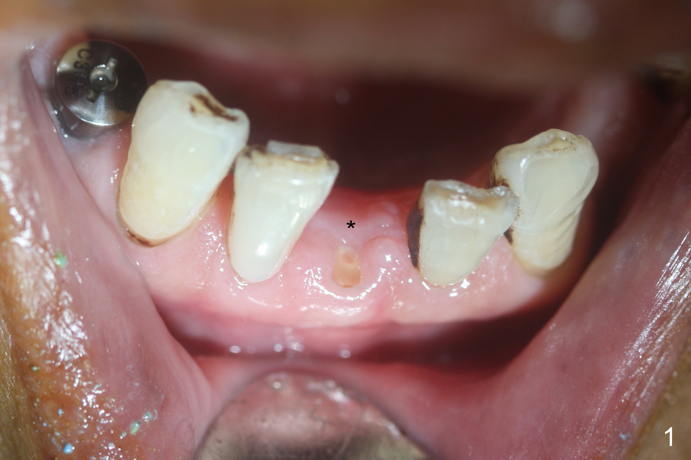
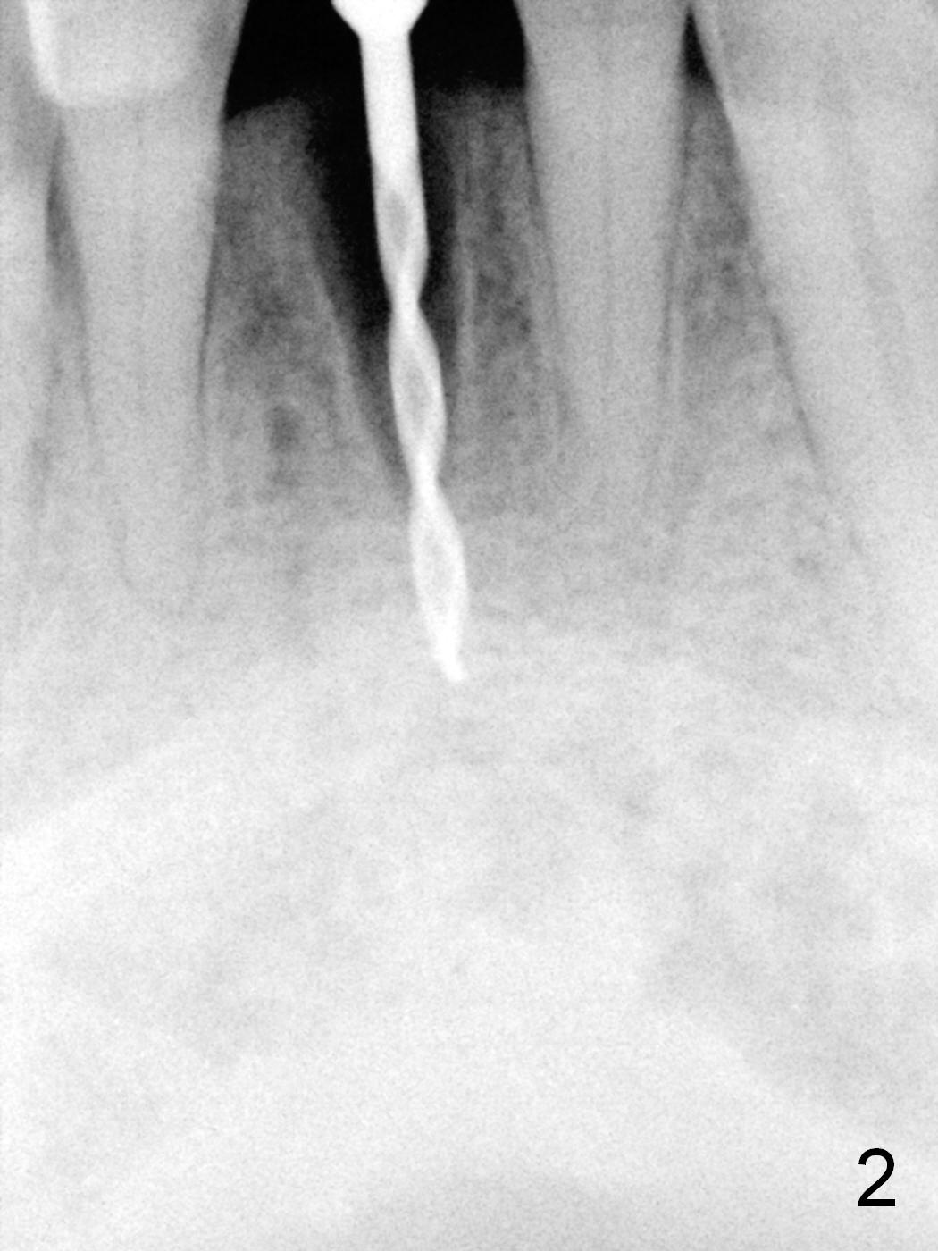
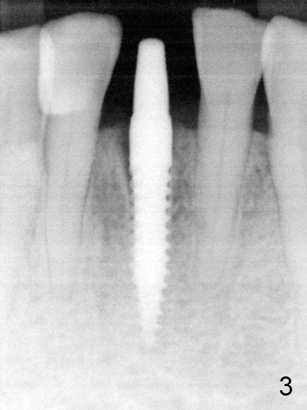
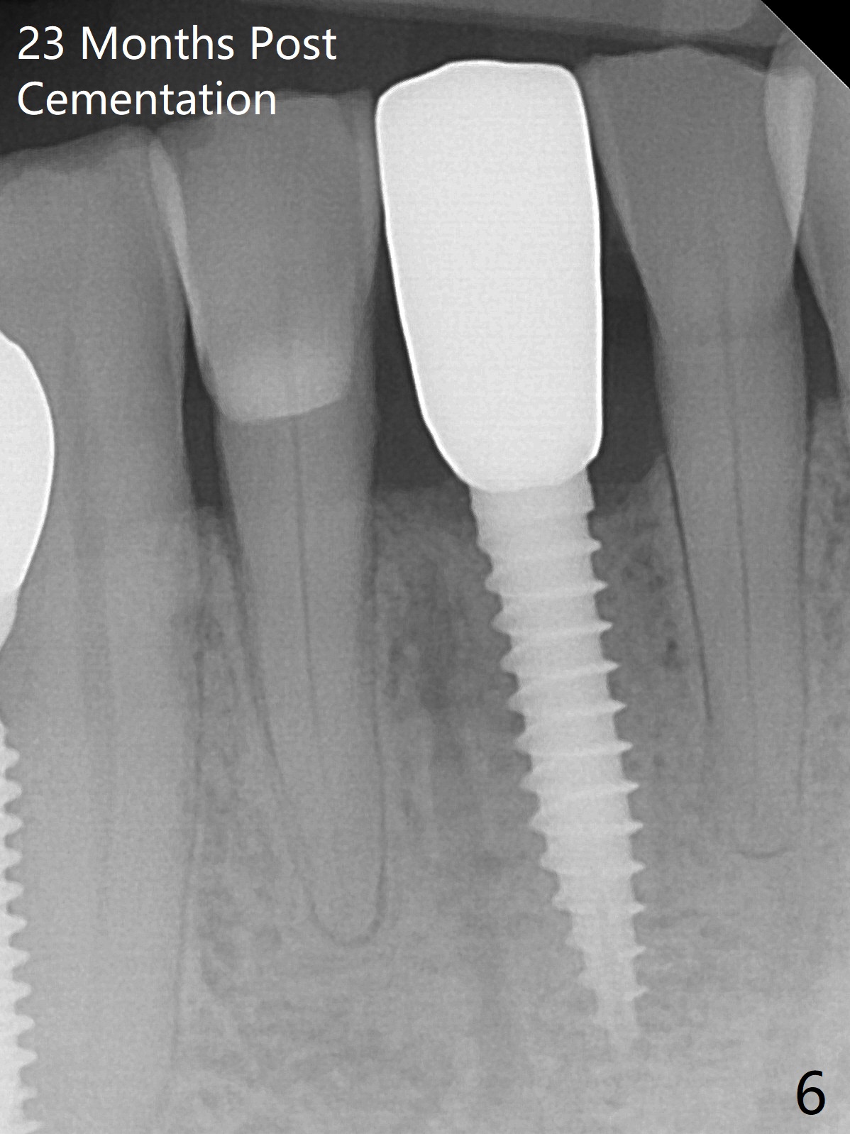
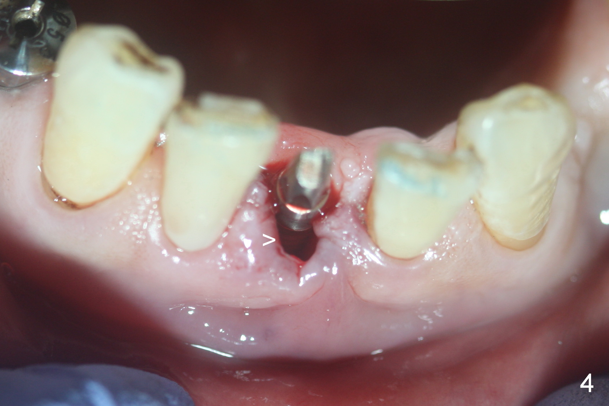
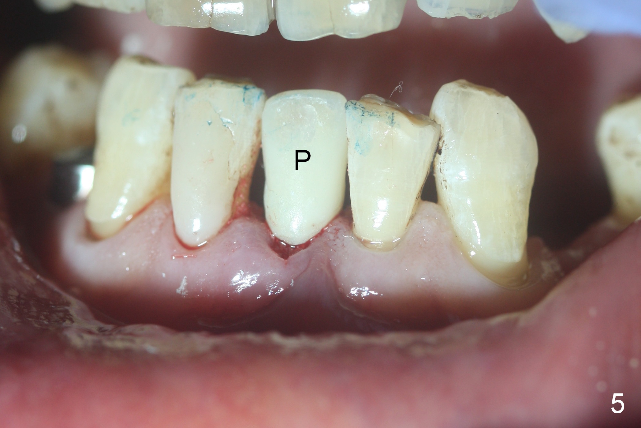
A few days after removal of the fractured crown, the residual root is partially covered by the lingual gingiva (Fig.1 *). The patient reveals that he has diabetes, which may be associated with re-placement of the implant at #28 (Fig.1). A 1.2 mm pilot drill is 3-4 mm apical to the socket (Fig.2). After increase in osteotomy depth by 2 mm, a 3x14 mm 1-piece implant is placed with insertion torque ~ 35 Ncm (Fig.3). After preparing the abutment (Fig.4), an immediate provisional is fabricated and allograft is placed in the buccal gap (Fig.4 >). Finally the provisional (Fig.5 P) is cemented to keep the graft in place. The permanent crown is cemented 4 months postop. Although the socket gap closes 23 months post cementation, there is 1-2 mm crestal bone loss (Fig.6). The latter may be related to the oversized implant (3 mm) relative to the small socket.
Return to Lower Incisor Immediate Implant
4
13
28
21
Xin Wei, DDS, PhD, MS 1st edition 05/03/2016, last revision 08/06/2018